This Journal Club highlights the landmark study that provided the first evidence of regeneration in mammals, demonstrating that neonatal mice can regenerate damaged hearts and influencing regenerative medicine until today.
Goat and sheep meat are highly regarded in the market for their unique flavor qualities. In this study, lipidomics technology was employed to compare the lipid differences in the longissimus dorsi muscle among 8-month-old Saanen dairy goats (DYG, n = 6), Shaanbei white cashmere goats (CAG, n = 6), and Tan sheep (TSH, n = 6). Electronic nose analysis was performed solely on the DYG samples, while the E-nose data for the CAG and TSH groups were referenced against previously published literature. The results indicated that aldehydes, ketones, methyl derivatives, and inorganic sulfides constituted the predominant stable volatile flavor compounds in the DYG muscle. Principal component analysis (PCA) and partial least square discriminant analysis (PLS-DA) revealed clear breed-dependent clustering and separation. Cluster analysis revealed that CAG and TSH groups exhibited highly similar lipid expression patterns, whereas the DYG group showed a distinct profile. Specifically, in the DYG group, SM (d18:1/20:3) and S1P (t17:0) were significantly increased, while most triglycerides (TG) were decreased and diglyceride DG (18:0/16:0) was increased. Additionally, multiple phosphatidylcholines (PC) and phosphatidylethanolamines (PE) were significantly reduced in the DYG group. This study revealed unique lipid signatures in the DYG group muscle compared with CAG and TSH groups, providing a theoretical basis for deciphering the lipid regulatory mechanisms underlying meat quality and flavor formation in goats and sheep.
N6-methyladenosine (m6A), a predominant and reversible modification of mammalian RNA, plays a critical role in regulating growth, development, and metabolism. While methyltransferase-like 14 (METTL14) is an essential component of the m6A methyltransferase complex, its specific function in regulating milk fat metabolism in dairy goats remains unexplored. This study therefore aimed to elucidate the role of METTL14 in lipid metabolism within dairy goat mammary epithelial cells (GMECs). METTL14 overexpression significantly promoted the synthesis of TAG (Triacylglycerol) and TC (Total cholesterol), as well as lipid droplet accumulation in GMECs. Furthermore, METTL14 upregulated CCAAT enhancer binding protein beta (CEBPB) expression at both the mRNA and protein levels by directly inducing m6A modification on its transcripts. Finally, we confirmed that m6A modification occurs specifically at site 1662 of CEBPB mRNA, and the "Readers" YTH N6-methyladenosine RNA binding protein F1 and F3 (YTHDF1/3) were found responsible for the m6A site recognition and interpretation. This study demonstrated that METTL14 facilitates lipid synthesis and deposition in GMECs. Mechanistically, METTL14 installs the m6A modification at site 1662 of CEBPB transcripts. This m6A mark is specifically recognized by the readers YTHDF1 and YTHDF3, which promote the translation of CEBPB mRNA, thereby upregulating its expression.
The growth of the global older population has necessitated the development of advanced models to investigate biological processes in aging and to explore viable anti-aging strategies. This review focuses on two objectives: First, to explore organoid technology as a paradigm for observing and intervening in human aging, and second, to examine the possible overlaps and crossroads between organoid-based biomedical paradigms and traditional Chinese medicine (TCM). Recent advancements in this field have increased the physiological relevance of organoids, incorporating both a dynamic microenvironment and multi-organ interactions. Such accomplishments have enhanced the accuracy of models of neurodegeneration, cardiovascular deterioration, metabolic diseases, and age-related conditions. In addition to pharmacological and genetic treatments, organoid models offer new opportunities to study well-known anti-aging techniques, such as those that rely on the TCM system, which aims to balance the body using natural substances with antioxidant and repair properties. Together, organoid-based aging research and mechanistic evaluation of TCM-derived compounds may help identify interventions that support healthy aging.
Supplemental Figure S2 describes that oxidative stress in microglia promotes the immunosuppressive functions of microglia.
Distinguishing tumor maintenance genes from initiation, progression, and passenger genes is critical for developing effective therapies. We employed a functional genomic approach using the Lazy Piggy transposon to identify tumor maintenance genes in vivo and applied this to sonic hedgehog (SHH) medulloblastoma (MB). Combining Lazy Piggy screening in mice and transcriptomic profiling of human MB, we identified the voltage-gated potassium channel KCNB2 as a candidate maintenance driver. KCNB2 governs cell volume of MB-propagating cells (MPCs), with KCNB2 depletion causing osmotic swelling, decreased plasma membrane tension, and elevated endocytic internalization of epidermal growth factor receptor (EGFR), thereby mitigating proliferation of MPCs to ultimately impair MB growth. KCNB2 is largely dispensable for mouse development and KCNB2 knockout synergizes with anti-SHH therapy in treating MB. These results demonstrate the utility of the Lazy Piggy functional genomic approach in identifying cancer maintenance drivers and elucidate a mechanism by which potassium homeostasis integrates biomechanical and biochemical signaling to promote MB aggression.
The remodeling of mammary glands during pregnancy is essential for initiating lactation. In dairy animals, the overlap of pregnancy and mammary involution triggers a unique process, regenerative remodeling, which is critical for extending lactation duration and enhancing milk production. Unlike the complete regression of lobuloalveolar structures during involution, the regenerative remodeling preserves alveolar structures and promotes rapid mammary gland renewal. However, the cellular and molecular mechanisms underlying such process remain elusive. Here, taking dairy goats (Capra hircus) as a ruminant model, we identified four luminal cell populations through single-cell RNA-sequencing and found a significant reduction in luminal hormone-responsive (LumHR) cells and an increase in luminal secretory precursors (LumSecP) during regenerative remodeling. A reduction of LumHR cells during regenerative remodeling is essential for promoting the accumulation of LumSecP. Goat mammary organoids and in vivo genetic ablation assays suggested that LumHR cells function as a crucial switch for the differentiation of LumSecP to LumSec cells through the prolactin receptor pathway. Furthermore, high levels of IRF1 inhibited while downregulation of IRF1 stimulated the proliferation of LumHR cells. We showed that IRF1 regulated the dynamics of LumHR cells through hormonal signaling targets, including ESRRB. Our findings identified a key cell type responsible for the dynamics of luminal lineages during regenerative remodeling in large mammals and highlighted the potential for accelerating tissue regeneration through targeted modulation of lineage stage-specific regulators.
Mammals display prominent diversity in the ability to regenerate damaged ear pinna, but the genetic changes underlying the failure of regeneration remain elusive. We performed comparative single-cell and spatial transcriptomic analyses of rabbits and mice recovering from pinna damage. Insufficient retinoic acid (RA) production, caused by the deficiency of rate-limiting enzyme Aldh1a2 and boosted RA degradation, was responsible for the failure of mouse pinna regeneration. Switching on Aldh1a2 or RA supplementation reactivated regeneration. Evolutionary inactivation of multiple Aldh1a2-linked regulatory elements accounted for the deficient Aldh1a2 expression upon injury in mice and rats. Furthermore, the activation of Aldh1a2 by a single rabbit enhancer was sufficient to improve ear pinna regeneration in transgenic mice. Our study identified a genetic switch involved in the evolution of regeneration.
Supplemental Figure S3 describes that inhibiting nuclear translocation of NR4A2 hampers immunosuppressive functions and promotes antigen presentation capacities of microglia.
Table S2 describes information of key research agents, data deposition and software versions.
Table S1 describes glioma patients and tissue samples information for evaluating the relationship of oxidative stress in microglia and prognosis
Supplemental Figure S7 describes that oxidative stress in microglia as an independent predictor for clinical outcome in glioma patients.
Supplemental Figure S6 describes that inhibition of NR4A2 or SQLE improves the therapeutic efficacy of immune checkpoint blockade.
Supplemental Figure S4 describes that NR4A2-knockdown affects lipid metabolism and promotes antigen presentation of microglia.
Table S4 describes list of literature-based immune marker panel selected to identity cell types of obtained clusters.
The short-lived African killifish Nothobranchius furzeri is an attractive genetic model for vertebrate aging and regeneration studies. The utilization of genetically modified animals is a common strategy for unveiling molecular mechanisms responsible for a biological phenomenon. Here, we report a highly efficient protocol for generating transgenic African killifish using the Tol2 transposon system, which creates random insertions in the genome. Transgenic vectors carrying gene-expression cassettes of interest and an eye-specific marker for transgene identification can be quickly assembled through Gibson assembly. The development of this new pipeline will facilitate transgenic reporter assays and gene-expression-related manipulations in African killifish.
Tumorigenesis involves transcriptomic alterations at both gene and isoform levels. Short-read RNA-seq struggles to capture isoform-level changes, but long-read RNA sequencing (lrRNA-seq) can address this limitation. We analyzed 144 tumor-normal tissue pairs from ten organs using lrRNA-seq. Our pan-cancer analysis revealed widespread gene expression convergence related to oncogenic dedifferentiation. However, isoform expression varied significantly across different organs and did not show a similar convergence. Isoform-level alterations, including aberrant (fusion) transcripts and altered isoform expression ratios, were prevalent in tumor transcriptomes and were partly due to changes in splicing factor expression. Many genes showed significant isoform expression ratio changes without corresponding gene expression differences between normal and tumor tissues. Our approach enabled a pan-cancer biomarker assessment system that values both gene and isoform alterations, leading to the identification of many potentially important but previously overlooked pan-cancer susceptibility genes.
Dexmedetomidine (DEX) is a highly selective α2-adrenoceptor agonist with sedative effects on sleep homeostasis. Oxytocin-expressing (OXT) neurons in the paraventricular nucleus (PVN) of the hypothalamus (PVNOXT) regulate sexual reproduction, drinking, sleep-wakefulness, and other instinctive behaviors. To investigate the effect of DEX on the activity and signal transmission of PVNOXT in regulating the sleep-wakefulness cycle. Here, we employed OXT-cre mice to selectively target and express the designer receptors exclusively activated by designer drugs (DREADD)-based chemogenetic tool hM3D(Gq) in PVNOXT neurons. Combining chemogenetic methods with electroencephalogram (EEG) /electromyogram (EMG) recordings, we found that cannula injection of DEX in PVN significantly increased the duration of non-rapid eye movement (NREM) sleep in mice. Furthermore, the chemogenetic activation of PVNOXT neurons using i.p. injection of clozapine N-oxide (CNO) after cannula injection of DEX to PVN led to a substantial increase in wakefulness. Electrophysiological results showed that DEX decreased the frequency of action potential (AP) and the spontaneous excitatory postsynaptic current (sEPSC) of PVNOXT neurons through α2-adrenoceptors. Therefore, these results identify that DEX promotes sleep and maintains sleep homeostasis by inhibiting PVNOXT neurons through the α2-adrenoceptor.
 Xin Wang (王鑫)合作论文数Department of Surgery, Faculty of Medicine, The Chinese University of Hong Kong21
 Shengtao Zhou (周圣涛)合作论文数West China Second University Hospital, Sichuan University;State Key Laboratory of Biotherapy, Sichuan University14
 Linjie Zhao (赵林杰 Zhao Linjie)合作论文数13
 Zhengnan Yang合作论文数12
 Hao Huang (黄昊)合作论文数School of Biological Science and Medical Engineering, Southeast University10
 Michael Taylor合作论文数Department of Medical Biophysics, Temerty Faculty of Medicine, University of Toronto;Departments of Pediatrics, Baylor College of Medicine;Texas Children’s Hospital10
 Alejandro Sánchez Alvarado合作论文数The Graduate School, Stowers Institute for Medical Research10
 Lunzhi Dai (戴伦治)合作论文数State Key Laboratory of Biotherapy, Sichuan Univerisity10Chaoxin Xiao合作论文数9
 Huairui Yuan (袁怀瑞)合作论文数Center for Excellence in Molecular Cell Science, Chinese Academy of Sciences;University of Chinese Academy of Sciences9Zengpanpan Ye合作论文数9Jing Yue合作论文数9Fan Fei合作论文数9Liang Huang合作论文数9
 Ryan C Gimple合作论文数Department of Medicine, University of California San Diego9Yanqiu Gong合作论文数9Xiaolin Ai合作论文数9Peidong Zhang合作论文数9Yang Kailin合作论文数Department of Radiation Oncology, Dana-Farber Cancer Institute;Department of Radiation Oncology, Dana-Farber Cancer Institute9
 Olivier Saulnier合作论文数From Sorbonne Universités, UPMC Univ Paris 06;Institut du Cerveau et de la Moelle épinière (ICM) (B.B., Y.M.), Plateforme de Génotypage Séquençage, Paris;and Groupe Hospitalier Pitié-Salpêtrière, Groupe Hospitalier Pitié Salpêtrière;National Neurological Institute C. Mondino (A.-L.D.S.),;Department of Neuroscience (M.R., University of Padova;and Unit of Molecular Neuro-Oncology (R.P., G.F.),, Fondazione IRCCS Carlo Besta;UPMC Univ Paris 06, From Sorbonne Universités9
 Chao You (游潮)合作论文数Department of Neurosurgery,West China Hospital,Sichuan University9
 Jianguo Xu (徐建国)合作论文数West China Hospital, Sichuan University9Xiaoling Liao合作论文数8
 Xiuxing Wang (汪秀星)合作论文数School of Basic Medical Sciences, Nanjing Medical University8Xiaoxiao Wan合作论文数Washington Univ, Sch Med, Dept Pathol & Immunol, St Louis, MO 63110 USA8
 Jeremy Rich合作论文数School of Medicine, University of North Carolina8
 Zhixin Qiu (邱志新)合作论文数8Young Ha Ahn合作论文数8
 John I. Yoder合作论文数Department of Plant Sciences, University of California, Davis7
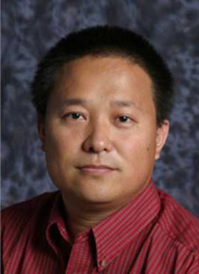 Feng Gao合作论文数Mary Culver Department of Surgery, Washington University School of Medicine in St. Louis5Xin Duan (段鑫)合作论文数5Augusto Ortega Granillo合作论文数4
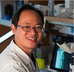 Yongfu Wang合作论文数Stowers Institute for Medical Research4Lian Xu (徐炼)合作论文数4
 Xia Zhao (赵霞)合作论文数West China Second University Hospital, Sichuan University4
 Jun Luo (罗军)合作论文数College of Animal Science and Technology, Northwest A&F University4Tao Yi合作论文数4Juqing Zhang合作论文数3Nianxin Zhou合作论文数3Qiuhong Shen合作论文数3Wei Yue合作论文数3Qiaoyan Shen合作论文数3QiLian Yang (杨其莲)合作论文数3Lau Bonnie合作论文数3
 Anne Brunet合作论文数Paul F. Glenn Laboratories for the Biology of Aging, Stanford University;Department of Genetics, Stanford University3
 Xiaolong wu合作论文数3Shuang Huang合作论文数3Yan Shun合作论文数3
 Eric D Ross合作论文数3




