ABSTRACTBackgroundOur study evaluated long‐term morbidities in patients and compared subjective morbidities to those measurable objectively.MethodsPatients completed a questionnaire regarding long‐term morbidity, filled out the Pain Sensitivity Questionnaire by Ruscheweyh et al. and were examined physically.ResultsEighty‐two patients were included in the study, 31 patients returned to the clinic for further evaluation and testing. The most common morbidities were weakened dorsiflexion of the great toe (34 patients), reduced ambulatory status (33), and muscle weakness (31). A significant correlation (p < 0.01) was found between higher pain intensity and high pain sensitivity scores. However, donor‐site morbidities were not significantly associated with pre‐existing comorbidities like smoking, diabetes, or age over 70. Subjective morbidities were reported 2.3 times more frequently than objectively measured ones.ConclusionsOur findings suggest a notable discrepancy between subjective and objective morbidities, highlighting the importance of considering patients' subjective experiences when assessing postoperative outcomes and rehabilitation progress.
The presented study aimed to evaluate the effect of mandibular protrusion with a temporarily applied mandibular advancement device (MAD) on the posterior airway space and to determine a reliable metric constant based on a three-dimensional computed tomography (CT) evaluation. The study population consisted of patients with oral squamous cell carcinoma who were treated at least six months prior to the follow-up CT in supine position. Each patient received an individually adjusted MAD that was temporarily applied with three different protrusion distances (P0 = 0 mm, P4 = 4 mm, and P8 = 8 mm) during follow-up CT. The open-source software Slicer was used to calculate three parameters: minimum cross-sectional area (minCSA), mean cross-sectional area (meanCSA), and volume. The results showed a significant increase for all three parameters. The minCSA increased as follows: P0 = 236.4 mm2 ± 192.2; P4 = 309.2 mm2 ± 235.4; and P8 = 430.6 mm2 ± 265.3. The meanCSA increased significantly (p < 0.001) in all protrusion steps and all parts of the pharynx. The volume changed as follows: P0 = 24.0 cm3 ± 5.0; P4 = 29.6 cm3 ± 18.1; and P8 = 33.6 cm3 ± 19.0. The minCSA increased by 24.9 mm2 ± 13.0 per millimeter mandibular protrusion. The results are interesting for both conservative and surgical therapy and could find future application in dental, orthodontic, and combined oral surgical therapy. With the results of this study, surgeons and dentists may better predict the change of PAS parameter in order to better prepare for orthognathic surgery. They also could ensure the right protrusion distance for mandibular advancement devices in the case of obstructive sleep apnea.
IntroductionMandibular reconstruction with the free fibula flap (FFF) has become a standardized procedure. The situation is different with oral rehabilitation, so the purpose of this study was to investigate the frequency of implant placement and prosthetic restoration. Additionally, the patients’ situation, motivation, and treatment course were structurally assessed.Materials and methodsAll cases between January 2013 and December 2018 that underwent mandibular reconstruction in our department with a free fibula flap and gave written informed consent to participate were interviewed with two structured questionnaires about their restoration and quality of life. Additionally, medical records, general information, status of implants and therapy, and metric analyses of the inserted implants were performed.ResultsIn total 59 patients were enrolled and analyzed in this monocentric study. Overall, oral rehabilitation was achieved in 23.7% at the time of investigation. In detail, implants were inserted in 37.3% of patients and showed an 83.3% survival of dental implants. Of these implanted patients, dental implants were successfully restored with a prosthetic restoration in 63.6. Within this subgroup, satisfaction with the postoperative aesthetic and functional result was 79.9% and with the oral rehabilitation process was 68.2%. Satisfaction with the implant-borne prosthesis was 87.5%, with non-oral-squamous-cell-carcinoma patients being statistically significantly more content with the handling (p=0.046) and care (p=0.031) of the prosthesis.DiscussionDespite the well-reconstructed bony structures, there is a need to increase the effort of achieving oral rehabilitation, especially looking at the patient’s persistent motivation for the procedure.
Currently, maxillary sinus floor (SF) elevation is based on off-the-shelf allogeneic, xenogeneic or synthetic bone augmentation materials (BAM) that are implanted via an open lateral sinus wall approach (OSFE). However, this invasive method is associated with postoperative complications caused by an inadequate blood supply of the alveolar ridge. Balloon-assisted procedures are minimal invasive alternatives with lower complication rates. The aim was to evaluate local new bone (NB) formation in the SF following the application of a particulate BAM (Easy graft) via two different SF elevation techniques in a split mouth mini-pig sinus augmentation model. Seven adult Goettingen minipigs were used for evaluation of a biphasic ceramic (PLGA/ß-TCP) BAM in the elevated SF region. Treatments were randomized to the contralateral sinus sites and included two procedures: OSFE (control group) versus minimally invasive SF elevation by a balloon-lift-control system (BLC) (treatment group). The animals were euthanized after 28 and 56 days for analysis of new bone (NB) formation. The biphasic synthetic BAM implanted via BLC increased more NB formation (5.2 ± 1.9 mm and 4.9 ± 1.6 mm vs. 2.6 ± 0.5 mm) and osseointegration of the particles (18.0 ± 6.0
BackgroundMandibular reconstruction with the fibula free flap (FFF) is performed freehand, CAD/CAM-assisted, or by using partially adjustable resection/reconstruction aids. The two latter options represent the contemporary reconstructive solutions of the recent decade. The purpose of this study was to compare both auxiliary techniques with regard to feasibility, accuracy, and operative parameters.Methods and materialsThe first twenty consecutively operated patients requiring a mandibular reconstruction (within angle-to-angle) with the FFF using the partially adjustable resection aids between January 2017 and December 2019 at our department were included. Additionally, matching CAD/CAM FFF cases were used as control group in this cross-sectional study. Medical records and general information (sex, age, indication for surgery, extent of resection, number of segments, duration of surgery, and ischemia time) were analyzed. In addition, the pre- and postoperative Digital Imaging and Communications in Medicine data of the mandibles were converted to standard tessellation language (.stl) files. Conventional measurements – six horizontal distances (A–F) and temporo-mandibular joint (TMJ) spaces – and the root mean square error (RMSE) for three-dimensional analysis were measured and calculated.ResultsIn total, 40 patients were enrolled (20:20). Overall operation time, ischemia time, and the interval between ischemia time start until end of operation showed no significant differences. No significant difference between the two groups were revealed in conventional measurements of distances (A–D) and TMJ spaces. The Δ differences for the distance F (between the mandibular foramina) and the right medial joint space were significantly lower in the ReconGuide group. The RMSE analysis of the two groups showed no significant difference (p=0.925), with an overall median RMSE of 3.1 mm (2.2–3.7) in the CAD/CAM and 2.9 mm (2.2–3.8) in the ReconGuide groups.ConclusionsThe reconstructive surgeon can achieve comparable postoperative results regardless of technique, which may favor the ReconGuide use in mandibular angle-to-angle reconstruction over the CAD/CAM technique because of less preoperative planning time and lower costs per case.
BACKGROUND:The aim of this study was to investigate the clinical course and to redefine an optimized algorithm for OE cases until epithetic restoration. METHODS:Indication, defect type according to Kesting, reconstructive technique, incidence of postoperative complications and peri-implantitis, patients' quality of life, timing of periorbital implant insertion, incidence of and interval to implant loss, and time until epithetic restoration were analyzed in 43 patients. RESULTS:A significant correlation was detected between wound dehiscence and defect type. Out of 24 patients, 7 were implanted secondarily with a median time interval of 399 days (270-2015) after OE. Eleven out of 83 placed implants were lost in 8 patients with a median time interval of 586 days (264-4485) after insertion. The majority of epithesis carriers had no or few restrictions in their quality of life. CONCLUSIONS:We recommend our modified treatment algorithm to further improve and shorten the clinical course.
BACKGROUND:Flap reconstruction is commonly used in advanced tongue carcinoma in order to compensate for the loss of tongue tissue and function. Surprisingly, a large number of reconstructed early-stage tongue cancer can be found. Survival or functional benefits in these cases remain unclear.METHODS:A retrospective data analysis of 384 surgically treated tongue carcinoma patients was conducted aiming to find epidemiologic and survival differences between patients with (n = 158) or without flap reconstruction (n = 226). A prospective functional analysis was performed on 55 early-stage tongue cancer patients, 33 without and 22 with radial-forearm flap reconstruction, focusing on post-therapeutic swallowing function as the primary endpoint, speech as the secondary endpoint, xerostomia, quality of life, and mouth opening.RESULTS:Consistent with the current literature, we demonstrated the significantly more frequent use of flap grafts in advanced tongue carcinomas. For the first time, we depicted a higher female ratio in the reconstructed group (p = 0.02). There were no significant differences in survival or functional outcomes between the groups. The none-reconstructed group showed more frequent use of adjuvant C/RT despite presenting fewer N+ stages.CONCLUSIONS:The higher female ratio in the reconstruction group is plausible due to the anatomically smaller oral cavity and relatively larger carcinoma in women. A higher presence of close margins in the none-reconstruction group may explain the more frequent use of adjuvant C/RT. Since we found no survival or functional differences between the groups, we propose a critical approach toward flap reconstruction in T1/2 tongue carcinoma. At the same time, proportional aspects and adequate resection margins should be taken into account.
BackgroundMicrovascular reconstruction remains challenging in previously operated and irradiated patients, especially when double flaps seem to be the only solution due to osteoradionecrosis. An alternative reconstructive option could be microvascular anastomosis to the temporal vessels to avoid the obligatory cervical incision.Methods and MaterialsAll consecutive cases between January 2013 and 2020 that underwent either mandibular resection and reconstruction with a free fibula flap (FFF) and another soft tissue flap (group I) or pure intraoral resection and reconstruction with an FFF or radial forearm flap (RFF) with temporal microvascular anastomosis (group II) were included. Patients’ general information, indication and extent of surgery, time of ischemia, time of total surgery, and duration of hospital stay as well as incidence of complications were retrospectively recorded and analyzed.ResultsSeventeen (group I) and 11 (group II) cases were included. In group I, FFF was combined with RFF (n = 9), anterolateral thigh flap (ALT, n = 7), or latissimus dorsi flap (n = 1). Group II consisted of six FFFs and five RFFs. Operation time and hospitalization duration were significantly shorter in group II (p < 0.001 and p = 0.025), whereas ischemic time of FFF was significantly shorter in group I (p = 0.002). All patients in group I required a tracheostomy, while only four cases in group II did (p = 0.004). The complication rate regarding hematoma removal, revision of anastomosis, flap loss, delirium, sepsis, pleural effusion, pneumonia, and pulmonary artery embolism showed no significant differences between the two groups.ConclusionsThe superficial temporal vessels served as versatile recipient vessels for intraoral mandibular and soft tissue reconstruction and led to reduced operation time, hospitalization duration, and indication for a primary tracheostomy. Thus, this approach may help to avoid cervical incision for reconstruction in irradiated patients.
Cracked tooth syndrome (CTS) is a common clinical finding for teeth, it affects about 5% of all adults each year. The finding of CTS is favored by several risk factors such as restorations, bruxism, occlusion habits, and age. Treatment options range, depending on the severity, from no treatment at all to tooth extraction. Early diagnosis of CTS is crucial for optimal treatment and symptom reduction. There is no standard procedure for an evidence-based diagnosis up to date. The diagnosis is a challenge by the fact that the symptoms, including pain and sensitivity to temperature stimuli, cannot be clearly linked to the disease. Commonly used visual inspection does not provide in-depth information and is limited by the resolution of human eyes. This can be overcome by magnifying optics or contrast enhancers, but the diagnosis will still strongly rely on the practicians experience. Other methods are symptom reproduction with percussions, thermal pulp tests or bite tests. Dental X-ray radiography, as well as computed tomography, rarely detect cracks as they are limited in resolution. Here, we investigate X-ray dark-field tomography (XDT) for the detection of tooth microcracks. XDT simultaneously detects X-ray small-angle scattering (SAXS) in addition to the attenuation, whereas it is most sensitive to the micrometer regime. Since SAXS originates from gradients in electron density, the signal is sensitive to the sample morphology. Microcracks create manifold interfaces which lead to a strong signal. Therefore, it is possible to detect structural changes originating from subpixel-sized structures without directly resolving them. Together with complementary attenuation information, which visualizes comparatively large cracks, cracks are detected on all length-scales for a whole tooth in a non-destructive way. Hence, this proof-of principle study on three ex-vivo teeth shows the potential of X-ray scattering for evidence-based detection of cracked teeth.
Background Mandibular reconstruction is conventionally performed freehand, CAD/CAM-assisted, or by using partially adjustable resection aids. CAD/CAM-assisted reconstructions are usually done in cooperation with osteosynthesis manufacturers, which entails additional costs and longer lead time. The purpose of this study is to analyze an in-house, open-source software-based solution for virtual planning. Methods and Materials All consecutive cases between January 2019 and April 2021 that underwent in-house, software-based (Blender) mandibular reconstruction with a free fibula flap (FFF) were included in this cross-sectional study. The pre- and postoperative Digital Imaging and Com munications in Medicine (DICOM) data were converted to standard tessellation language (STL) files. In addition to documenting general information (sex, age, indication for surgery, extent of resection, number of segments, duration of surgery, and ischemia time), conventional measurements and three-dimensional analysis methods (root mean square error [RMSE], mean surface distance [MSD], and Hausdorff distance [HD]) were used. Results Twenty consecutive cases were enrolled. Three-dimensional analysis of preoperative and virtually planned neomandibula models was associated with a median RMSE of 1.4 (0.4–7.2), MSD of 0.3 (-0.1–2.9), and HD of 0.7 (0.1–3.1). Three-dimensional comparison of preoperative and postoperative models showed a median RMSE of 2.2 (1.5–11.1), MSD of 0.5 (-0.6–6.1), and HD of 1.5 (1.1–6.5) and the differences were significantly different for RMSE (p < 0.001) and HD (p < 0.001). The difference was not significantly different for MSD (p = 0.554). Three-dimensional analysis of virtual and postoperative models had a median RMSE of 2.3 (1.3–10.7), MSD of -0.1 (-1.0–5.6), and HD of 1.7 (0.1–5.9). Conclusions Open-source software-based in-house planning is a feasible, inexpensive, and fast method that enables accurate reconstructions. Additionally, it is excellent for teaching purposes.
In this study, top ology optimized, patient specific os teosynthesis plates (TOPOS-implants) are evaluated for the mandibular reconstruction using fibula segments. These shape optimized implants are compared to a standard treatment with miniplates (thickness: 1.0 mm, titanium grade 4) in biomechanical testing using human cadaveric specimen. Mandible and fibula of 21 body donors were used. Geometrical models were created based on automated segmentation of CT-scans of all specimens. All reconstructions, including cutting guides for osteotomy as well as TOPOS-implants, were planned using a custom-made software tool. The TOPOS-implants were produced by electron beam melting (thickness: 1.0 mm, titanium grade 5). The fibula-reconstructed mandibles were tested in static and dynamic testing in a multi-axial test system, which can adapt to the donor anatomy and apply side-specific loads. Static testing was used to confirm mechanical similarity between the reconstruction groups. Force-controlled dynamic testing was performed with a sinusoidal loading between 60 and 240 N (reconstructed side: 30% reduction to consider resected muscles) at 5 Hz for up to 5 · 10 5 cycles. There was a significant difference between the groups for dynamic testing: All TOPOS-implants stayed intact during all cycles, while miniplate failure occurred after 26.4% of the planned loading (1.32 · 10 5 ± 1.46 · 10 5 cycles). Bone fracture occurred in both groups (miniplates: n = 3, TOPOS-implants: n = 2). A correlation between bone failure and cortical bone thickness in mandible angle as well as the number of bicortical screws used was demonstrated. For both groups no screw failure was detected. In conclusion, the topology optimized, patient specific implants showed superior fatigue properties compared to miniplates in mandibular reconstruction. Additionally, the patient specific shape comes with intrinsic guiding properties to support the reconstruction process during surgery. This demonstrates that the combination of additive manufacturing and topology optimization can be beneficial for future maxillofacial surgery.
Die klinische S3-Leitlinie „Diagnostik und Therapie des Mundhöhlenkarzinoms“ stellt seit ihrer Etablierung 2012 eine Handlungsgrundlage dar, um die bestmögliche Prognose und Lebensqualität für jede individuelle Befundkonstellation zu erreichen. Um dem stetigen Erkenntnisgewinn Rechnung zu tragen, ist nach dem Regelwerk der Arbeitsgemeinschaft der Wissenschaftlichen Medizinischen Fachgesellschaften (AWMF) eine Aktualisierung der Leitlinie nach 5 Jahren erforderlich. Ziel dieser Aktualisierung ist es, Evidenz und Nutzen bestehender Empfehlungen zu überprüfen, aktuelle Änderungen der klinischen Praxis mit der größtmöglichen Evidenz zu bewerten und auf dem Boden wissenschaftlich gesicherter Erkenntnisse erreichte Fortschritte mit entsprechenden Empfehlungen in die Handlungsgrundlage der Leitlinie zu übernehmen. Im November 2017 trat die Leitliniengruppe unter Federführung der Deutschen Gesellschaft für Mund‑, Kiefer- und Gesichtschirurgie (DGMKG) und mit Förderung im Rahmen des Leitlinienprogramms Onkologie erneut zusammen, um die Aktualisierung auf den Weg zu bringen. Zu von der Leitliniengruppe priorisierten Schlüsselfragen erfolgte eine systematische Literaturrecherche, bei der relevante Studien mittels Titel‑/Abstract- sowie Volltext-Screening identifiziert, kritisch bewertet und in Evidenztabellen zusammengefasst wurden. Für den Update-Prozess wurden Fragen zur Rolle des Wächterlymphknotens, zur Erweiterung der zervikalen Halslymphknotenausräumung („neck dissection“, ND) bei Lymphknotenbefall in Level IIb oder III, eines möglichen „Wait-and-see-Konzepts“ bei cT1/2-Karzinomen des Oberkiefers, eines neoadjuvanten Therapiekonzepts bei cT3/4 und zur Frage des Vorgehens bezüglich einer adjuvanten Radio(chemo)therapie bei pT1/2pN1-Situation einer systematischen Literaturrecherche unterzogen. Darüber hinaus wurden Fragen zur der Indikation der PET/CT, dem Einsatz der CAD/CAM-Technologie im Rahmen der knöchernen Rekonstruktion sowie der Differenzialindikation für die Panendoskopie und der Immuntherapie in Arbeitsgruppen geprüft. Alle bereits bestehenden Statements und Empfehlungen wurden primär auf Änderungsbedarf geprüft; geänderte und neue Statements und Empfehlungen wurden mittels formaler Konsensustechniken (nominaler Gruppenprozess, Konsensuskonferenz) überprüft und abgestimmt. Es wurden 88 Statements oder Empfehlungen geprüft und bestätigt sowie 21 modifiziert oder gänzlich neu hinzugefügt. Die Indikation zur ND beim invasiven Mundhöhlenkarzinom bleibt trotz neuer Studien hinsichtlich der Bildgebung und Sentinelbiopsie nach wie vor bestehen und kann nur bei sehr oberflächlichen cT1cN0-Karzinomen des Oberkiefers durch eine engmaschige Nachsorge ersetzt werden. Auch kann bei negativem FDG-PET-Befund nach primärer Radio(chemo)therapie auf eine Salvage-ND verzichtet werden. Eine Differenzialindikation zwischen modifiziert radikaler und supraomohyoidaler ND bei cN1-Befund ist aufgrund vorhandener Studien weiterhin nicht zu begründen. Die Indikation zur adjuvanten Radio(chemo)therapie bei pN1-Befund ist optional. Die Anwendung eines Checkpoint-Inhibitors wird bei geeigneten Patienten anstelle des bisher favorisierten EXTREME-Schemas als Erstlinientherapie beim rezidivierenden oder metastasierenden Plattenepithelkarzinom der Mundhöhle empfohlen. Die Schnittstellen von MKG, HNO, Strahlentherapie und Onkologie wurden bestätigt. Die neue TNM-Klassifikation und Einteilung der Tumorstadien wurden eingearbeitet. Zur prospektiven Erfassung der Häufigkeit von R1-Situationen bei kurativ intendierter Resektion wurde ein neuer Qualitätsindikator eingeführt. Der Qualitätsindikator zur Erfassung der prätherapeutischen HNO-Vorstellung konnte wegen der bereits erreichten konsequenten vollständigen Umsetzung wegfallen. Durch ein strukturiertes, unabhängig von der Deutschen Krebsgesellschaft gefördertes Update-Verfahren wurde die bestehende Leitlinie nicht nur vollständig aktualisiert, sondern besonders zu Fragen der multimodalen Therapie auch weiterentwickelt. Aufgrund einer fehlenden oder nicht ausreichenden Datenlage konnten allerdings zahlreiche Fragen nach wie vor lediglich auf dem Boden eines Expertenkonsenses beantwortet werden. Die konsequente Durchführung prospektiver Therapiestudien ist zur zukünftigen Weiterentwicklung der Leitlinie daher zwingend erforderlich. Entscheidend für den Behandlungserfolg bleibt die interdisziplinäre Therapieplanung und -durchführung unter Berücksichtigung der individuellen Befundkonstellation und des Patientenwunsches.
Objectives Mandibular reconstruction after segmental mandibulectomy can be challenging without virtual surgical planning and osteotomy guides. The purpose of this study was to analyze anatomic parameters to facilitate the evaluation of ideal fibula wedge osteotomies to reconstruct the neomandibula in a simple and cost-effective manner without the need for preoperative virtual planning. Materials and methods Computed tomography scans were acquired from randomly selected patients, and all images were obtained from routine clinical diagnostics, e.g., tumor staging, or preoperatively before reconstruction. Data was used to calculate stereolithographic models of the mandible for length and angle measurements. Statistical analysis was performed ( p < 0.05). Results CT scans of 100 patients were analyzed: 39 were female and 61 were male patients, mean age was 59.08a. The mandibular arch angle proved to be constant with 241.07 ± 2.39°. The outside B-segment length was 80.05 ± 5.16 mm; the anterior S-segment length was 27.69 ± 3.16 mm. The angle of the mandibular arch showed differences in means ( p = 0.004) between age groups, but effect was proved low. No relevant statistical significances were detected. Conclusions The development of a mandible reconstruction template tool would benefit the majority of head and neck patients, which is due to a constant mandibular arch angle and symphysis segment length throughout the general patient population, allowing the mimicking of a harmonic mandibular arch with up to three fibula segments. Clinical relevance The developed mandible reconstruction template tool can facilitate the fibula wedge osteotomies necessary for reconstruction of an ideal neomandibula providing a novel approach which is simple and cost-effective.
Combined scalp and skull deficiency due to malignant scalp tumors or sequelae of intracranial surgery present challenging entities for both neurosurgeons and reconstructive treatment. In complex cases, an interdisciplinary approach is needed between neurosurgeons and cranio-maxillofacial surgeons. We present a considerably large series for which we identify typical complications and pitfalls and provide evidence for the importance of an interdisciplinary algorithm for chronic wound healing complications and malignomas of the scalp and skull. We retrospectively reviewed all patients treated by the department of neurosurgery and cranio-maxillofacial surgery at our hospital for complex scalp deficiencies and malignant scalp tumors affecting the skull between 2006 and 2019, and extracted data on demographics, surgical technique, and perioperative complications. Thirty-seven patients were treated. Most cases were operated simultaneously (n: 32) and 6 cases in a staged procedure. Nineteen patients obtained a free flap for scalp reconstruction, 15 were treated with local axial flaps, and 3 patients underwent full thickness skin graft treatment. Complications occurred in 62% of cases, mostly related to cerebrospinal fluid (CSF) circulation disorders. New cerebrospinal fluid (CSF) disturbances occurred in 8 patients undergoing free flaps and shunt dysfunction occurred in 5 patients undergoing local axial flaps. Four patients died shortly after the surgical procedure (perioperative mortality 10.8%). Combined scalp and skull deficiency present a challenging task. An interdisciplinary treatment helps to prevent severe and specialty-specific complications, such as hydrocephalus. We therefore recommend a close neurological observation after reconstructive treatment with focus on symptoms of CSF disturbances.
Background: Temporary tracheotomies are often used in oral microvascular flap reconstruction surgery to secure postoperative airway management and avoid emergency tracheotomies. Even when planned electively, a tracheotomy can cause severe and life-threatening complications. The aim of this study was to evaluate the complications of tracheotomies performed on oral cancer patients with microvascular flap reconstructions and differentiated patterns, which could lead to postoperative complications. Methods: 150 patients, treated in the Department of Oral and Maxillofacial Surgery from March 2017 to August 2018, were included in this study. Patient records and perioperative data were analysed and the following specific items were evaluated: time after surgery until removal of the tracheal cannula, complications, cause and point of time of reinsertion of the cannula, anticoagulative treatment, ASA grade (American Society of Anaesthesiologists), TNM stage, and patient-specific data. Results: 30 patients (20%) developed tracheotomy-associated complications, most commonly pneumonia (50%). There was a significant correlation between the time period until removal of the cannula and the occurrence of complications such as pneumonia and bleeding. Conclusion: The results of our study lead us to recommend continuing to perform temporary tracheotomies in oral cancer surgery with microvascular flap reconstruction. The overall complication rate is low and postoperative airway management can be performed in a safe and controlled manner. Nevertheless, the time period for the inserted cannula should be kept as short as possible. (C) 2019 European Association for Cranio-Maxillo-Facial Surgery. Published by Elsevier Ltd. All rights reserved.
Reconstructions of complex scalp after ablative resection or by post-traumatic tissue loss, can present difficulties regarding recipient vessel selection, functional, and aesthetic outcome. The harvesting method for many microvascular free flaps requires a need for changing patients position during surgery and makes a simultaneous interdisciplinary two-team approach complicated, which is a major disadvantage regarding safety and operation time. The ideal flap for scalp reconstruction has yet to be described, although the microvascular latissimus dorsi flap is frequently referred to as the first choice in this context, especially after resection of large defects. The purpose of this study is to compare two different microvascular free flaps for a simultaneous scalp reconstruction in an interdisciplinary two-team approach applying a standardized algorithm. All consecutively operated complex scalp defects after ablative surgery from April 2017 until August 2018 were included in this retrospective study. The indications were divided into neoplasm or wound healing disorder. Two microvascular flaps (latissimus dorsi or parascapular flap) were used to cover the soft tissue component of the resulting defects. Seventeen patients met the inclusion criterion and were treated in an interdisciplinary two-team approach. Skull reconstruction with a CAD/CAM implant was performed in 10 cases of which four were in a secondary stage. Nine patients received a parascapular flap and eight patients were treated with latissimus dorsi flap with split thickness skin graft. Anastomosis was performed with no exception to the temporal vessels. One parascapular flap had venous insufficiency after 1 week followed by flap loss. One latissimus dorsi flap had necrosis of the serratus part of the flap. All other flaps healed uneventful and could be further treated with adjuvant therapy or CAD/CAM calvarial implants. Regarding overall complications, flap related complications, flap loss, and inpatient stay no statistical differences were seen between the diagnosis or type of reconstruction. The parascapular flap seems to be a good alternative for reconstruction of complex tumor defects of the scalp besides the latissimus dorsi flap. Stable long-term results and little donor site morbidity are enabled with good aesthetic outcomes and shorter operation time in an interdisciplinary two-team approach.
INTRODUCTION:Treatment decisions for fractures of the orbital floor are based on clinical appearance, ophthalmological examination, and computed tomography (CT) scans. In extensive fractures, decisions are easily made between conservative and surgical treatment. However, objective parameters are rare in inconclusive cases.MATERIALS AND METHODS:Our retrospective study included 106 patients with unilateral isolated orbital floor fractures. Correlations between preoperative ophthalmological examinations and specific CT parameters were performed.RESULTS:The defect size of the fracture appeared to be significantly associated with the presence of diplopia. CT-morphological parameters and preoperative ophthalmological results showed statistical significance for diplopia and incarceration of inferior rectus muscle (IRM), diplopia and displacement of IRM, decreased mobility and incarceration of IRM, and decreased mobility and displacement of IRM.DISCUSSION:Our clinical assessment scheme for CT scans of orbital floor fractures is aimed at facilitating treatment decision making using four CT-based variables. As critical size defects of the orbital floor of ≥2 cm2 are likely to cause clinically significant posterior displacement of the globe, resulting in enophthalmos, the proposed parameters offer a readily accessible and easy to evaluate scheme that helps to identify patients in need of surgical intervention.
INTRODUCTION:The authors developed a semi-standardised resection and cutting guide for mandibular reconstruction with free fibula flap based on data of mandible sizes and angles.METHODS:After analyzing the angles and lengths of mandibular angles and segments on computer tomography, a partly-adjustable resection guide for the mandible and cutting guide for the fibula were designed.RESULTS:After testing and optimizing the guides on plastic models and cadavers, the guides were successfully used for mandible resection and reconstruction with free fibula flap in 8 patients with segmental mandibulectomy. Application of the cutting and resection guides and functional results like occlusion and aesthetic appearence were satisfactory in all cases.CONCLUSIONS:The developed semi-standardised device is a helpful instrument for facilitating reconstruction of segmental mandibular defects with free fibula flaps. No extensive preoperative preparation and 3D printing is necessary which can avoid additional costs for virtual planning. Especially for lower budget health systems this can be an alternative to virtual planning.
Purpose: The free osteomyocutaneous fibular flap has become one of the primary options for mandibular reconstruction, because of the later introduction and development of virtual surgical planning (VSP). However, VSP is associated with high additional pre-operative effort and costs. Therefore, the purpose of the study was to develop a new individual cost-effective pre-operative planning concept for free fibula mandible reconstruction and to compare it with VSP regarding clinical parameters and post-operative outcome.Materials and methods: 31 patients undergoing mandibular reconstruction with a microvascular free fibular flap were divided into two groups and retrospectively reviewed. For the first group A (18 of 31 patients), an individual method with stererolithographic (STL) models, silicon templates and hand-made cutting guides was used (about 250 V planning costs/patient). For the second group B (13 of 31 patients), VSP including pre-fabricated cutting guides was used (about 2500 V planning costs/patient).Results: We found no statistically significant differences with respect to intra-operative time of mandibular reconstruction, duration of hospitalisation or post-operative complications between the two groups (p >= 0.05).Conclusion: The surgical outcomes and operative efficiency of this individual and cost-effective planning concept are comparable with the much more expensive complete VSP concept. (C) 2017 European Association for Cranio-Maxillo-Facial Surgery. Published by Elsevier Ltd. All rights reserved.
Background: The functional and aesthetic reconstruction of head and neck defects with microvascular or pedicled flaps is an established operative technique. However, in cases of large primary tumours, tumour recurrence or osteoradionecrosis, a single flap can be not sufficient because of extensive soft tissue and bone defects. Postoperative complications include intra- and extraoral fistula formation, tissue repair defects, dehiscence and necrosis, all of which could be very difficult to treat. Objectives: A solution to reducing postoperative complications is to make a wider range of tissues available with regard to skin, muscle and bone. For this very reason we have established the sandwich technique. This operative concept comprises two microvascular and/or pedicled flaps, which are taken from different regions depending on which tissues and vascular structures are needed. Methods: Between 2012 and 2016 the sandwich technique was used on 21 patients. Each operation strictly followed a detailed preoperative planning with the following flaps which were freely combined according to their desired features: fibula free flap; anterolateral thigh; radial forearm flap; tensor fascia lata; latissimus dorsi; deltopectoral flap and pectoralis major flap. Findings: The postoperative results show a sufficient functional and aesthetic reconstruction from complicated and extensive defects within the head and neck region. Conclusion: The sandwich technique proved suitable for complex reconstructions. The extensive variability and options for combinations of flaps is a huge advantage for complicated cases. A sufficient preoperative planning is without doubt important and necessary to achieve a successful outcome.
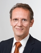 Klaus-Dietrich Wolff合作论文数22
 Marco Kesting合作论文数16
 Andreas Kolk合作论文数14
 Lucas M Ritschl合作论文数9
 Kilian Kreutzer合作论文数8
 K.-D. Wolff合作论文数7Rommel Niklas合作论文数6Christoph Pautke合作论文数6
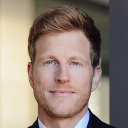 Andreas Fichter合作论文数Department of Oral and Maxillofacial Surgery, Technische Universität München5
 Thomas Tischer合作论文数Department of Orthopedic Surgery, Technische Universtität München5
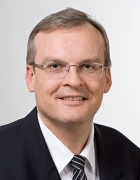 Herbert Deppe合作论文数Department of Oral and Craniomaxillofacial Surgery, Klinikum rechts der Isar, Technical University of Munich5
 Oliver Bissinger合作论文数4
 Florian A Probst合作论文数4
 Stephen Sturzenbaum合作论文数Department of Analytical, Environmental & Forensic Sciences, School of Cancer & Pharmaceutical Sciences, Faculty of Life Sciences & Medicine, King’s College London4Carolin Goetz合作论文数3
 Minli Niu合作论文数3
 Gesche Frohwitter合作论文数3Florian D Grill合作论文数3Rohleder Nils H合作论文数3
 Steffen Koerdt合作论文数3Andrea Rau合作论文数3
 Florian Bauer合作论文数3
 Bettina Hohlweg-Majert合作论文数3
 Sven Otto合作论文数3Frank Hölzle合作论文数3O. Bissinger合作论文数2Frank Gerhards合作论文数2Katharina Storck合作论文数2Matthias Schwarz合作论文数2rui miguel paz合作论文数2Wolff Regine合作论文数2Steiner T合作论文数2
 Christopher-Philipp Nobis合作论文数2C. Koch合作论文数2F. Bauer合作论文数2Christian Plank合作论文数Institute of Molecular Immunology and Experimental Oncology, Technische Universitat Munchen2
 Bernhard Meyer合作论文数School of Medicine and Health, Technical University of Munich;Neurosurgical Clinic and Policlinic, Technical University of Munich2
 Ralf Smeets合作论文数2Julia Dinges合作论文数2
 Stephan Vogt合作论文数2
 Alexander Hapfelmeier合作论文数School of Medicine and Health, Technical University of Munich;Institute of General Practice and Health Services Research, Technical University of Munich2Tim C Luth合作论文数Technische Universität Lehrstuhl für Mikro- und Medizingerätetechnik München Deutschland2
 Jamal M. Stein合作论文数2
 Klemens Scheidhauer合作论文数Technical University of Munich;Klinik und Poliklinik für Nuklearmedizin, Klinikum der Technischen Universität München2C. Götz合作论文数1N.H. Rohleder合作论文数1
 Nikolas Wilhelm合作论文数Klinikum Rechts der Isar, Technische Universität München;Abteilung für Sportorthopädie, Technische Universität München1




