Purpose: Evaluate the use of both single-echo gradient recalled echo (SE-GRE) and EPI approaches to creating temperature maps on a mid-field head-only scanner, both in vivo and on a tissue mimicking gel. Methods: Three 2D protocols were investigated (an SE-GRE, single-shot EPI, and an averaged single-shot EPI). The protocols used either a gradient recalled acquisition or an echo planar acquisition, with EPI parameters optimized for the longer T-2* at lower field-strengths. Phantom experiments were conducted to evaluate temperature tracking while cooling, comparing protocol to measurements from an optical fiber thermometer. Studies were performed on a 0.5T head only MR scanner. Temperature stability maps were produced in vivo for the various protocols to evaluate precision. Results: The use of an EPI protocol for thermometry improved temperature precision in a temperature control phantom and provided an 18% improvement in temperature measurement precision in vivo. Temperature tracking using a fast (<2 s) update rate EPI thermometry sequence provided a similar precision to the slower SE-GRE protocol. Conclusion: While SE-GRE PRF thermometry shows good performance, EPI methods offer improved tracking precision or update rate, making them a better option for thermometry in the brain at mid-field.
fMRI is typically not performed at field strengths < 1.5T due to low magnetic susceptibility contrast and inadequate gradient performance. Leveraging the high-performance gradient set of a head-only 0.5T MRI, the feasibility of motor task based fMRI was evaluated using a 4mm isotropic GRE-EPI acquisition. Activated regions within the PMC were consistent with expected behaviour. Furthermore, a significant change in signal during activation of 1.8+/-0.4% was measured within an ROI of activated voxels. These results suggest that motor task based BOLD fMRI is possible at 0.5T.
Thermal Mapping using MRI is a non-invasive method for probing the body and has both diagnostic and interventional applications. If conducted on more accessible low field strength systems, these can improve the accessibility of the method for wider use. In order to validate and compare thermal mapping, a temperature control phantom was developed with tools for heating the phantom and externally tracking the temperature of the phantom. A heating study was performed with the phantom at two temperatures, and the thermometry methods showed good connection to the change in temperature in the phantom.
This simulation and electromagnetic design work explored the development of an active second order spherical harmonic shim set for eddy current compensation in a shoulder cut-out head and neck imaging platform. While the small radius shoulder cut-out shims can be driven faster and stronger than the larger radius complete cylinder shims they suffer in homogeneity and eddy current compensation. This difference in compensation is minimized with the addition of two purpose designed eddy current compensation coils.
Preliminary evaluation of the feasibility to perform resting state BOLD fMRI at a low field (0.5 T) MR scanners equipped with a high-performance gradient system by investing the detection capability of RSNs at lower field strengths. There was an overall deterioration in the number of detected regions within all RSNs as the tSNR decreased. However, the 0.5 Evry system that will be used for experimental studies offer a better SNR efficiency than the modelled cases considered here suggesting reasonable capability in extracting RSNs that can potentially provide clinical utility.
Purpose Delta relaxation-enhanced MR (dreMR) is a field-cycling quantitative method for molecular imaging. The dreMR method uses a B-0 insert coil to shift the magnitude of the main magnetic field as a magnetization preparation phase of the pulse sequence. Here, an improved coil design method is presented that minimizes field inhomogeneities and allows for explicit control of the ROI. Methods A solenoid produces the bulk field shift, and a boundary element method is employed to design in-series shim and shield layers. A design is presented and compared to the current generation dreMR coil design on field inhomogeneity maps, shield performance, and simulated dreMR image. A proof-of-concept design is also presented with an ROI shifted from isocenter. Results The new design is able to image a sphere of 8.5 cm in diameter with field inhomogeneity of < 1% versus the previous design's 5 cm. The new design presented an increase in shielding capabilities, whereas inductance and resistance increased. For a simulated dreMR image, the new design presented errors < 10% compared to an ideal field simulation, whereas the previous design had errors > 18%. The shifted ROI design produced a region of < 1% inhomogeneity much larger than a design with no shim layer. Conclusion The new design method was found to greatly improve the insert coil field homogeneity and reduce errors in dreMR imaging in simulation without detriment to shielding. This method's capability to increase ROI and control its location will be used to design human dreMR coils going forward.
In this work a 21 MHz radiofrequency (RF) exposure system was designed, constructed and evaluated as a testing platform for MRI device safety. The exposure system consisted of a birdcage coil powered in quadrature by a power system consisting of a hybrid coupler, a power splitter, a phase shifter and two baluns. The birdcage coil was designed as a low-pass 16 rung coil. Both the coil and the power system were fabricated as printed circuit boards with a copper thickness of 205.7 mu m and a G10 substrate thickness of 581.7 mu m. The birdcage was assembled by wrapping the copper clad G10 inside of an acrylic tube. A shield for the coil was created as a cylindrical ground plane with a diameter of 39.7 cm. The birdcage and the power system were simulated in ADS and EMPro, Keysight Technologies Inc. both as lumped elements and using full wave 3D simulations. Simulation results were compared to measurements of the produced components to validate each component and entire system. In addition, a 3D magnetic field probe was developed to measure and compare the fields to analytical and numerical calculations.
A gradient coil with integrated second and third order shims has been designed and constructed for use inside an actively shielded 310 mm horizontal bore 9.4 T small animal MRI. An extension of the boundary element method, to minimise the power deposited in conducting surfaces, was used to design the gradients, and a boundary element method with a constraint on mutual inductance was used to design the shims. The gradient coil allows for improved imaging performance and was optimized for an imaging region appropriate for marmoset imaging studies. Efficiencies of 1.5 mT m−1 A−1 were achieved in a 15 cm wide bore while maintaining gradient uniformity ≤5% over the 8 cm region of interest. Two new cooling methods were implemented which allowed the gradient coil to operate at 100 A RMS, 25 % of max current with a temperature rise below 30 C.
This paper presents the design and construction of a magnetic field probe with an active balun for conditional testing of medical devices within a magnetic resonance imaging (MRI). The magnetic field probe was designed using a small loop antenna with 2 mm radius to have high spatial resolution and accuracy. It was tuned and matched at a center frequency of 128 MHz, which corresponds to 3 T MRI systems or equivalent radio frequency (RF) exposure systems for MRI-conditional testing of medical devices. An active balun with a differential transistor topology and a passive low-profile transformer were employed to boost the detected magnetic field signal level lead to higher sensitivity. It also has high input impedance that improves the decoupling of the probe to nearby medical devices. The designed magnetic field probe and active balun have been fabricated on a double-sided printed circuit board, FR4 thickness of 1.57 mm and a copper thickness of 35 mu m, with overall footprint of 22 mm x 11 mm. A verification test setup was developed to generate a known field and calibrate the probe based upon an analytic calculation of field, FDTD simulation and a 10-mm radius passive tuned/matched loop antenna.
This paper presents the design, construction, and testing of an RF injection network for MR-conditional medical testing of devices for use within 1.5 T MRI scanners (i.e., frequency of 63.4 MHz). The system was developed to meet the requirements of ISO/TS 10974:2018(E). A directional lumped element coupler, power splitter, an attenuator/isolator, low-pass filter, and high-pass filter were designed and implemented as part of the network. The RF injection network was developed in both a compact version implemented in a single PCB and discrete PCB version for use in different situations. The performance of each designed component was simulated and compared to measurement results. As an application example, a neuromodulation system was tested using the developed RF injection network for conductive emission testing.
As a part of this work a small patch antenna probe was developed to measure the variation in the electric field produced by gradient coils within an MRI in the presence of any active implantable medical devices (AIMDs). This probe was designed, fabricated, and tested within a gradient coil mimicking dB/dt exposure platform. A 2×1 cm small patch antenna followed by an instrumentational amplifier was chosen to measure the electric fields. Probe was fabricated using a 4-layer PCB. The fabricated probe was used to monitor the electric fields within the phantom in the gradient coil environment. To verify the observed behavior of the probe a simulation study was performed using Sim4Life. This study aims to assess the performance of this probe in a tissue mimicking environment within the coil.
In this article, an ultralow-frequency electric field probe capable of measuring time-varying gradient coil induced electric fields in a tissue-mimicking material was constructed, calibrated, and validated. This probe consisted of a 2 cm short dipole antenna followed by an instrumentation amplifier. The designed amplifier had a gain of 100 and a cutoff frequency of 31 kHz. The probe was fabricated on a four-layer printed circuit board (PCB), FR4 of thickness 1.57 mm and a copper thickness of 35 mu m. A waterproofed 3-D probe holder was used to cover the electronics and expose the dipole tip to the saline solution. To calibrate this probe in a known environment, a parallel-plate conductor was used. The calibrated probe was validated by measuring the electric field in a saline-filled cylindrical phantom placed within a cylindrically symmetric test coil. In order to do the validation, simulations were performed using an in-house tool developed in MATLAB and a validated tool in Sim4Life.
This paper presents the design and validation of a tuned time-domain electric field probe for mapping of radio frequency (RF) exposures used during testing of magnetic resonance imaging (MRI) conditional medical devices. The probes were 5 and 10 mm short dipole antenna, developed as a tradeoff between spatial resolution, linearity, and sensitivity. The probes were tuned and matched at a center frequency of 127.6 MHz, which corresponds to the RF frequency for 3T MRI scanners. To improve the accuracy and sensitivity, an RF low noise amplifier with high gain and very low noise figure was developed, followed by distributed lambda / 4 baluns along a triaxial cable to reduce the electric field pickup in the MRI environment. The probe was fabricated on a double-sided printed circuit board, FR4 thickness of 1.57 mm and a copper thickness of 35 mu m. Theoretical analysis was performed to calculate the exposed electric field from the real-time receive signals. To verify the probe performance finite-difference time-domain method simulations were compared to the actual measured electric fields. Developed probe was tested in a commercially available 3T RF exposure system to determine the probe dynamic range and linearity.
In this work, a modified low pass filter with 20 dB improved rejection at the center frequency of the RF injection network was developed to monitor the RF rectification of an active implantable medical devices (AIMD) during conductive emission testing. Transfer function of the designed filter was calculated, verified and validated by ADS simulation and S-parameter measurement using RF network analyzer. Analytical calculation, simulation and measurement results of the filter transfer function are in good agreement.
In this Letter ultra-low frequency probes for measuring time-varying magnetic field produced in gradient coils were designed, fabricated, and tested. A 1 cm radius induction loop antenna followed by an instrumentational amplifier with a gain of 40 dB and a cutoff frequency of 30 KHz was designed to measure the magnetic field. The probe was fabricated on a single-sided PCB, FR4 of thickness 1.57 mm and copper of thickness 35 mu m was employed in a dB/dt exposure system within a saline phantom using 3D printed waterproofed cover. Simulation and measurement results of the magnetic field were in good agreements.
In this work ultra-low frequency probes for measuring time-varying electric field produced in gradient coils were designed, fabricated and tested. A 2 cm short dipole antenna followed by an instrumentation amplifier was chosen to measure the electric field. The designed amplifier has a gain of 100 and a cutoff frequency of 31 KHz. Probes were fabricated on a 4-layers Printed Circuit Board (PCB), FR4 of thickness 1.57 mm and a copper thickness of 35 μm. Verification and validation of the probes were performed using a parallel plate capacitor with a known field which was calculated analytically and numerically. These calculations were compared against measurement results.
This work presents the design and construction of a H-field probe with active balun for MRI (magnetic resonance imaging) conditional testing of medical devices. The H-field probe was designed using a small loop antenna with 2 mm radius to have high spatial resolution and sensitivity. It was tuned and matched at a center frequency of 128 MHz, which corresponds to 3 T MRI systems or equivalent RF exposure systems. An active balun with a gain 18.28 dB and noise figure of 0.5 dB was employed to boost the detected H-field signal level. The designed Hfield probe and active balun have been fabricated on a doublesided printed circuit board (PCB), FR4 thickness of 1.57 mm and a copper thickness of 35 μm, with overall footprint of 22 mm × 11 mm.
A new RF B/H field probe for time-domain monitoring of RF electromagnetic-radiated exposure during medical device testing inside the lossy tissue-mimicking material is presented. The probe was tuned and matched for a centre frequency of 127.6 MHz to optimise the response for testing within typical 3T MRI scanners or RF exposure platforms. A 5 mm radius loop was chosen as a trade-off between spatial resolution and sensitivity and fabricated on a double-sided FR4 PCB. In the presented scheme, a low-noise RF amplifier was employed to reduce the sensitivity of the probe to induce voltage on the transmission lines and to improve accuracy. The implemented probe was found to provide acceptable spatial resolution and sensitivity for reliable B/H field mapping for this application.
Radio frequency (RF) heating of leads on medically implanted devices is a critical patient safety matter in Magnetic Resonance Imaging (MRI). Safety is generally assessed via large-scale computer simulations, which relies on the use of a "transfer function" (TF) approach in order to make the simulations sufficiently efficient to allow very large numbers of lead trajectories to be considered. In this work, a method to measure the transfer function of a simple stainless-steel wire with insulator was developed, which serves as proof-of-principle for use of the method in more realistic devices. A Finite-difference time-domain (FDTD) method was employed for comparison and to determine the induced electric field near a test wire which was then compared to the measured values. The TF method was applied to 127.6 MHz RF exposure (corresponding to a 3 T MRI system) using a custom developed RF probe in order to improve the accuracy and sensitivity of the measurements. Hydroxyethylcellulose (HEC) gel was used to mimic the lossy tissue environment of the human body. Reasonable agreement between the simulations and measurement were obtained and the method is under development for use at other frequencies of interest.
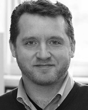 Blaine A. Chronik合作论文数64
 Timothy James Scholl合作论文数Department of Physics and Astronomy, University of Western Ontario20
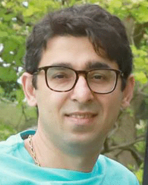 Ali Attaran合作论文数13
 Chad T. Harris合作论文数Synaptive Medical12
 Kyle M Gilbert合作论文数Department of Physics and Astronomy;U niversity of Western Ontario;Department of Physics and Astronomy, U niversity of Western Ontario9
 geron a bindseil合作论文数8
 Arjama Halder合作论文数6Alford Jamu K合作论文数Department of Physics and Astronomy, University of Western Ontario5T. C. Harris合作论文数4Brian Dalrymple合作论文数4Araya Yonathan合作论文数4Curtis N Wiens合作论文数3Van Sas Frank合作论文数3
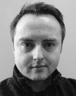 Krzysztof Wawrzyn合作论文数Department of Physics, Ryerson University3Ryan Chaddock合作论文数2
 Rebecca Feldman合作论文数Department of Computer Science, Mathematics, Physics and Statistics, Irving K. Barber Faculty of Science, The University of British Columbia2Harris Chad T合作论文数2
 Ravi S. Menon合作论文数Centre of Functional and Metabolic Mapping, Western University;Department of Medical Biophysics, Schulich School of Medicine & Dentistry, Western University;Robarts Research Institute, Schulich School of Medicine & Dentistry, Western University2
 Shaun P. Salisbury合作论文数2Stephen D. Hudson合作论文数235 H.L. Hunter Laboratories2Parisa Hudson合作论文数2K. Craig Goodrich合作论文数2Christine Wawrzyn合作论文数1James Pardy合作论文数1BA Chronik合作论文数1Tiejun Zhao合作论文数1Paul Christiaans合作论文数1
 Matthew A. McCready合作论文数Dept Phys & Astron, Univ Western Ontario1
 Yang Liu合作论文数Department of Physics and Astronomy, The University of Western Ontario1
 William Dominguez Viqueira合作论文数1
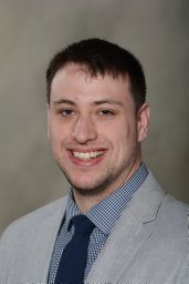 E. Lessard合作论文数Department of Medical Biophysics, The University of Western Ontario1Hai Zheng合作论文数Dept Bioengn, Univ Pittsburgh1Hao Peng合作论文数1
 Cameron C. Hopkins合作论文数Okinawa Inst Sci & Technol Grad Univ1
 Giles Santyr合作论文数Department of Medical Biophysics, Temerty Faculty of Medicine, University of Toronto1
 Parra-Robles Juan合作论文数1Odegaard J W合作论文数1Odegaard James合作论文数1
 Wyenberg, C.合作论文数1
 Peter Caravan合作论文数Center for Interdisciplinary Innovation in Imaging, Massachusetts General Hospital;Institute for Innovation in Imaging, Massachusetts General Hospital;Harvard Medical School1
 Matthew S. Fox合作论文数1
 Joshua Kaggie合作论文数Department of Radiology, University of Cambridge1Krishnamurthy Narayanan合作论文数1
 Raval Shailesh B合作论文数1
 Yujuan Zhao合作论文数1
 Tamer S. Ibrahim合作论文数Swanson School of Engineering, University of Pittsburgh;School of Medicine, University of Pittsburgh1Van Sass Frank合作论文数1




