Conventional color flow processing is associated with a high degree of operator dependence, often requiring the careful tuning of clutter filters and priority encoding to optimize the display and accuracy of color flow images. In a companion paper, we introduced a novel framework to adapt color flow processing based on local measurements of backscatter spatial coherence. Through simulation studies, the adaptive selection of clutter filters using coherence image quality characterization was demonstrated as a means to dynamically suppress weakly-coherent clutter while preserving coherent flow signal in order to reduce velocity estimation bias. In this study, we extend previous work to evaluate the application of coherence-adaptive clutter filtering (CACF) on experimental data acquired from both phantom and in vivo liver and fetal vessels. In phantom experiments with clutter-generating tissue, CACF was shown to increase the dynamic range of velocity estimates and decrease bias and artifact from flash and thermal noise relative to conventional color flow processing. Under in vivo conditions, such properties allowed for the direct visualization of vessels that would have otherwise required fine-tuning of filter cutoff and priority thresholds with conventional processing. These advantages are presented alongside various failure modes identified in CACF as well as discussions of solutions to mitigate such limitations.
The appropriate selection of a clutter filter is critical for ensuring the accuracy of velocity estimates in ultrasound color flow imaging. Given the complex spatio-temporal dynamics of flow signal and clutter, however, the manual selection of filters can be a significant challenge, increasing the risk for bias and variance introduced by the removal of flow signal and/or poor clutter suppression. We propose a novel framework to adaptively select clutter filter settings based on color flow image quality feedback derived from the spatial coherence of ultrasonic backscatter. This framework seeks to relax assumptions of clutter magnitude and velocity that are traditionally required in existing adaptive filtering methods to generalize clutter filtering to a wider range of clinically-relevant color flow imaging conditions. In this study, the relationship between color flow velocity estimation error and the spatial coherence of clutter filtered channel signals was investigated in Field II simulations for a wide range of flow and clutter conditions. This relationship was leveraged in a basic implementation of coherence-adaptive clutter filtering (CACF) designed to dynamically adapt clutter filters at each imaging pixel and frame based on local measurements of spatial coherence. In simulation studies with known scatterer and clutter motion, CACF was demonstrated to reduce velocity estimation bias while maintaining variance on par with conventional filtering.
Ultrasound is an essential tool for diagnosing and monitoring diseases, but it can be limited by poor image quality. Lag-one coherence (LOC) is an image quality metric that can be related to signal-to-noise ratio and contrast-to-noise ratio. In this study, we examine matched LOC and B-mode images of the liver to discern patterns of low image quality, as indicated by lower LOC values, occurring beneath the abdominal wall, near out-of-plane vessels and adjacent to hyperechoic targets such the liver capsule. These regions of suppressed coherence are often occult; they present as temporally stable uniform speckle on B-mode images, but the LOC measurements in these regions suggest substantially degraded image quality. Quantitative characterization of the coherence suppression beneath the abdominal wall reveals a consistent pattern both in simulations and in vivo; sharp drops in coherence occurring beneath the abdominal wall asymptotically recover to a stable coherence at depth. Simulation studies suggest that abdominal wall reverberation clutter contributes to the initial drop in coherence but does not influence the asymptotic LOC value. Clinical implications are considered for contrast loss in B-mode imaging and estimation errors for elastography and Doppler imaging.
Increasing B-mode ultrasound transmit intensity improves signal to noise ratio (SNR) by increasing backscattered echo magnitude relative to thermal noise. To ensure ultrasound remains safe, acoustic output limits exist, and regulatory bodies advise observing the ALARA (As Low As Reasonably Achievable) principle. Despite this, studies show sonographers rarely adjust intensity, resulting in unnecessary acoustic exposure or sub-optimal image quality. We have developed a framework for automated transmit intensity adjustment which we demonstrate on a Verasonics Vantage ultrasound system and C5-2v transducer. The coherence of signals received by neighboring ultrasound array elements, the lag-one coherence (LOC), quantifies clutter and temporally varying noise and serves as the automation feedback parameter. In the automated sequence, receive data are quickly acquired over a region of interest (ROI) for nine intensities ranging from mechanical indices (MI) of 0.08 to 1.4. LOC asymptotically increases with acoustic intensity as the effect of thermal noise decreases until intensity increases minimally improve SNR; the intensity at 98% of the maximum LOC is used for B-mode scanning. In preliminary hepatic imaging studies, a ROI of 7 lateral lines extending 30λ axially achieves temporally stable intensity updates. The optimization time for this ROI is 0.7 seconds, enabling real-time intensity adaptation.
Lag-one coherence (LOC) estimates local levels of acoustic noise by measuring the spatial coherence between backscattered echo signals received by neighboring pairs of transducer elements. LOC can be directly related to signal to noise ratio (SNR) and contrast to noise ratio (CNR). We have acquired B-mode images and matched pixelwise LOC estimates in the livers of 10 healthy volunteers using a C5-2v probe on a Verasonics Vantage system and in Fullwave simulations with six abdominal walls over uniform speckle. We present evidence of temporally stable regions of suppressed LOC beneath the abdominal wall which recover to a stable asymptotic value at depth. Fullwave simulation results suggest that reverberation determines the initial amount of coherence suppression and aberration determines the asymptotic LOC value. The in vivo LOC values beneath the abdominal wall range from roughly 0.4 to 0.85, corresponding to SNRs of −3.5 dB to 15 dB, and the length of coherence suppression ranges from 0.5cm to 2.5 cm. These regions are occult; they present as temporally stable uniform liver on B-mode images. This is significant because clinicians will not be aware that lesions may be much more difficult to detect in this region, potentially leading to missed diagnoses.
Ultrasonic image quality is often challenged by speckle noise and acoustic clutter which reduces the anatomical conspicuity of medically relevant features. Deep learning approaches show promise in correcting these sources of noise, but there currently lacks a large dataset that i) contains enough feature variation to prevent model over-fitting and ii) captures higher-order effects such as reverberation, aberration, and harmonic generation. To address these needs, we simulated a 180-channel linear transducer acquiring 850,000 transmit-receive events to beamform 10,000 ultrasound images on unique scatter field maps. We fit fully convolutional neural networks (CNNs) to this dataset as a proof-of-concept for noise reduction tasks. We show that CNNs are able to translate from in silico to in vivo images for speckle reduction. However, naive fully convolutional networks are challenged to correct for clutter and aberration simultaneously, even within an in silico training dataset. We hope that this dataset, termed UltraNet, will provide analogous benefits to ultrasound image reconstruction as the ImageNet dataset did for image recognition. These data and tooling will be made available at: https://github.com/ouwen/ultranet.
The magnitudes by which aberration and incoherent noise sources, such as diffuse reverberation and thermal noise, contribute to degradations in image quality in medical ultrasound are not well understood. Theory predicting degradations in spatial coherence and contrast in response to combinations of incoherent noise and aberration levels is presented, and the theoretical values are compared to those from simulation across a range of magnitudes. A method to separate the contributions of incoherent noise and aberration in the spatial coherence domain is also presented and applied to predictions for losses in contrast. Results indicate excellent agreement between theory and simulations for beamformer gain and expected contrast loss due to incoherent noise and aberration. Error between coherence-predicted aberration contrast loss and measured contrast loss differs by less than 1.5 dB on average, for a -20 dB native contrast target and aberrators with a range of root-mean-square time delay errors. Results also indicate in the same native contrast target the contribution of aberration to contrast loss varies with channel signal-to-noise ratio (SNR), peaking around 0 dB SNR. The proposed framework shows promise to improve the standard by which clutter reduction strategies are evaluated.
The lag-one coherence (LOC), derived from the correlation between the nearest-neighbor channel signals, provides a reliable measure of clutter which, under certain assumptions, can be directly related to the signal-to-noise ratio of individual channel signals. This offers a direct means to decompose the beamsum output power into contributions from speckle and spatially incoherent noise originating from acoustic clutter and thermal noise. In this study, we applied a novel method called lag-one spatial coherence adaptive normalization (LoSCAN) to locally estimate and compensate for the contribution of spatially incoherent clutter from conventional delay-and-sum (DAS) images. Suppression of incoherent clutter by LoSCAN resulted in improved image quality without introducing many of the artifacts common to other adaptive imaging methods. In simulations with known targets and added channel noise, LoSCAN was shown to restore native contrast and increase DAS dynamic range by as much as 10–15 dB. These improvements were accompanied by DAS-like speckle texture along with reduced focal dependence and artifact compared with other adaptive methods. Under in vivo liver and fetal imaging conditions, LoSCAN resulted in increased generalized contrast-to-noise ratio (gCNR) in nearly all matched image pairs ( ${N} =366$ ) with average increases of 0.01, 0.03, and 0.05 in good-, fair-, and poor-quality DAS images, respectively, and overall changes in gCNR from −0.01 to 0.20, contrast-to-noise ratio (CNR) from −0.05 to 0.34, contrast from −9.5 to −0.1 dB, and texture $\mu /\sigma $ from −0.37 to −0.001 relative to DAS.
Image post-processing is used in clinical-grade ultrasound scanners to improve image quality (e.g., reduce speckle noise and enhance contrast). These post-processing techniques vary across manufacturers and are generally kept proprietary, which presents a challenge for researchers looking to match current clinical-grade workflows. We introduce a deep learning framework, MimickNet, that transforms conventional delay-and-summed (DAS) beams into the approximate Dynamic Tissue Contrast Enhanced (DTCE™) post-processed images found on Siemens clinical-grade scanners. Training MimickNet only requires post-processed image samples from a scanner of interest without the need for explicit pairing to DAS data. This flexibility allows MimickNet to hypothetically approximate any manufacturer's post-processing without access to the pre-processed data. MimickNet post-processing achieves a 0.940 ± 0.018 structural similarity index measurement (SSIM) compared to clinical-grade post-processing on a 400 cine-loop test set, 0.937 ± 0.025 SSIM on a prospectively acquired dataset, and 0.928 ± 0.003 SSIM on an out-of-distribution cardiac cine-loop after gain adjustment. To our knowledge, this is the first work to establish deep learning models that closely approximate ultrasound post-processing found in current medical practice. MimickNet serves as a clinical post-processing baseline for future works in ultrasound image formation to compare against. Additionally, it can be used as a pretrained model for fine-tuning towards different post-processing techniques. To this end, we have made the MimickNet software, phantom data, and permitted in vivo data open-source at https://github.com/ouwen/MimickNet.
The coherence of signals received by neighboring transducer array elements, the lag-one coherence (LOC), provides a local measure of clutter and temporally varying noise. Increasing transmit intensity decreases the impact of temporal noise on image quality, but does not eliminate stationary clutter. By assessing changes in LOC with acoustic intensity, acoustic exposure can be minimized without compromising image quality, achieving the ALARA (As Low As Reasonably Achievable) principle. In this study, adaptive intensity adjustment based on LOC is integrated into B-mode imaging. Hepatic imaging tests in two volunteers provide preliminary support for the stability and specificity of intensity updates, potentially relieving the physical and workload demands placed on sonographers while ensuring both high-quality imaging and limited acoustic exposure.
Image post-processing is used in clinical-grade ultrasound scanners to improve image quality (e.g., reduce speckle noise and enhance contrast). These post-processing techniques vary across manufacturers and are generally kept proprietary, which presents a challenge for researchers looking to match current clinical-grade workflows. We introduce a deep learning framework, MimickNet, that transforms conventional delay-and-summed (DAS) beamformed images into the approximate post-processed images found on clinical-grade scanners. Training MimickNet only requires post-processed image samples from a scanner of interest without the need for explicit pairing to DAS data. Unpaired image flexibility allows MimckNet to hypothetically approximate any manufacturer's post-processing without hacking into commercial machines for pre-processed data. MimickNet generates images with an average similarity index measurement (SSIM) of 0.930±0.0892 on a 300 cineloop test set, and it generalizes to cardiac cineloops achieving an SSIM of 0.967±0.002 despite using no cardiac data in the training process. To our knowledge, this is the first work to approximate current clinical-grade ultrasound post-processing under realistic black-box constraints where before and after post-processing data is unavailable. MimickNet can be used out of the box or retrained to serve as a clinical post-processing baseline to compare against for future works in ultrasound image formation. To this end, we have made the MimickNet software open source at https://github.com/ouwen/mimicknet.
Reliable assessment of image quality is an important but challenging task in complex imaging environments such as those encountered in vivo. To address this challenge, we propose a novel imaging metric, known as the lag-one coherence (LOC), which leverages the spatial coherence between nearest-neighbor array elements to provide a local measure of thermal and acoustic noise. In this paper, we derive the theory that relates LOC and the conventional image quality metrics of contrast and contrast-to-noise ratio (CNR) to channel noise. Simulation and phantom studies are performed to validate this theory and compare the variability of LOC to that of conventional metrics. We further evaluate the performance of LOC using matched measurements of contrast, CNR, and temporal correlation from in vivo liver images formed with varying mechanical index (MI) to assess the feasibility of adaptive acoustic output selection using LOC feedback. Simulation and phantom results reveal a lower variability in LOC relative to contrast and CNR over a wide range of clinically relevant noise levels. This improved stability is supported by in vivo measurements of LOC which show an increased monotonicity with changes in MI compared to matched measurements of contrast and CNR (88.6% and 85.7% of acquisitions, respectively). The sensitivity of LOC to stationary acoustic noise is evidenced by positive correlations between LOC and contrast (r = 0.74) and LOC and CNR (r = 0.66) at high acoustic output levels in the absence of thermal noise. Results indicate that LOC provides repeatable characterization of patient-specific trends in image quality, demonstrating feasibility in the selection of acoustic output using LOC and its application for in vivo image quality assessment.
The US Food and Drug Administration (FDA) provides guidelines for acoustic output to ensure that ultrasound remains a safe imaging modality used widely in applications such as obstetrics. However, even within these limits, additional acoustic output that does not lead to improved image quality represents an unnecessary risk to patient safety. Ultrasound manufacturers and professional societies advise users to observe the ALARA (As Low As Reasonably Achievable) principle with regard to acoustic exposure, but studies have shown that most ultrasound users do not adjust the transmit power. In this study, an adaptive ultrasound tool was implemented to automatically adjust acoustic exposure in response to real-time image quality feedback based on lag-one coherence (LOC). LOC is the average correlation between backscattered echoes received on neighboring array elements. Previous work has shown that LOC is predictive of local signal-to-noise ratio and sensitive to incoherent acoustic clutter and temporally-incoherent noise. LOC as a function of MI was assessed in seven volunteers. While imaging the placenta or the fetal abdomen, the system swept through 18 transmit voltages that correspond to MIs ranging from 0.08 to 1.21 and automatically selected the optimum MI based on reaching 95% of the maximum LOC. In this study, the optimal MI values were between 0.48 and 0.89, depending on the acoustic window. For all acquisitions, we saw a steady increase that approached an asymptote in LOC with increasing MI. Contrast, contrast to-noise ratio (CNR), and LOC followed similar trends. These results suggest that maximum image quality can be achieved with acoustic output levels lower than the FDA limits and an automated tool can be employed in real-time to find this optimal MI for specific imaging conditions. Our results support the feasibility of an automated, LOC-based implementation of the ALARA principle with regard to acoustic exposure for obstetric ultrasound.
Tissue harmonic image quality has been shown to increase with elevated pressure output. The Peak Rarefaction Pressure (PRP) for a given transmit, however, is limited by the Mechanical Index (MI) guideline. We have previously demonstrated that the MI overestimates in situ PRP for tightly focused beams such as F/1.5 because it does not account for phase aberration. In this study, we propose a new spatial coherence based metric to estimate phase aberration and predict in vivo PRP. We show harmonic Short Lag Spatial Coherence (SLSC) to linearly correlate with in situ PRP with a 0.86 coefficient of determination across 11 different body walls. This correlation suggests the potential for more accurate PRP estimation in situ than the current MI scheme, allowing patient specific acoustic output.
Hepatic image quality has been shown to be degraded by the clutter-generating effects of the abdominal wall, and while beamforming and signal processing methods exist to minimize clutter, there is currently no automated method by which parameters such as imaging frequency are selected. Adaptive imaging seeks to optimize these parameters using lag-one coherence (LOC), a direct estimate of clutter levels in vivo, as a real-time feedback metric. The aim of this study is to determine the viability of using LOC to adaptively select imaging frequencies in the presence of clutter. Across in silico, ex vivo, and in vivo setups, a monotonic decrease in LOC was observed as imaging frequency was increased, indicating increased clutter, but the lesion conspicuity metric (LCM), which includes the expected increase in resolution with frequency, is optimized. The cases in which no abdominal wall is present, and therefore without a near-field source of clutter, had the highest measured LOC values and allowed for the highest optimized frequencies. The results suggests that an optimal frequency can be automatically selected for lesion detection. The findings of this study also match the clinical experience, in which lower frequencies are used for difficult-to-image patients.
Ultrasound imaging is the product of numerous pulse sequence and processing design choices, but it is impossible to optimize for every clinical imaging case with a single set of parameters. Many sonographers do not take advantage of even the limited set of controls provided to them during an exam. We have demonstrated that lag-one coherence (LOC), an aperture-domain signal quality metric, reflects image quality in the presence of in vivo clutter effects. This feedback could be useful in adaptively selecting patient- and view-specific optimal parameters. Increased system bandwidth and computing power now make it possible to estimate LOC in real-time. We demonstrate a Verasonics-based platform for real-time temporal correlation and spatial coherence estimation during B-mode imaging at 18 frames per second. Demonstrations of abdominal and cardiac imaging under varying common scanning conditions are presented using the tool.
In this study, we evaluate the clinical utility of fetal short-lag spatial coherence (SLSC) imaging. Previous work has documented significant improvements in image quality with fetal SLSC imaging as quantified by measurements of contrast and contrast-to-noise ratio (CNR). The objective of this study was to examine whether this improved technical efficacy is indicative of the clinical utility of SLSC imaging. Eighteen healthy volunteers in their first and second trimesters of pregnancy were scanned using a modified Siemens SC2000 clinical scanner. Raw channel data were acquired for routinely examined fetal organs and used to generate fully matched raw and post-processed harmonic B-mode and SLSC image sequences, which were subsequently optimized for dynamic range and other imaging parameters by a blinded sonographer. Optimized videos were reviewed in matched B-mode and SLSC pairs by three blinded clinicians who scored each video based on overall quality, target conspicuity and border definition. SLSC imaging was highly favored over conventional imaging with SLSC scoring equal to (28.2 ± 10.5%) or higher than (63.9 ± 12.9%) B-mode for video pairs across all examined structures and processing conditions. Multivariate modeling revealed that SLSC imaging is a significant predictor of improved image quality with p ≤ 0.002. Expert-user scores for image quality support the application of SLSC in fetal ultrasound imaging.
Poor quality ultrasound images and inadequate or suboptimal visualization of imaging targets is a common problem in individuals that are overweight or obese. Acoustic reverberation is an incoherent noise source that is a common factor in overweight and obese individuals and is a significant contributor to the poor image quality. Specifically, diffuse acoustic reverberation is problematic because it appears similar to common tissue texture in ultrasound images, thereby exacerbating the inadequate and suboptimal visualization. We describe the coherence imaging technique called the short-lag spatial coherence (SLSC) beamformer and its related imaging methods as potential solutions to the inadequate and suboptimal visualization problem. The SLSC beamformer detects the spatial similarity of the backscattered ultrasound waves, with a greater emphasis on the spatial similarity at closely-spaced positions. Because diffuse reverberation is spatially incoherent in the wavefield, noise can be differentiated from tissue and other desired imaging targets. Applications of the SLSC beamformer to in vivo imaging and adaptations of the technique to other imaging modalities, including flow imaging, molecular ultrasound imaging, and photoacoustic imaging are reviewed. Although computationally more intensive than conventional delay-and-sum beamforming, we describe several techniques for fast computation of coherence, which enable real-time imaging. The challenges and criticisms of spatial coherence beamforming are reviewed, including the loss in phase information and the nonlinear behavior of the technique.
Current clinical abdominal imaging arrays are designed to maximize angular field of view rather than the extent of the coherent aperture. We illustrate, in ex vivo experiments, the use of a large effective aperture to perform high-resolution imaging, even in the presence of abdominal wall-induced acoustic clutter and aberration. Point and lesion phantom targets were imaged through a water path and through three excised cadaver abdominal walls to create different clinically relevant clutter effects with matched imaging targets. A 7.36-cm effective aperture was used to image the targets at a depth of 6.4 cm, and image quality metrics were measured over a range of aperture sizes using synthetic aperture techniques. In all three cases, although degradation compared with the control was observed, lateral resolution improved with increasing aperture size without loss of contrast. Spatial compounding of the large-aperture data drastically improved lesion detectability and produced contrast-to-noise ratio improvements of 83%-106% compared with the large coherent aperture. These studies indicate the need for the development of large arrays for high-resolution abdominal diagnostic imaging. (C) 2018 World Federation for Ultrasound in Medicine & Biology. All rights reserved.
 Gregg Trahey合作论文数Department of Biomedical Engineering, Pratt School of Engineering, Duke University23
 Nick Bottenus合作论文数Dept. of Biomed. Eng., Duke Univ.;c;Dept. of Biomed. Eng., Duke Univ.18
 James Long合作论文数Duke Univ, Dept Biomed Engn, Durham, NC 27706 USA9
 David Bradway合作论文数Duke Univ.;c;Duke Univ.7
 Ouwen Huang合作论文数Duke University4
 Matthew Huber合作论文数Duke Univ, Dept Biomed Engn, Durham, NC 27706 USA4
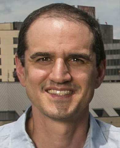 Mark L. Palmeri合作论文数Department of Biomedical Engineering, Pratt School of Engineering, Duke University;Department of Anesthesiology, School of Medicine, Duke University4
 Katelyn Flint合作论文数Department of Biomedical Engineering, Duke University3
 Sarah Catherine Ellestad合作论文数Department of Obstetrics and Gynecology, Duke University School of Medicine3
 Patricia Mcnally合作论文数3
 Dongwoon Hyun合作论文数3
 Gianmarco Pinton合作论文数Joint Department of Biomedical Engineering, College of Arts and Sciences, The University of North Carolina at Chapel Hill3
 Katelyn Offerdahl合作论文数2
 Rifat Ahmed合作论文数Hajim School of Engineering and Applied Sciences, University of Rochester, Rochester, NY, USA2
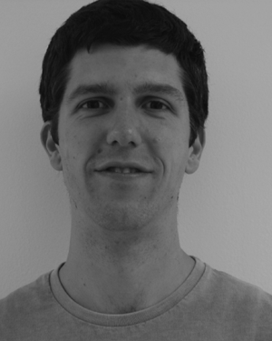 Jakovljevic, M.合作论文数Dept. of Biomed. Eng., Duke Univ.;c;Dept. of Biomed. Eng., Duke Univ.2
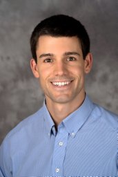 Morgan Matthew R合作论文数Department of Biomedical Engineering, Duke University2
 Kingshuk Roy Choudhury合作论文数University College2S. Farsiu合作论文数Lots of important places2Julien Jouganous合作论文数1
 Aaron P Ragsdale合作论文数Dept Integrat Biol, Univ Wisconsin1
 Marcelo Lerendegui合作论文数Imperial College London1You Li合作论文数Stanford Univ, Dept Radiol, Stanford, CA 94305 USA1Bofeng Zhang合作论文数1
 Vaibhav Kakkad合作论文数Dept. of Biomed. Eng., Duke Univ.;c;Dept. of Biomed. Eng., Duke Univ.1
 Haichong K. Zhang合作论文数1
 Kathryn Radabaugh Nightingale合作论文数Department of Biomedical Engineering, Pratt School of Engineering, Duke University1
 Simon Gravel合作论文数Department of Human Genetics, School of Biomedical Sciences, Faculty of Medicine and Health Sciences, McGill University;Victor Phillip Dahdaleh Institute of Genomic Medicine, School of Biomedical Sciences, Faculty of Medicine and Health Sciences, McGill University1
 Sina Farsiu合作论文数Department of Biomedical Engineering, Pratt School of Engineering, Duke University;Department of Ophthalmology, Duke University School of Medicine1
 Muyinatu A. Lediju Bell合作论文数Photoacoustic & Ultrasonic Systems Engineering Lab, Whiting School of Engineering, Johns Hopkins University1Brita K. Boyd合作论文数1
 Rendon C. Nelson合作论文数Division of Abdominal Imaging, Department of Radiology, Duke University School of Medicine;Duke Cancer Institute, Duke University;Pharmaceutical & Biotechnical Industry;RadMD1
 Jeremy Dahl合作论文数Department of Radiology, School of Medicine, Stanford University1
 Emad Boctor合作论文数Division of Medical Imaging Physics, Department of Radiology and Radiological Science, Johns Hopkins University;Department of Computer Science, Whiting School of Engineering, Johns Hopkins University;Laboratory for Computational Sensing and Robotics, The Malone Center for Engineering in Healthcare, Johns Hopkins University1




