A major challenge in treating AML with the BCL-2 inhibitor venetoclax is the frequent development of drug resistance, which diminishes therapeutic efficacy and leads to patient death. The fundamental mechanisms underlying this resistance are not fully understood. Here, we established venetoclax-resistant cell models of AML that propagate even when the levels of BCL-2, MCL-1, cleaved PARP, and cleaved caspase-9 are reduced, suggesting a BCL-2-independent resistance mechanism. Compared to sensitive cells, resistant Kasumi-1 (VENK) and MV4-11 (VENM) cells exhibit enhanced proliferation both in vitro and in vivo, forming larger and more numerous spheroids and colonies, and displaying higher tumorigenicity in mice. RNA sequencing and KEGG pathway analysis identified the neuroactive ligand-receptor interaction (NLRI) pathway as a key vulnerability in both resistant cell lines. While the NLRI pathway contains numerous altered genes, CHRNB4 is the only gene commonly shared and significantly downregulated in both VENK and VENM cells and tumors. Enforced expression of CHRNB4 in resistant cells with low basal expression impaired cell adhesion and colony formation. Clinically, CHRNB4 downregulation is associated with poor AML patient overall survival and predicts a diminished response to venetoclax treatment. This study identifies the NLRI pathway as a crucial vulnerability in venetoclax resistance and unveils CHRNB4 as a promising predictive biomarker for treatment response. These results suggest that targeting the NLRI pathway represents a novel strategy for developing next-generation therapies to improve the poor outcomes of current combination treatments.
Background: The clinical efficacy of the BCL-2 inhibitor venetoclax in acute myeloid leukemia (AML) is significantly undermined by the frequent emergence of drug resistance, which precipitates disease progression and poor patient outcomes. However, the molecular landscape of this resistance remains insufficiently understood. Methods: To address this, we developed venetoclax-resistant AML cell models and utilized transcriptomic profiling integrated with comprehensive in vitro and in vivo functional assays. Results: Resistant cells demonstrated sustained proliferation even under the suppression of BCL-2, MCL-1, and key intrinsic apoptotic markers, including cleaved PARP and caspase-9, indicating a bypass mechanism independent of classical BCL-2 signaling. Compared to their sensitive counterparts, resistant Kasumi-1 (VENK) and MV4-11 (VENM) cells exhibit aggressive growth phenotypes in vitro and in vivo, characterized by larger, more numerous spheroids and colonies, alongside heightened tumorigenicity in murine models. Transcriptomic profiling and KEGG analysis identified the neuroactive ligand-receptor interaction (NLRI) pathway as a significant signaling node shared between these resistant lines. While multiple NLRI-associated genes were altered, CHRNB4 was consistently and significantly downregulated in both VENK and VENM cells and tumors. Re-expression of CHRNB4 in resistant cells, a primary gain-of-function approach, significantly impaired colony formation, and tumor growth in vivo. Clinically, CHRNB4 downregulation correlates with shortened overall survival and diminished response to venetoclax. Conclusions: Our findings implicate the NLRI pathway in venetoclax resistance and identify CHRNB4 as a robust prognostic indicator and a promising therapeutic target for developing next-generation AML strategies.
mRNA therapeutics are transitioning from transient anti-viral vaccines into precise cancer immunotherapies capable of orchestrating potent antigen-specific T-cell and humoral responses. However, therapeutic resistance within immunologically "cold" tumors remains a formidable barrier, necessitating multiaxial optimization across transcript architecture, neoantigen selection, delivery vector engineering, and tumor microenvironment (TME) reprogramming. This review synthesizes critical breakthroughs in mRNA biochemistry-including chemical nucleotide modifications, optimized untranslated regions, structural codon adjustments, and stringent purification methodologies-that extend transcript longevity while limiting off-target reactogenicity to maximize functional antigen expression. We evaluate multiomic neoantigen discovery workflows leveraging genomics, transcriptomics, immunoproteomics, and computational HLA-binding algorithms to refine patient-specific target selection. Next, we dissect advanced lipid nanoparticles, surface-functionalized biomaterials, and engineered extracellular vesicles optimized to enhance antigen-presenting cell tropism and lymphoid homing. We further detail how vaccine-induced cytokine fluxes actively remodel the TME, successfully reversing local immune tolerance and driving robust effector leukocyte infiltration into the tumor stroma. Specifically, we highlight the convergence of mRNA-mediated cytokine signaling and epigenetic imprinting, which cooperatively induce trained immunity for durable preventive surveillance. Finally, we delineate rational combinations with immune checkpoint blockades while addressing translational challenges: identifying predictive biomarkers, mapping presentation kinetics, and structuring adaptive clinical trial frameworks.
[This corrects the article DOI: 10.1016/j.gendis.2022.11.024.].
BACKGROUND:Enhanced protein expression of ALL1-fused gene from chromosome 1q (AF1Q) after (chemo)radiotherapy has been described in vitro, but is largely understudied in gastrointestinal cancer. We aimed to investigate AF1q expression in rectal cancer (RC) patients treated with short-term radiation therapy and a possible correlation with markers crucial for RC prognosis. METHODS:A cohort of 75 RC patients scheduled for surgery was defined and patients with moderately locally advanced tumors (cT3Nx) received preoperative hyperfractionated short-term radiation therapy (cumulative dose 25 Gy). Immunohistochemical analysis was conducted to assess AF1q, STAT1, IDO1 and other prognostic markers (CD3/CD8-Immunoscore, PD-L1) and marker correlations were evaluated. RESULTS:Irradiated tumors exhibited significantly higher AF1q expression than treatment-naïve samples (n = 60: AF1q + to AF1q+++ 98.3% (n = 59), AF1q- 1.7% (n = 1) vs. n = 15: AF1q + 78.6% (n = 11), AF1q- 21.4% (n = 4); p < 0.001). Specifically, irradiated tumors showed high STAT1, but low IDO1 expression compared to treatment-naïve samples (p = 0.019 and p = 0.015, respectively). Overall, enhanced tumoral AF1q expression was associated with negative lymph node stage (p = 0.012) as well as with diminished expression of STAT1 (rs = -0.468, p = 0.038) and IDO1 (rs = -0.246, p = 0.020). CONCLUSION:AF1q is expressed in RC, especially after short-term radiation therapy. Here, AF1q may support tumor suppression, possibly through the involvement of the pro-apoptotic STAT1 axis. Further mechanistic evidence and investigation involving a larger patient cohort are needed to validate a radiation-induced, AF1q-driven tumor-suppressing effect, which may impact RC patient outcomes.
Long non-coding RNAs (lncRNAs) and RNA N⁶-methyladenosine (m6A) have been linked to leukemia drug resistance. However, whether and how lncRNAs and m6A coordinately regulate resistance remain elusive. Here, we show that many differentially expressed lncRNAs enrich m6A, and more lncRNAs tend to have higher m6A content in CML cells resistant to tyrosine kinase inhibitors (TKIs). We demonstrate the broad clinical relevance of our findings, showing that upregulation of top-ranked lncRNAs (e.g., SENCR, PROX1-AS1, LINC00892) in TKI-resistant cell lines occurs in CML patients at the diagnostic stage, blast crisis phase, or not responding to TKIs compared to the chronic phase or TKI responders, respectively. Higher lncRNAs predict drug resistance and shorter survival duration. The knockdown of SENCR, PROX1-AS1, or LINC00892 restores TKI sensitivity. Mechanistically, upregulation of PROX1-AS1, SENCR, and LINC00892 results from FTO-dependent m6A hypomethylation that stabilizes lncRNA transcripts and empowers resistant cell growth through overexpression of PI3K signaling mediators (e.g., ITGA2, F2R, COL6A1). Treatment with PI3K inhibitor alpelisib eradicates resistant cells in vitro and in vivo, with prolonged survival of leukemic mice through downregulation of F2R, ITGA2, and COL6A1. Thus, the lncRNA-m6A-PI3K cascade represents a new non-genetic predictor for drug resistance and poorer prognosis in cancer, and a pan-cancer mechanism underlying TKI resistance.
Despite the initial responses to the tyrosine kinase inhibitor (TKI) for cancer therapy, many patients often relapse with no curative regimens available. Further, the ability to target therapeutic agents to cancer cells with appropriate doses remains challenging in the clinic, especially for leukemia. Here, we show that naïve CML cells are dynamically heterogeneous in colony formation. Larger clones expand while smaller ones diminish and eventually disappear. Compared to resistant cells, parental populations, including CD44+ stem cells, form a greater number of larger, solid spheroids. Upregulation of fat mass and obesity associated protein (FTO), an RNA N6-methyladenosine demethylase, and stem cell markers (e.g., CD44, CD133, CD25) is more obvious in resistant cells compared to parental cells. FTO inhibitors (e.g., CS1, FB23-2) appreciably impair the growth of resistant cells either alone or in combination with nilotinib. FTO protein expression is unexpectedly upregulated by CS1 or FB23-2 treatment in multiple leukemia cell lines. We then constructed RNA nanoparticles encapsulating FTO siRNAs and conjugated with anti-CD133 RNA aptamers. We showed that, compared to negative control, these nanoparticles were taken up much more efficiently by resistant cells that highly express CD133. Treatment with the CD133-guided FTO siRNA nanoparticles efficiently silenced FTO expression in resistant cells, which leads to a significant reduction in their colony and spheroid formation. These findings offer new insights into cancer drug resistance and advance the application of RNA nanotechnology for treating leukemia. The research provides a foundation for developing novel, targeted therapies for resistant leukemia.
Introduction: Primary myelofibrosis (PMF), a myeloproliferative neoplasm (MPN), features bone marrow fibrosis, splenomegaly, and abnormal blood cell production. Aberrant JAK/STAT signaling, elevated cytokines or hyperactive NFκB are implicated in MPN pathogenesis; however, their interplay remains unclear. JAK inhibitors (JAKi) alleviate PMF symptoms, but they don't fully address disease progression or mitigate cytokine storms. NFκB inhibitors have shown promise in MPN models but are associated with off-target effects and toxicities. This study aims to elucidate the mechanisms of JAKi resistance and develop novel therapeutic strategies targeting hyperactive NFκB in vitro and in vivo, offering potential benefits beyond current JAKi therapies. Methods: To investigate JAKi resistance in PMF, ruxolitinib-resistant HEL cells were generated and NFκB/JAK2/STAT signaling was examined by Western blot. Biomarkers associated with JAKi response were identified in PMF patients treated with BMS-9115437 (Gangat et al, Blood Cancer J, 2023) via Western blot. Molecular mechanisms of MPN pathogenesis were further elucidated using Affymetrix HG-U133Plus2 assay on RNA from treatment-naïve PMF, essential thrombocythemia (ET), polycythemia vera (PV) patients and healthy donors, followed by KEGG and gene set enrichment (GSE) analysis. In vitro, NFκB was inactivated by shRNA, Ixazomib or emetine followed by Western blot for NFκB/JAK2/STAT phosphorylation as well as flow cytometry and clonogenic assays for cell growth. NFκB DNA binding activity was determined by electrophoretic mobility-shift assays (EMSA). The therapeutic and preventive effects of Ixazomib and emetine were evaluated in HEL-engrafted nude mice by assessing tumor formation, development, and underlying mechanisms using Western blot, H&E and IHC staining. Results: Short-term JAKi (BMS-911543, ruxolitinib) exposure altered JAK2 phosphorylation, increasing Tyr1008 phosphorylation and decreasing Tyr221, Tyr570, and STAT5 phosphorylation. Notably, JAK2, STAT3 and STAT5 were phosphorylated in ruxolitinib-resistant HEL cells or after treatment cessation, implying sustained signaling activity. Western blot analysis and clinical data in PMF patients (n=3) receiving BMS-911543 showed no hyperphosphorylation of JAK2 Tyr1008 with diverse responses independent of JAK2V617F status. A lack of correlation between JAK2 and STAT5 expression/phosphorylation suggests that other factors contribute to hyperactive STAT5 in PMF. Symptomatic improvement in a JAK2V617F-positive patient correlated with decreased STAT5 protein expression and phosphorylation, potentially serving as a predictor for JAKi response. Transcriptomic analysis in MPN patients (PMF (n=11), ET (n=6), PV (n=4), HD (n=5)) pinpointed the transcriptomic dissimilarities between PMF and PV or ET, and highlighted the higher NFκB activity in PMF compared to PV or ET. The importance of NFκB in PMF was further evidenced by the network analysis for the overproduction of circulating cytokines in PMF patients (Tefferi et al, JCO, 2011). In vitro studies revealed that NFκB overexpression promotes cell proliferation and inhibits apoptosis through enhanced IL-6/IL-6R expression and JAK2/STAT5 phosphorylation. Inactivating NFκB using shRNA, emetine, or ixazomib reversed these effects. Further, IL-6 treatment increased JAK2 phosphorylation. In vivo studies showed that Ixazomib significantly reduces tumor volume (734 ±133 to 98 ±14 mm3; P <0.001) and weight (582 ±156 to 87 ±22 mg; P =0.0005) compared to controls. Emetine similarly suppressed tumor growth dose-dependently without affecting spleen weight and even prevented tumor formation when administered prior to cell engraftment. Mechanistically, both Ixazomib and emetine inhibited NFκB and JAK2 phosphorylation while increasing cleaved caspase-3 in tumors. Conclusions: We discovered that STAT inactivation independently predicts improved outcomes in PMF and that NFκB hyperactivity holds particular significance for PMF compared to other MPN subtypes. We identified a NFκB-IL-6-JAK2-STAT axis in regulating PMF growth regardless of JAK2V617F status. Targeting NFκB with novel inhibitors Ixazomib and emetine disrupts the NFκB-IL-6-JAK2-STAT axis, suppressing PMF growth and offering potential therapeutic alternatives to overcome current JAKi limitations.
Background/Objectives: Glucagon-like peptide-1 receptor agonists (GLP-1 RAs) effectively manage type 2 diabetes mellitus (T2DM) but may impair gastrointestinal motility, increasing the risk of small intestinal bacterial overgrowth (SIBO). Diagnostic evaluation of SIBO commonly involves breath testing and clinical assessment. This study aimed to assess the association between GLP-1 RAs or dual GLP-1/glucose-dependent insulinotropic polypeptide (GIP) RAs are associated with incident SIBO. Methods: We conducted a retrospective cohort study using the TriNetX global database, identifying adult T2DM patients initiating GLP-1 RA or dual GLP-1/GIP RA therapy versus other second-line T2DM agents (OSLT2DM) from 1 January 2006 to 2 December 2024. Patients with major abdominal surgery, connective tissue disorders, gastroparesis, or other high-risk conditions for SIBO were excluded. 1:1 Propensity score matching was applied. Short-term (<1 year) and long-term (up to 5 years) risks were evaluated with Kaplan–Meier curves and univariable Cox models. Results: After matching, 216,173 patients per cohort were analyzed. Short-term analysis demonstrated a higher incidence of diagnostically confirmed SIBO in patients treated with GLP-1 RA/GIP (0.177 per 1000 patient-years) compared to OSLT2DM (0.083 per 1000 patient-years; HR 2.14, 95% CI 1.13–4.07; p = 0.0491). Long-term analysis indicated a non-significant trend toward increased risk in the GLP-1 RA/GIP group (HR 2.02, 95% CI 0.98–4.12), though Kaplan–Meier analysis revealed a sustained divergence (p = 0.017). Conclusions: GLP-1 RA and dual GLP-1/GIP RA therapy are associated with increased short-term SIBO risk. Symptom-driven SIBO breath-test evaluation may be warranted in patients initiating these agents.
 Kevin Bunting合作论文数Department of Pediatrics, Division of Hematology and Oncology, Emory University27
 Reggie Duerst合作论文数26
 Jennifer Schneiderman合作论文数Hematology, Oncology and Stem Cell Transplantation, Department of Pediatrics, Feinberg School of Medicine, Northwestern University23
 Morris Kletzel合作论文数21
 Sonali Chaudhury合作论文数Hematology, Oncology and Stem Cell Transplantation, Department of Pediatrics, Feinberg School of Medicine, Northwestern University20Scot C. Remick合作论文数16Michael Craig合作论文数14
 Hillard M. Lazarus合作论文数Department of Medicine, School of Medicine, Case Western Reserve University14
 Zhengqi Wang合作论文数12Jino Park合作论文数West Virginia University, West Virginia University12
 Richard Moriggl合作论文数Institute of Molecular Pathology, Dr. Bohr-Gasse 7, A-1030 Vienna, Austria12
 Lukas Kenner合作论文数Department of Pathology, Medical University Vienna12
 David Wald合作论文数Department of Pathology, School of Medicine, Case Western Reserve University;Case Comprehensive Cancer Center, Case Western Reserve University;Kure.ai11David A. Jacobsohn合作论文数11Brenda W. Cooper合作论文数10Cumpston Aaron合作论文数10
 Mary J. Laughlin合作论文数Cleveland Cord Blood Center10Geqiang Li合作论文数10Gang Huang合作论文数Memorial Sloan-Kettering Cancer Center, Sloan-Kettering Institute8
 Shujun Liu合作论文数Case Comprehensive Cancer Center, Case Western Reserve University;Center for Cancer Research, MetroHealth Medical Center, Case Western Reserve University8Zizhen Kang合作论文数Department of Immunology, Lerner Research Institute8
 Mehdi Hamadani合作论文数Department of Hematology-Oncology, Oklahoma University Health Sciences Center8Krem Maxwell M合作论文数Medical Oncology Program, Institute for Stem Cell &Regenerative Medicine, University of Washington7
 Stanton L. Gerson合作论文数Case Comprehensive Cancer Center, Case Western Reserve University;School of Medicine, Case Western Reserve University7
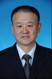 Chunyan Ji (纪春岩)合作论文数Qilu Hospital of Shandong University7Laura F. Gibson合作论文数7
 Xiaonan Han合作论文数7
 Yon Rojanasakul合作论文数Department of Pharmaceutical Sciences, School of Pharmacy, West Virginia University;Cancer Institute, West Virginia University7
 Alexander Chi合作论文数7
 Gengqing Song合作论文数Tongji Medical College, Huazhong University of Science and Technology7Yue Zhang (张悦)合作论文数6
 Qianfei Wang (王前飞)合作论文数Beijing Institute of Genomics, Chinese Academy of Sciences6
 Chengkui Qu (瞿成奎)合作论文数Department of Pediatrics, School of Medicine, Emory University;Aflac Cancer and Blood Disorders Center, Emory University;Winship Cancer Institute, Emory University6
 Stephen D. Nimer合作论文数Miller School of Medicine, University of Miami;Nimer Lab, Sylvester Comprehensive Cancer Center, University of Miami6
 Zhijian Xiao (肖志坚)合作论文数SKLEH, Chinese Academy of Medical Sciences;Chinese Academy of Medical Sciences Peking Union Medical College6
 Jame Abraham合作论文数6
 Irene Helenowski合作论文数Department of Preventive Medicine, Northwestern University;Loyola Medicine6
 Soumit K Basu合作论文数5Sicheng Bian合作论文数5Soojin Kim合作论文数5Yoshihiro Hayashi合作论文数5Yan Xiaomei合作论文数5
 Aili Chen (陈艾莉)合作论文数Beijing Institute of Genomics, Chinese Academy of Sciences5
 Alfred W Rademaker合作论文数Feinberg School of Medicine, Northwestern University5
 Nam Nguyen合作论文数Department of Radiation Oncology, University of Arizona5
 Jiasheng Wang合作论文数5
 Ashok Raj合作论文数5P Bunner合作论文数5




