COVID orchitis (testicular pain) is reported in 10-15% of men with long COVID. We identified 2 siblings with COVID orchitis and hypothesized that genetic mutations are associated with susceptibility. Blood samples from 5 COVID-19 (+) men, three of whom had orchitis were evaluated by whole-exome-sequencing. A rare deletion on chromosome 7 was found in NACAD among the 3 men with orchitis. Interestingly, circulating ACE2 levels was decreased in men with COVID orchitis. This pilot study generated the hypothesis that men who develop COVID orchitis could have underlying genetic variants and altered levels in circulating ACE2 that may increase their risk.
You have accessJournal of UrologyInfections/Inflammation/Cystic Disease of the Genitourinary Tract: Prostate & Genitalia (MP35)1 Sep 2021MP35-17 NOVEL PATHOGENIC VARIANRTS IN ALMS1 GENE IS ASSICIATED WITH PEYRONIE’S DISEASE AND DEPUYTREN’S DISEASE Iakov Efimenko, Willy Chertman, Thomas Masterson, Anthony Griswold, and Ranjith Ramasamy Iakov EfimenkoIakov Efimenko More articles by this author , Willy ChertmanWilly Chertman More articles by this author , Thomas MastersonThomas Masterson More articles by this author , Anthony GriswoldAnthony Griswold More articles by this author , and Ranjith RamasamyRanjith Ramasamy More articles by this author View All Author Informationhttps://doi.org/10.1097/JU.0000000000002044.17AboutPDF ToolsAdd to favoritesDownload CitationsTrack CitationsPermissionsReprints ShareFacebookLinked InTwitterEmail Abstract INTRODUCTION AND OBJECTIVE: Peyronie's disease (PD) is a fibrotic disease of the penis affecting up to 13% of men leading to debilitating penile curvature and inability to have intercourse. Depuytren’s disease is a fibro-proliferative condition of the palmar fascias in the hand, typically resulting in progressive contracture of one or more fingers. PD and DD are reported to occur together in 3–15% of men with PD. Although an underlying genetic relationship has been suggested, the specific genetic factors that predispose men to PD and DD are unknown. We hypothesized that men with PD and DD will have genetic mutations involved in fibrosis. The aim of this study was to find a possible genetic etiology in three non-related patients with both PD and DD. METHODS: We performed a university-wide database search of patients with diagnoses of PD and DD. To identify causative mutations of PD and DD, we performed whole-genome sequencing on three unrelated patients with both PD and DD. Assuming a genetic variant with Mendelian heritability, we prioritized protein-coding variants below population frequency threshold of 5%. RESULTS: We identified 16 candidate genes in which the 3 men with PD and DD each carried at least one rare protein-coding variant. Many of these variations were novel and considered as pathogenic or likely pathogenic. After searching the literature for each of their functions, the AMLS1 gene had the highest probability of involvement in the phenotype. The ALMS1 gene contained a heterozygous nonsynonymous mutation in all three patients, though the exact location varied. The mutations were located in exon 8, 15, and 16, on chromosome 2. The mutations for all three individuals were identified at less than 2% prevalence in ExACT. Interestingly multiorgan fibrosis, a common phenotype related to ALMS, is well-known to be associated with aberrant transforming growth factor beta/bone morphogenetic protein (TGF-β/BMP) signaling and alterations in the TGF-β pathway seem to be a pathogenetic factor in the development of PD and DD. CONCLUSIONS: We identified rare nonsynonymous mutations in AMLS1 gene in 3 unrelated men with both PD and DD. Our results support a genetic basis for PD and DD. Our future studies will evaluate how genetic defects in ALMS could lead to alterations in TGF- β signaling leading to excessive ECM production and a failure to eliminate myofibroblasts. Source of Funding: Hayward Foundation, Hussman Institute for Human Genomics, University of Miami to Iakov Efimenko and Ranjith Ramasamy © 2021 by American Urological Association Education and Research, Inc.FiguresReferencesRelatedDetails Volume 206Issue Supplement 3September 2021Page: e633-e633 Advertisement Copyright & Permissions© 2021 by American Urological Association Education and Research, Inc.MetricsAuthor Information Iakov Efimenko More articles by this author Willy Chertman More articles by this author Thomas Masterson More articles by this author Anthony Griswold More articles by this author Ranjith Ramasamy More articles by this author Expand All Advertisement Loading ...
ObjectiveWe hypothesized that REI fellows had predominantly trained at OBGYN programs with REI fellowships. We investigated the training pedigrees of current reproductive endocrinology and infertility (REI) fellows.Materials and MethodsWe identified 140 current or recent REI fellows at all currently accredited REI fellowship programs. Using publicly available information from program websites or LinkedIn, we obtained the medical school, residency program, and fellowship program that the fellows attended. All information was input into a database and anonymized. Of the forty-nine accredited fellowship programs, forty-eight currently have fellows. Of those, forty-one programs had sufficient information available for inclusion.ResultsAll fellows included started residency between 2017 to 2021. 21 out of 140 (15%) fellows stayed at the same institution they attended for residency for fellowship. Ninety-one (65%) of fellows graduated from an OBGYN residency program that had an REI fellowship. 49 out of 140 (35%) of fellows graduated from OBGYN programs that did not have an REI fellowship. 55 out of 140 (39%) fellows graduated from a medical school with an REI fellowship program. Of the 140 fellows, 49 had both chronological information on residency graduation and fellowship commencement publicly available. For those fellows, the gap between residency and fellowship ranged from 0 to 5 years. 41 out of 49 entered fellowship the same year they graduated residency. The average gap between residency completion and fellowship was < 5 months.ConclusionsA majority (65%) of REI fellows ascertained through publicly available data graduated from OBGYN residency programs with REI programs. Given the competitive nature of REI fellowship applications and previously reported data on REI applicant characteristics, these findings underscore the need for exposure to REI as a specialty in OBGYN residency programs that do not have an REI division.Impact StatementThese findings provide some insight into REI fellowship training paths. ObjectiveWe hypothesized that REI fellows had predominantly trained at OBGYN programs with REI fellowships. We investigated the training pedigrees of current reproductive endocrinology and infertility (REI) fellows. We hypothesized that REI fellows had predominantly trained at OBGYN programs with REI fellowships. We investigated the training pedigrees of current reproductive endocrinology and infertility (REI) fellows. Materials and MethodsWe identified 140 current or recent REI fellows at all currently accredited REI fellowship programs. Using publicly available information from program websites or LinkedIn, we obtained the medical school, residency program, and fellowship program that the fellows attended. All information was input into a database and anonymized. Of the forty-nine accredited fellowship programs, forty-eight currently have fellows. Of those, forty-one programs had sufficient information available for inclusion. We identified 140 current or recent REI fellows at all currently accredited REI fellowship programs. Using publicly available information from program websites or LinkedIn, we obtained the medical school, residency program, and fellowship program that the fellows attended. All information was input into a database and anonymized. Of the forty-nine accredited fellowship programs, forty-eight currently have fellows. Of those, forty-one programs had sufficient information available for inclusion. ResultsAll fellows included started residency between 2017 to 2021. 21 out of 140 (15%) fellows stayed at the same institution they attended for residency for fellowship. Ninety-one (65%) of fellows graduated from an OBGYN residency program that had an REI fellowship. 49 out of 140 (35%) of fellows graduated from OBGYN programs that did not have an REI fellowship. 55 out of 140 (39%) fellows graduated from a medical school with an REI fellowship program. Of the 140 fellows, 49 had both chronological information on residency graduation and fellowship commencement publicly available. For those fellows, the gap between residency and fellowship ranged from 0 to 5 years. 41 out of 49 entered fellowship the same year they graduated residency. The average gap between residency completion and fellowship was < 5 months. All fellows included started residency between 2017 to 2021. 21 out of 140 (15%) fellows stayed at the same institution they attended for residency for fellowship. Ninety-one (65%) of fellows graduated from an OBGYN residency program that had an REI fellowship. 49 out of 140 (35%) of fellows graduated from OBGYN programs that did not have an REI fellowship. 55 out of 140 (39%) fellows graduated from a medical school with an REI fellowship program. Of the 140 fellows, 49 had both chronological information on residency graduation and fellowship commencement publicly available. For those fellows, the gap between residency and fellowship ranged from 0 to 5 years. 41 out of 49 entered fellowship the same year they graduated residency. The average gap between residency completion and fellowship was < 5 months. ConclusionsA majority (65%) of REI fellows ascertained through publicly available data graduated from OBGYN residency programs with REI programs. Given the competitive nature of REI fellowship applications and previously reported data on REI applicant characteristics, these findings underscore the need for exposure to REI as a specialty in OBGYN residency programs that do not have an REI division. A majority (65%) of REI fellows ascertained through publicly available data graduated from OBGYN residency programs with REI programs. Given the competitive nature of REI fellowship applications and previously reported data on REI applicant characteristics, these findings underscore the need for exposure to REI as a specialty in OBGYN residency programs that do not have an REI division.
To characterise trends and interest in selective androgen receptor modulators (SARMs). SARMs are androgen receptor ligands that bind androgen receptors selectively. SARMs have anabolic effects on muscle and bone and were originally synthesised for treatment of muscle wasting conditions, osteoporosis and breast cancer. To date, no SARM has been clinically approved and little is known about their beneficial effects and other adverse effects on users. We examined Google Trend searches of SARMs. Using Google Trends, we analysed how interest in SARMs has evolved over the last 15 years and compared it to trends in testosterone. Comparing 'TRT', 'SARMs' and 'Low Testosterone' together, we see low search interest in SARMs compared to TRT until February 2018, when the interest in both SARMs and TRT terms appear to be the same. Since February 2018, search interest for SARMs has surpassed search interest for both 'TRT' and 'Low Testosterone'. Trends for SARMs demonstrate a continuous increase over the years which has to date surpassed interest for both 'TRT' and 'low testosterone'. The rising interest in SARMs is concerning as the adverse effects of its usage, including its potential effects on fertility, have not been explored. Further investigation is necessary.
Male factor infertility is an important clinical problem whose most severe phenotype, severe oligospermia or azoospermia, has a variety of genetic causes. Some, like Klinefelter syndrome or cystic fibrosis, are well understood, but most are still unknown, and Y chromosome microdeletions only explain a fraction of the remaining cases. In consanguineous and nonconsanguineous families, whole exome sequencing (WES) has been successfully used to identify likely causal mutations in severe oligospermia (1Chertman W. Arora H. Griswold A.J. Lopategui D.M. Ramasamy R. whole exome sequencing identifies a rare nonsense mutation in FAM47C as a possible cause of severe oligospermia in brothers with varicocele.Urology. 2019; 129: 71-73Abstract Full Text Full Text PDF PubMed Scopus (2) Google Scholar, 2Ramasamy R. Bakırcıoğlu M.E. Cengiz C. Karaca E. Scovell J. Jhangiani S.N. Whole-exome sequencing identifies novel homozygous mutation in NPAS2 in family with nonobstructive azoospermia.Fertil Steril. 2015; 104: 286-291Abstract Full Text Full Text PDF PubMed Scopus (41) Google Scholar). In this issue, Tu et al. (3Tu C. Meng L. Nie H. Yuan S. Wang W. Du J. et al.A homozygous RPL10L missense mutation associated with male factor infertility and severe oligozoospermia.Fertil Steril. 2020; 113: 561-568Abstract Full Text Full Text PDF PubMed Scopus (3) Google Scholar) use WES in a set of Han Chinese brothers with idiopathic severe oligospermia and identify a rare recessive mutation in RPL10L that may account for the phenotype. They then demonstrated decreased protein levels compared with the wild type in an in vitro model, examined a cohort of fertile and infertile men for the incidence of RPL10L mutations, and demonstrated a higher frequency in infertile men with oligospermia. Previous studies have not directly examined the incidence of identified variants in other infertile men, therefore this study sheds light on how generalizable the WES studies can be. Identifying this variant at higher frequencies in infertile men should make researchers hopeful that even rare mutations can be validated. Because differentiating between causes of male factor infertility has clinical utility, WES may eventually become a part of the workup for men with idiopathic severe oligospermia or azoospermia in whom the usual tests, such as Y chromosome microdeletion and karyotype, have not yielded any abnormalities. Future directions of this work may include [1] pooling large cohorts of men with azoospermia for WES, [2] the use of WES and analysis of genomic structural variation in cases where WES fails to yield a result, [3] more in vitro confirmatory work when possibly causal variants are identified, and [4] identifying a future male factor infertility diagnostic gene panel that has sound evidence to support the gene-disease relationship. A homozygous RPL10L missense mutation associated with male factor infertility and severe oligozoospermiaFertility and SterilityVol. 113Issue 3PreviewTo identify the genetic cause of male factor infertility characterized by severe oligozoospermia. Full-Text PDF
You have accessJournal of UrologyInfertility: Basic Research & Pathophysiology (MP75)1 Apr 2019MP75-08 WHOLE EXOME SEQUENCING IDENTIFIES A RARE NONSENSE MUTATION IN FAM47C AS A POSSIBLE CAUSE OF SEVERE OLIGOSPERMIA IN BROTHERS WITH VARICOCELE Willy Chertman, Diana M Lopategui, Anthony J Griswold, Himanshu Arora*, and Ranjith Ramasamy Willy ChertmanWilly Chertman More articles by this author , Diana M LopateguiDiana M Lopategui More articles by this author , Anthony J GriswoldAnthony J Griswold More articles by this author , Himanshu Arora*Himanshu Arora* More articles by this author , and Ranjith RamasamyRanjith Ramasamy More articles by this author View All Author Informationhttps://doi.org/10.1097/01.JU.0000557236.06494.54AboutPDF ToolsAdd to favoritesDownload CitationsTrack CitationsPermissionsReprints ShareFacebookLinked InTwitterEmail Abstract INTRODUCTION AND OBJECTIVES: The pathologic enlargement of veins of the pampiniform plexus within the spermatic cord, or varicocele, is a common cause of impaired semen parameters. Varicocele is prevalent among adolescent males (15%), nevertheless only a small proportion (1%) of men with varicoceles have impaired semen parameters. Yet, varicocele remains the most common correctable cause of male infertility. We investigated genetic variants as possible causes of varicocele with decreased sperm count in two brothers, using targeted exon/whole-exome sequencing (WES). METHODS: We extracted DNA from the blood of 2 affected brothers (with severe oligospermia) and 1 unaffected brother (normozoospermia) using QIAamp DNA Blood Maxi Kit. Extracted DNA was used for the whole exome sequencing (WES). All 3 brothers had left grade II varicocele. Two of them had decreased sperm count (< 1 million/cc) while the other brother had normal count (>20 million/cc) on at least 2 semen analyses. The raw sequence data were aligned to the reference genome (hg19) and filtered for quality control including coverage and genotype quality. Potentially pathogenic variants were identified by the following method: homozygous variants shared by the two brothers with varicocele and severe oligospermia but not by the brother with varicocele and normal sperm count, rare (below 0.05 allele frequency) according to ExAC and the 1000 Genomes database, and missense, nonsense, or frameshift variants. Sanger sequencing was performed to confirm the variant in the 2 affected brothers and determine segregation in the parents. RESULTS: A premature stop codon alteration (nonsense) was identified on Chromosome X (37028866 C>T) in the gene FAM47C. The affected brothers were found to be hemizygous for the variant, while the mother was a heterozygous carrier. The unaffected brother and the father both carried the reference allele. Sanger sequencing confirmed the variant. The gene FAM47C has been previously identified with male infertility in a copy-number variant that included the FAM47C gene and is also highly expressed in testis tissue, though its function is unknown [2]. CONCLUSIONS: Using WES we were able to identify a nonsense mutation in FAM47C that segregates with the oligospermia phenotype. The mutation was present in the severely oligospermic brothers and not in the normozoospermic brother despite all 3 brothers having a similar varicocele clinical grade. Because varicocele is prevalent (15%) in the population, identifying men that would have impaired spermatogenesis using approaches like WES can be paradigm shifting. Source of Funding: Supported by the American Urological Association Research Scholar Award and Stanley Glaser Award to RR. MIAMI, FL; Miami, FL© 2019 by American Urological Association Education and Research, Inc.FiguresReferencesRelatedDetails Volume 201Issue Supplement 4April 2019Page: e1084-e1085 Advertisement Copyright & Permissions© 2019 by American Urological Association Education and Research, Inc.MetricsAuthor Information Willy Chertman More articles by this author Diana M Lopategui More articles by this author Anthony J Griswold More articles by this author Himanshu Arora* More articles by this author Ranjith Ramasamy More articles by this author Expand All Advertisement PDF downloadLoading ...
Varicocele is a common cause of impaired semen parameters in men with infertility. Here, we investigated genetic variants as possible causes of varicocele with impaired semen parameters using whole exome sequencing in a family with 2 brothers with severe oligospermia, 1 unaffected brother, father, and the mother. Results showed a premature stop codon alteration on Chromosome X (37028866 CT) in the gene FAM47C. The affected brothers were found to be hemizygous for the variant, while the mother was a heterozygous carrier. In conclusion, identifying men with varicocele that would have impaired spermatogenesis, using approaches like whole-exome sequencing, can be paradigm shifting.
 Ranjith Ramasamy合作论文数Jumeirah American Clinic8
 Iakov Efimenko合作论文数4
 Anthony J. Griswold合作论文数Department of Biology, Indiana University4
 Himanshu Arora合作论文数Geetanjali Institute of Technical Studies Udaipur3
 Thomas Masterson合作论文数2 Department of Urology, University of Miami3Diana M. Lopategui合作论文数2Ayodele Omotoso合作论文数1
 Justin M Dubin合作论文数Dept Urol, Univ Miami1
 R. Reddy合作论文数Univ Texas Southwestern Med Ctr1
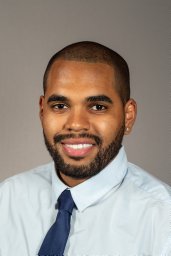 Parris Diaz合作论文数Miller School of Medicine, University of Miami1
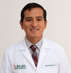 Alex P. Sanchez-Covarrubias合作论文数Sylvester Comprehensive Cancer Center, University of Miami Miller School of Medicine1
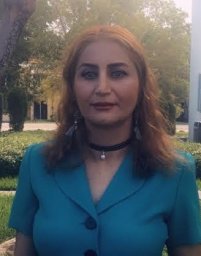 Kajal Khodamoradi合作论文数Desai Sethi Urology Institute, University of Miami1
 Andre Pinto合作论文数1
 Sophia H. L. George合作论文数1
 Matthew P Schlumbrecht合作论文数Department of Gynecologic Oncology, The University of Texas M. D. Anderson Cancer Center1




