This review examines challenges in post-stroke dysphagia management, including access to swallowing imaging, enteral feeding decisions, and barriers to reassessment and rehabilitation. It evaluates how healthcare systems and practice patterns influence swallowing outcomes and stroke recovery. Recent literature emphasizes that post-stroke dysphagia is a dynamic condition requiring longitudinal management rather than a temporary acute complication. Evidence supports validated dysphagia screening followed by clinical swallowing evaluation and swallowing imaging for accurate diagnosis and treatment planning. Studies highlight variability in gastrostomy tube practices, post-acute access to swallowing imaging, continuity of care, and the psychosocial burden of oral restriction and feeding decisions after stroke. Dysphagia management systems remain fragmented, contributing to inconsistent diagnosis, reassessment, access to intervention, and prolonged unnecessary oral intake restrictions. Future efforts should prioritize rehabilitation pathways that support recovery-oriented dysphagia management.
Cranial radiation is a cornerstone in management of glioma though is associated with increased stroke risk. Hyperlipidemia has previously been found to be associated with increased stroke risk in this population. This study aimed to evaluate whether statin use is associated with reduced stroke risk in patients with glioma and hyperlipidemia who underwent cranial radiation. We conducted a retrospective study of adult patients with glioma and hyperlipidemia who received cranial radiation between 2005 and 2021. Data were collected from time of cranial radiation until last follow-up. Patients diagnosed with hyperlipidemia through review of medical records at time of stroke were included in the analysis. Cox-proportional hazards modeling was performed to evaluate incident stroke with death as a competing event to evaluate the impact of statin exposure on stroke. In a cohort of 297 patients that received cranial radiation and had hyperlipidemia, 65 (21.9
Over the past 2 decades, transcranial direct current stimulation has attracted substantial interest as an adjunctive strategy to enhance poststroke motor recovery. In addition to its potential neuromodulatory effects on motor cortical networks, transcranial direct current stimulation devices are low-cost, easy to use, and compatible with concurrent rehabilitation. Yet, despite its promise, several barriers hinder translation into routine clinical practice, including neutral results from several recently completed multicenter trials, such as TRANSPORT2 (Transcranial Direct Current Stimulation for Post-Stroke Motor Recovery). Moving forward, progress will depend on addressing issues in 3 broad domains: device-related (stimulation parameters and montage), disease-related (patient characteristics and timing), and trial design (outcomes, analytical approaches, adjunctive therapy, and trial infrastructure). In this topical review, we critically examine these challenges and outline strategies to refine transcranial direct current stimulation application, with the goal of more effectively leveraging its neuromodulation properties to promote neuroplasticity and enhance motor recovery after stroke.
BACKGROUND:Normobaric hyperoxia (NBO) is a simple neuroprotective strategy that may augment endovascular thrombectomy (EVT) in acute ischemic stroke. We evaluated the safety and preliminary efficacy of NBO plus EVT in patients with large-vessel occlusion presenting 6 to 24 hours after onset. METHODS:In this phase IIb, randomized, assessor-blinded, controlled trial conducted at 2 academic comprehensive stroke centers in China, patients aged ≥18 years with anterior circulation large-vessel occlusion presenting 6 to 24 hours after acute ischemic stroke onset were assigned 1:1 to EVT+NBO or EVT alone. The NBO group received 100% oxygen via a face mask at 10 L/min for 4 hours, starting before recanalization. The primary end point was early neurological improvement (≥30% reduction in the National Institutes of Health Stroke Scale score at 24 hours). Primary analyses used adjusted regression models controlling for prespecified prognostic covariates. Secondary end points included infarct volume at 24 to 48 hours and the modified Rankin Scale score at 90 days. Safety outcomes were mortality, intracranial hemorrhage, and symptomatic intracranial hemorrhage. Analyses followed the intention-to-treat principle. Early neurological improvement was analyzed using an adjusted binomial regression model, and the 90-day modified Rankin Scale shift was analyzed using an adjusted ordinal logistic regression model, controlling for age, sex, intravenous thrombolysis, and occlusion site. RESULTS:Between October 2021 and October 2023, 324 patients were screened, and 120 were randomly assigned to NBO+EVT or EVT alone (60 patients per group; intention-to-treat population). The median baseline National Institutes of Health Stroke Scale score was 12 (interquartile range [IQR], 8-15) in the NBO+EVT group and 12 (IQR, 9-16) in the EVT-alone group. The median time from stroke onset to randomization was 10.0 hours (IQR, 7.0-14.4) and 9.9 hours (IQR, 8.4-14.8), respectively. The EVT+NBO group demonstrated a significantly higher rate of early neurological improvement compared with EVT alone (35% versus 19%; adjusted odds ratio, 2.86 [95% CI, 1.12-7.45]). The median infarct volume at 24 to 48 hours was significantly smaller in the EVT+NBO group (20.5 [IQR, 13.6-31.8] mL versus 32.3 [IQR, 22.7-44.5] mL; P=0.001). At 90 days, the modified Rankin Scale distribution numerically favored NBO+EVT but was not statistically significant (median modified Rankin Scale, 2 [IQR, 1-3] versus 3 [IQR, 1-4]; adjusted common odds ratio, 1.52 [95% CI, 0.87-2.63]). Mortality, symptomatic intracranial hemorrhage, early neurological deterioration, and recurrent stroke did not differ between groups. CONCLUSIONS:In patients with acute ischemic stroke treated 6 to 24 hours after onset, adjunctive NBO with EVT was safe and improved early neurological outcomes and infarct volume, supporting further evaluation in larger trials. REGISTRATION:URL: https://www.clinicaltrials.gov; Unique identifier: NCT05128422.
Cerebrospinal fluid circulation through the glymphatic system plays a crucial role in removing metabolic waste from the central nervous system. However, the mechanism underlying the brain-wide glymphatic dynamics is not yet fully understood, in part due to the lack of glymphatic imaging technologies on deep brains. Here, we report a hybrid imaging technology that integrates three-dimensional photoacoustic tomography and ultrasound localization microscopy (3D-PAULM), enhanced by a photoacoustic dye with strong optical absorption in the second near-infrared window (NIR-II). 3D-PAULM allows for continuous, noninvasive, whole-brain imaging in mice through intact skull, providing superresolution mapping of the brain vasculature and highly sensitive tracing of the NIR-II dye in the glymphatic system. Using 3D-PAULM, we investigated the glymphatic function impaired by ischemic stroke, aging, and anesthesia. Our results provide insights into glymphatic transport under various physiological as well as pathological conditions and establish 3D-PAULM as a valuable tool for preclinical glymphatic research.
BACKGROUND:Stroke leads to complex chronic structural and functional brain changes that specifically affect motor outcomes. The brain predicted age difference (PAD) has emerged as a sensitive biomarker of both sensorimotor and cognitive function after stroke. Our previous study showed a higher global brain PAD associated with poorer motor function after stroke. However, the association between local stroke lesion load, regional brain age, and motor impairment is unclear. This study aimed to investigate the associations between focal lesion damage, regional brain PAD in both hemispheres, and motor outcomes in chronic stroke, and to identify key predictors of motor impairment. METHODS:In this multicohort, retrospective, observational study, we included individuals with chronic unilateral stroke (>180 days post stroke) from the ENIGMA Stroke Recovery Working Group dataset and used individuals from the UK Biobank cohort to train the regional brain age prediction model. Structural T1-weighted MRI scans were used to estimate regional brain PAD in 18 predefined functional subregions via a graph convolutional network algorithm. Lesion load for each region was calculated on the basis of lesion overlap. Linear mixed-effects models assessed associations between lesion size, local lesion load, and regional brain PAD. Machine learning classifiers predicted motor outcomes using lesion loads and regional brain PADs. Structural equation modelling examined directional relationships among corticospinal tract lesion load, ipsilesional brain PAD, motor outcomes, and contralesional brain PAD. FINDINGS:We included 501 individuals from the ENIGMA Stroke Recovery Working Group dataset (34 cohorts in eight countries) and 17 791 individuals from the UK Biobank dataset. Larger total lesion size was positively associated with higher ipsilesional regional brain PADs (older brain age) across most regions (β=0·5420 to 0·9458 across significantly correlated regions, false discovery rate [FDR]-corrected p<0·05), and with lower brain PAD in the contralesional ventral attention and language network region (β=-0·3747, 95% CI -0·6961 to -0·0534, FDR-corrected p<0·05). Higher local lesion loads showed similar patterns. Specifically, lesion load in the salience network significantly influenced regional brain PADs across both hemispheres. Machine learning models identified corticospinal tract lesion load (adjusted mean difference -0·0905, 95% CI -0·1221 to -0·0589, p<0·0001), salience network lesion load (-0·0632, -0·0906 to -0·0358, p<0·0001), and regional brain PAD in the contralesional frontoparietal network (0·9939, 0·4929 to 1·4950, p=0·0001) as the top three predictors of motor outcomes. Structural equation modelling revealed that higher corticospinal tract lesion load was associated with poorer motor outcomes (β=-0·355, 95% CI -0·446 to -0·267, p<0·0001), which were further linked to younger contralesional brain age (0·204, 0·111 to 0·295, p<0·0001), suggesting that severe motor impairment is linked to compensatory decreases in contralesional brain age. INTERPRETATION:Our findings reveal that larger stroke lesions are associated with accelerated ageing in the ipsilesional hemisphere and paradoxically decelerated brain ageing in the contralesional hemisphere, suggesting compensatory neural mechanisms. Assessing regional brain age might serve as a biomarker for neuroplasticity and inform targeted interventions to enhance motor recovery after stroke. FUNDING:US National Institutes of Health.
BACKGROUND: Stroke is a possible complication after bioprosthetic aortic valve replacement (AVR) for severe aortic stenosis (AS), impacting morbidity and mortality. Accurate estimates of the proportion of individuals who experience stroke within and beyond the periprocedural period after transcatheter AVR (TAVR), surgical AVR, and valve-in-valve (ViV) replacement are essential for management and prognostication. The objective was to determine the proportion of adults aged >18 who experienced an ischemic stroke after bioprosthetic AVR for AS METHODS: A systematic search of MEDLINE, Embase, and Web of Science was conducted from database inception through March 2024. Studies reporting on stroke rates at least 90 days after bioprosthetic AVR for severe AS, including VIV procedures, and meeting predefined eligibility criteria were included. The pooled proportion of individuals experiencing a stroke was estimated for TAVR and ViV procedures, whereas comparative analyses between TAVR and surgical AVR were performed using mixed-effects models in studies directly comparing both procedures. RESULTS: Twenty-seven studies were included in the native AS treatment cohort, and 5 in the ViV subanalysis. In native AS, the pooled 30-day proportion of individuals who had a stroke after TAVR was 3.0% (95% CI, 2.5-3.9), with different studies reporting major and minor stroke proportions of 1.7% each. At 1 year, all stroke proportion was 5.0% (95% CI, 4.0-6.0), major stroke was 3.0%, and minor stroke was 2.0%. Comparative analysis demonstrated that TAVR was associated with significantly lower odds of all stroke at 30 days compared with surgical AVR (odds ratio, 0.73 [95% CI, 0.57-0.93]). No significant difference in the proportions of individuals who had a stroke was observed in TAVR versus surgical AVR at 1, 2, or 5 years. In the ViV cohort, the pooled 30-day and 1-year all stroke proportion after ViV was 2.0% (95% CI, 1.0-3.0) and 3.0% (95% CI, 2.0-6.0), respectively. CONCLUSIONS: This meta-analysis provides updated estimates of stroke after bioprosthetic AVR for AS, capturing risk beyond the early periprocedural period. Future studies should investigate the causes of long-term stroke post-AVR, the effects of different antithrombotic therapies on the risk of stroke, as well as the potential impact of these procedures on short and long-term cognitive function.
Transcranial focused ultrasound (tFUS) is a non-invasive neuromodulatory tool that holds promise for various neuropsychiatric disorders. While it offers several distinct advantages, it also faces notable technical challenges. The irregular shape and inhomogeneous acoustic properties of the human skull impede efficient acoustic energy transmission through the skull. So far, clinical semi-spherical (hemispherical) arrays still suffer from strong wave reflection and refraction at the skull interface, especially with steering. We propose a flexible ultrasound array that conforms to individual skull shapes and can be optimized to target vertex-accessible subcortical regions. The impact of flexible array configuration was investigated by comparing the flexible array with a semi-spherical array commonly used in the clinical setting. Numerical results show that the random-patterned flexible array reduces the z-axis -6 dB full width at half maximum (FWHM) by 29.4% and enhances the focal peak pressure by 44.4% when compared to the semi-spherical array without steering. In addition, it achieves a wide steering range over a 30 × 20 mm2 region while maintaining the focusing performance. We expect that our proposed tFUS stimulation with a flexible array may provide a theoretical framework for improving the therapeutic efficiency for various neuropsychiatric conditions.
INTRODUCTION:Post-stroke dysphagia (PSD) is a common complication following acute ischemic stroke (AIS). Predicting the recovery of swallow function remains challenging. The Predictive Swallow Score (PRESS) model, derived and validated in a Swiss cohort, sought to predict the recovery of PSD after AIS. We aimed to validate the PRESS model in a US-cohort, conducting a two-center retrospective review of 149 patients with AIS and functional oral intake scale (FOIS) ≤ 4. METHODS:We collected the predictors of recovery of PSD according to PRESS (age, NIH Stroke Scale (NIHSS), any2 score, stroke location, FOIS score), with a primary outcome of impaired swallow at day 7 (FOIS ≤ 4). Model validation was completed using the Hosmer-Lemeshow (HL) test, calibration plots, and AUC analysis. RESULTS:Median (IQR) age was 74 (61-86); 53% were female. Median (Q1-Q3) NIHSS at presentation was 14 (7-20). HL test demonstrated that the PRESS model did not fit the validation data (p < 0.00001, x2=48.343, df=5), and the calibration curve analysis (intercept = -0.80 (95% CI: -1.21 to -0.38), slope = 0.60 (95% CI: 0.37 to 0.82)) also demonstrated a poor calibration of the model. Area under the curve analyses demonstrated a C statistic of 0.75 (95% CI 0.67-0.82), indicating suboptimal model discrimination in predicting the recovery of swallow 7 days following AIS. In particular, the model overpredicted dysphagia severity at day-7 in patients with higher PRESS scores and more severe strokes. CONCLUSION:Further validation of the PRESS score in prospective cohorts is warranted. The suboptimal model performance could be attributed to temporal advances in stroke care, as the original PRESS cohort was derived between 2011 and 2014. Geographic variability in acute stroke care practice could also be a factor, as the PRESS score was derived solely from a European cohort. This study, however, is limited by its retrospective design and a lack of generalizability.
BackgroundRehabilitation is an essential health service that should be available for all with health conditions affecting functioning in daily life. Initiatives for equitable distribution of scarce and limited rehabilitation resources are frequently guided by burden of diseases measures. Most studies on burden of diseases have laid greater emphasis on disability adjusted life years (DALYs) and rarely compared these burdens among conditions. This present study aims to systematically review and compare the burden of the top 10 GBD ranking of disorders.MethodThis review was pre-registered with PROSPERO (CRD42022316091). PubMed and Google Scholar were systematically searched as well as a manual search of grey literatures from 01/01/1990 to 29/02/2022. The results of this review were reported based on PRISMA guideline. Meta-analysis results were presented using forest plots and summary tables.ResultsA total of 11,367 studies were obtained from the searches, while the findings of 55 studies (17,753,434 participants) were reviewed. Majority of the studies were conducted in high-income-countries (56.1%) though, all the studies on neonatal disorders (100.0%) and congenital birth disorders (100.0%) came from the low-and-middle-income-countries. Neonatal disorders, stroke and ischaemic heart disease (IHD) topped the rank of disease burden. Overall, the burden of neurological disorders and their associated risk factors (aRFs), in terms of disease prevalence, were evaluated to be 36.75% while their mortality rate was 29.90%. Neurological conditions and aRFs accounted for 61.83% of total economic burden.ConclusionNeonatal disorders, stroke and IHD are the three most burdensome disorders. There is therefore need for greater focus of rehabilitation attention and resources in the coming decades.
Transcranial ultrasonic stimulation (TUS) is emerging as a non-invasive neuromodulatory technique capable of delivering millimeter-precision stimulation at whole-brain depths. Research efforts have increasingly focused on its translational potential. Promising data have been reported across several disease populations, including Parkinson's disease and stroke, paving the way for clinical applications of TUS. Clinical studies to date, however, show substantial variability in transducer fixation, targeting approaches, and acoustic parameters. This limits the interpretability and comparability of results. Existing methodological guides address human TUS in general but do not focus on applications in neurological populations. This experimental protocol presents a standardized yet adaptable framework for applying TUS to neurological cohorts such as stroke. It offers detailed guidance on: (1) essential and optional hardware components in the context of therapy-oriented TUS; (2) hardware settings and parameter selection, including strategies to minimize auditory confounds; (3) calibration and quality assurance procedures to ensure the transducer delivers waveforms as specified; (4) targeting approaches based on simulation or non-simulation methods for accurate localization of TUS focus/foci to the intended anatomical region(s); (5) methodology adaption for clinical populations; and (6) outcome measures for clinical TUS, encompassing safety assessments and surrogate outcome measures such as corticospinal excitability and motor sequence learning. This protocol is designed as a replicable, modular resource. It accommodates both novice users (seeking a practical entry point into patient-based TUS) and experienced researchers (aiming to align with emerging scientific and methodological standards). The goal is to support the growing clinical interest in TUS and to facilitate clinically translatable, reproducible, and comparable results across research groups and patient populations.
The prevalence of Alzheimer’s Disease (AD) is increasing worldwide, with more emergency providers and neurologists expecting to encounter these patients. The paradigm of management of AD is expected to change given the recent approval of anti-amyloid therapies (AATs). The most concerning complication of these therapies is amyloid-related imaging abnormalities (ARIA), which can lead to an increased risk of cerebrovascular complications. Given a growing population of patients with AD and growing use of AATs, providers must be prepared to manage patients at risk of cerebrovascular disease and those presenting with neurologic deficits. This subpopulation warrants a unique approach given the risk of ischemic stroke and the associated risk of hemorrhage present in the use of AATs. In this narrative review, we present and propose management considerations in the acute stroke setting and patients at risk of cerebrovascular disease, including patients with indications for anticoagulation, to most appropriately manage this special population. Future cross-disciplinary collaboration and use of registry data will be essential to narrow management approaches and develop safety data.
Background Endovascular treatment improves the recanalisation rate for patients with acute ischaemic stroke; however, even with endovascular treatment, approximately half of patients do not have a favourable functional outcome. We aimed to evaluate the effect of normobaric hyperoxia combined with endovascular treatment on functional outcomes up to 90 days after treatment in patients who had an acute ischaemic stroke with large-vessel occlusion. Methods In this multicentre, randomised, single-blind, sham-controlled trial, patients aged 18-80 years presenting within 6 h of acute ischaemic stroke attributed to large-vessel occlusion in anterior circulation, who were candidates for endovascular treatment, were recruited from 26 comprehensive stroke centres in China. Eligible patients were randomly assigned (1:1), with an Interactive Web Response System on the basis of a minimisation process to balance assignment at each participating site both overall and according to age, sex, occlusion location, and use of intravenous thrombolytics, to receive either normobaric hyperoxia combined with endovascular treatment or sham normobaric hyperoxia combined with endovascular treatment. Participants and assessors were blinded to treatment assignment. Normobaric hyperoxia treatment involved inhaling 100% oxygen at a flow rate of 10 L/min through a non-rebreather mask for 4 h, or an inspiratory oxygen fraction (FiO(2)) of 10 in participants for whom intubation was necessary. Sham treatment was 100% oxygen delivered at a flow rate of 1 L/min or an FiO(2) of 03. The primary outcome was the comparison of the ordinal scores on the modified Rankin Scale (mRS) at 90 days assessed in the intention-to-treat population (including all patients randomly assigned to treatment). Safety was assessed in all patients who received any oxygen therapy. This trial is registered with ClinicalTrials.gov, NCT04681651, and is now complete. Findings Between April 22, 2021, and Feb 5, 2023, 473 patients were screened, of whom 282 were randomly assigned to either normobaric hyperoxia plus endovascular treatment (n=140) or sham normobaric hyperoxia plus endovascular treatment (n=142; intention-to-treat population). The median age was 65 years (IQR 57-71), 75 (27%) of 282 participants were female, 207 (73%) were male, and 282 (100%) of participants were of Chinese Han ethnicity. At 90 days, the median score on the mRS for the normobaric hyperoxia group was 2 (IQR 1-4) and it was 3 (1-4) in the sham normobaric hyperoxia group (adjusted common odds ratio 165 [95% CI 109-250]; p=0018). At 90 days, 14 (10%) of 140 patients in the normobaric hyperoxia group and 17 (12%) of 142 in the sham normobaric hyperoxia group died (adjusted risk difference -002 [95% CI -009 to 006]) and 28 (20%) and 33 (23%) had serious adverse events (adjusted risk difference -003 [-012 to 007]). Interpretation In patients with acute ischaemic stroke caused by large-vessel occlusion in the anterior circulation who were candidates for endovascular treatment, normobaric hyperoxia yielded superior functional outcomes at 90 days compared with the sham normobaric hyperoxia, without raising safety concerns.
BACKGROUND:Motor impairments contribute substantially to long-term disability following stroke. Studies of transcranial direct current stimulation (tDCS), combined with various rehabilitation therapies, have shown promising results in reducing motor impairment. We aimed to evaluate the safety and efficacy of three doses of tDCS in combination with modified constraint-induced movement therapy (mCIMT) in people who have had their first ischaemic stroke in the preceding 1-6 months. METHODS:We conducted a phase 2, multicentre, randomised, triple-blind, sham-controlled study with a blinded centrally scored primary outcome. The trial was conducted at 15 medical centres in the USA. Eligible participants were enrolled between 1 month and 6 months after their first ischaemic stroke. Inclusion criteria required participants to have a persistent motor deficit, defined as a Fugl-Meyer Upper-Extremity (FM-UE) score of 54 or lower (out of 66), and two consecutive baseline visits (separated by 7-14 days) with an absolute difference of 2 or fewer points on the FM-UE scale. Participants were randomly assigned to treatment groups by an adaptive randomisation algorithm hosted on the TRANSPORT2 WebDCU study website. Participants received either sham, 2 mA, or 4 mA of bi-hemispheric tDCS for the first 30 min and mCIMT with 120 min of active therapy time per session, administered over ten sessions during a 2-week period. The primary endpoint was the change in FM-UE score from baseline to day 15, which was analysed in all participants who have data both at baseline and post-baseline (modified intention-to-treat group). Safety outcomes were analysed in all participants. TRANSPORT2 is registered at clinicaltrials.gov (NCT03826030) and its status is completed. FINDINGS:129 participants were recruited between Sept 9, 2019, and June 14, 2024, and 43 participants were randomly assigned to each group. 54 (42%) of 129 participants were female, and 69 (53%) were White. Two participants in the sham plus mCIMT group withdrew consent before the day 15 assessment and were excluded from the primary analysis. The median baseline FM-UE score was 39·0 (IQR 30·0-46·0) in the sham plus mCIMT group, 39·0 (27·0-48·0) in the 2 mA plus mCIMT group, and 40·0 (27·0-48·0) in the 4 mA plus mCIMT group. For the primary outcome, the adjusted mean change from baseline to day 15 in FM-UE was 4·91 (3·00-6·82) for sham plus mCIMT, 3·87 (2·00-5·74) for 2 mA plus mCIMT, and 5·53 (3·64-7·42) for 4 mA plus mCIMT (p=0·39). No clinically important adverse events were observed in any group and no deaths were reported. INTERPRETATION:tDCS at doses of 2 mA or 4 mA, in addition to mCIMT, did not lead to further reduction in motor impairment in patients 1-6 months after stroke, but it was safe, well tolerated, and feasible for clinical practice. tDCS at higher doses (ie, >4 mA) might be a consideration for future trials in addition to balancing known covariates affecting stroke recovery during the group allocation. FUNDING:National Institute of Neurological Disorders and Stroke.
Objective: We aimed to determine the maximum safe spatial-peak pulse-average intensity (ISPPA) of low-intensity focused ultrasound stimulation (LIFUS) in stroke patients and explore its effect on motor learning and corticospinal excitability. Methods: We adopted the classic 3 + 3 design to escalate ISPPA (estimated in-vivo transcranial value) from 0, 1, 2, 4, 6, to 8 W/cm2. Stopping rules were pre-defined: 2nd-degree scalp burn, clinical seizure, new lesion on diffusion-weighted imaging or major reduction in apparent diffusion coefficient, and participant discontinuation due to any reason. We applied 12-min LIFUS over the ipsilesional motor cortex while participants were concurrently practicing 3 blocks of a motor sequence learning (MSL) task using the affected hand. We measured MSL (response time) and corticospinal excitability (motor evoked potential) pre- and post-stimulation and compared MSL and corticospinal excitability between the LOW (0, 1, and 2 W/cm2) and HIGH (4, 6, and 8 W/ cm2) groups. Results: ISPPA was escalated to 8 W/cm2 with 18 stroke participants without meeting the stopping rules. Compared to the LOW, more participants in the HIGH performed better on MSL (6/9 vs. 0/9, p = 0.009) and showed a sign of greater corticospinal excitability (7/9 vs. 5/9, p = 0.62). Interpretation: Our phase-I safety study suggests that one session of LIFUS up to 8 W/cm2 ISPPA is safe and feasible in stroke patients, and LIFUS at high intensity induces positive changes in both MSL and corticospinal excitability. The next logical step is to conduct a phase-II trial testing the efficacy of LIFUS and continuously monitoring its safety profiles.
Background:Stroke leads to complex chronic structural and functional brain changes that specifically affect motor outcomes. The brain-predicted age difference (brain-PAD) has emerged as a sensitive biomarker. Our previous study showed higher global brain-PAD associated with poorer motor function post-stroke. However, the relationship between local stroke lesion load, regional brain age, and motor impairment remains unclear. Methods:We studied 501 individuals with chronic unilateral stroke (>180 days post-stroke) from the ENIGMA Stroke Recovery Working Group dataset (34 cohorts). Structural T1-weighted MRI scans were used to estimate regional brain-PAD in 18 predefined functional subregions via a graph convolutional network algorithm. Lesion load for each region was calculated based on lesion overlap. Linear mixed-effects models assessed associations between lesion size, local lesion load, and regional brain-PAD. Machine learning classifiers predicted motor outcomes using lesion loads and regional brain-PADs. Structural equation modeling examined directional relationships among corticospinal tract lesion load (CST-LL), ipsilesional brain-PAD, motor outcomes, and contralesional brain-PAD. Findings:Larger total lesion size was positively associated with higher ipsilesional regional brain-PADs (older brain age) across most regions (p < 0.05), and with lower contralesional brain-PAD, notably in the ventral attention-language network (p < 0.05). Higher local lesion loads showed similar patterns. Specifically, lesion load in the salience network significantly influenced regional brain-PADs across both hemispheres. Machine learning models identified CST-LL, salience network lesion load, and regional brain-PAD in the contralesional frontoparietal network as the top three predictors of motor outcomes. Structural equation modeling revealed that larger stroke damage was associated with poorer motor outcomes (β = -0.355, p < 0.001), which were further linked to younger contralesional brain age (β = 0.204, p < 0.001), suggesting that severe motor impairment is linked to compensatory decreases in contralesional brain age. Interpretation:Our findings reveal that larger stroke lesions are associated with accelerated aging in the ipsilesional hemisphere and paradoxically decelerated brain aging in the contralesional hemisphere, suggesting compensatory neural mechanisms. Assessing regional brain age may serve as a biomarker for neuroplasticity and inform targeted interventions to enhance motor recovery after stroke. Fundings:Micheal J Fox Foundation, National Institutes of Health, Canadian Institutes of Health Research, National Health and Medical Research Council, Australian Brain Foundation, Wicking Trust, Collie Trust, and Sidney and Fiona Myer Family Foundation, National Heart Foundation, Hospital Israelita Albert Einstein, Australian Research Council Future Fellowship, Wellcome Trust, National Institute for Health Research Imperial Biomedical Research Centre, European Research Council, Deutsche Forschungsgemeinschaft, REACT Pilot, National Resource Center, Research Council of Norway, South-Eastern Norway Regional Health Authority, Norwegian Extra Foundation for Health and Rehabilitation, Sunnaas Rehabilitation Hospital HT, University of Oslo, and VA Rehabilitation Research and Development.
 Pratik Chhatbar合作论文数42
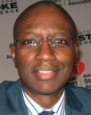 Bruce Ovbiagele合作论文数Weill Institute for Neurosciences, University of California, San Francisco;School of Medicine, University of California, San Francisco;San Francisco Veterans Health Care System38
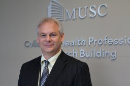 Steven A. Kautz合作论文数Department of Veteran Affairs Rehabilitation Research and Development Brain Rehabilitation Research Center, the Malcom Randall VA Medical Center36
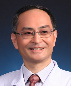 Xunming Ji (吉训明)合作论文数Department of Neurosurgery, Xuanwu Hospital, Capital Medical University;Graduate School, Capital Medical University25
 Haiqing Song (宋海庆)合作论文数Xuanwu Hospital, Graduate School, Capital Medical University25
 Robert Joseph Adams合作论文数College of Medicine, Medical University of South Carolina20
 Shimeng Liu合作论文数Neurol, Univ Calif Irvine14
 George Wittenberg合作论文数University of Pittsburgh14
 Mark S George合作论文数Magnetic Brain Stimulation Laboratory, Department of Psychiatry, Medical University of South Carolina;Brain Imaging Division, Department of Psychiatry, Medical University of South Carolina14
 Steven L. Wolf合作论文数Department Rehabilitation Medicine, School of Medicine, Emory University12
 Gottfried Schlaug合作论文数Neuroimaging Laboratory, Beth Israel Deaconess Medical Center;Music and Neuroimaging Laboratory, Harvard Medical School;Department of Neurology, University of Massachusetts Medical School12
 Xiaoning Jiang合作论文数11
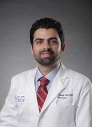 Alawieh Ali合作论文数Department of Neurosciences, Medical University of South Carolina;Department of Internal Medicine, American University of Beirut Medical Center;Medical University of South Carolina, American University of Beirut Medical Center11
 Dylan Ryan合作论文数10
 Jody Feld合作论文数Department of Orthopaedic Surgery, Duke University School of Medicine10
 Qingfeng Ma (马青峰)合作论文数Xuanwu Hospital Capital Medical University10
 Gerard E. Francisco合作论文数Department of Physical Medicine and Rehabilitation, McGovern Medical School, University of Texas Health Science Center at Houston10
 Taewon Kim合作论文数Neuroscience and Rehabilitation Laboratory, Washington University School of Medicine in St. Louis9
 Brian C. Mac Grory合作论文数School of Medicine, Duke University9
 Steven C. Cramer合作论文数University of California, Los Angeles9
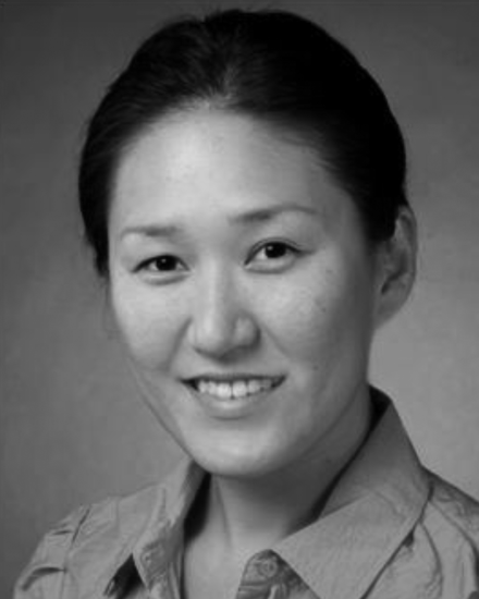 Na Jin J Seo合作论文数9
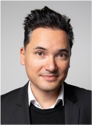 Surjo R. Soekadar合作论文数The Clinical Neurotechnology Laboratory, Charité –University Medicine Berlin9
 Borich Michael R合作论文数Department of Physical Medicine and Rehabilitation, University of Minnesota9
 Yuping Wang (王玉平)合作论文数Department of Neurology, Xuanwu Hospital, Capital Medical University;Graduate School of Capital Medical University9
 Daniel T. Lackland合作论文数Stroke Research and Education Center, Department of Neurology, College of Medicine, Medical University of South Carolina9Forrest Lowe合作论文数8
 Mohamed Salah H. Khlif合作论文数8
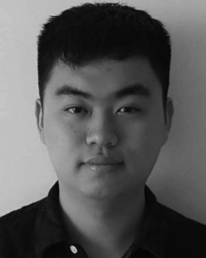 Mengyue Chen合作论文数8Cathrin M Buetefisch合作论文数8Qian Zhang合作论文数8
 Sook-Lei Liew合作论文数Brain and Creativity Institute, University of Southern California Los Angeles;Herman Ostrow School of Dentistry, University of Southern California Los Angeles;University of Southern California Los Angeles, University of Southern California Los Angeles8Liu Jingchun合作论文数8
 Hordacre Brenton合作论文数The Robinson Research Institute, University of Adelaide8
 Revill Kate Pirog合作论文数Center for Advanced Brain Imaging, Georgia Institute of Technology8
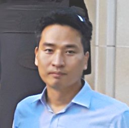 Hosung Kim合作论文数Montreal Neurological Institute and Hospital, McGill University8
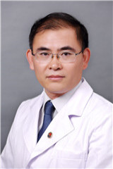 Chunshui Yu (于春水)合作论文数Tianjin Medical University General Hospital;Tianjin Medical University8
 Fabrizio Piras合作论文数IRCCS Santa Lucia Foundation8Martin Lotze合作论文数8
 Feroze B. Mohamed合作论文数Department of Radiology, Thomas Jefferson University8
 Colleen A Hanlon合作论文数Department of Neurobiology, Duke University8
 Amy Brodtmann合作论文数Department of Medicine (Neurology)8Tarun Girotra合作论文数Department of Neurology, University of New Mexico School of Medicine7
 Daniela Vecchio合作论文数7
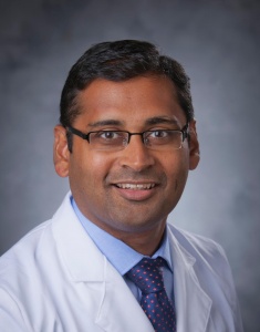 Shreyansh Shah合作论文数Division of Neurocritical Care, Department of Neurology, Duke University School of Medicine;Division of Stroke and Vascular Neurology, Department of Neurology, Duke University School of Medicine;Durham Veterans Affairs Medical Center7
 HaiQing Zheng (郑海清)合作论文数中山大学附属第三医院天河医院7
 Lars Tjelta Westlye合作论文数Department of Psychology, Faculty of Social Sciences, University of Oslo;Norwegian Center for Mental Disorders Research, Oslo University Hospital;Department of Mental Health and Addiction, Oslo University Hospital7
 Gregory T. Thielman合作论文数Physical Therapy Department, University of the Sciences in Philadelphia7
 Lara Boyd合作论文数Department of Physical Therapy, Faculty of Medicine, The University of British Columbia;Djavad Mowafaghian Centre for Brain Health7
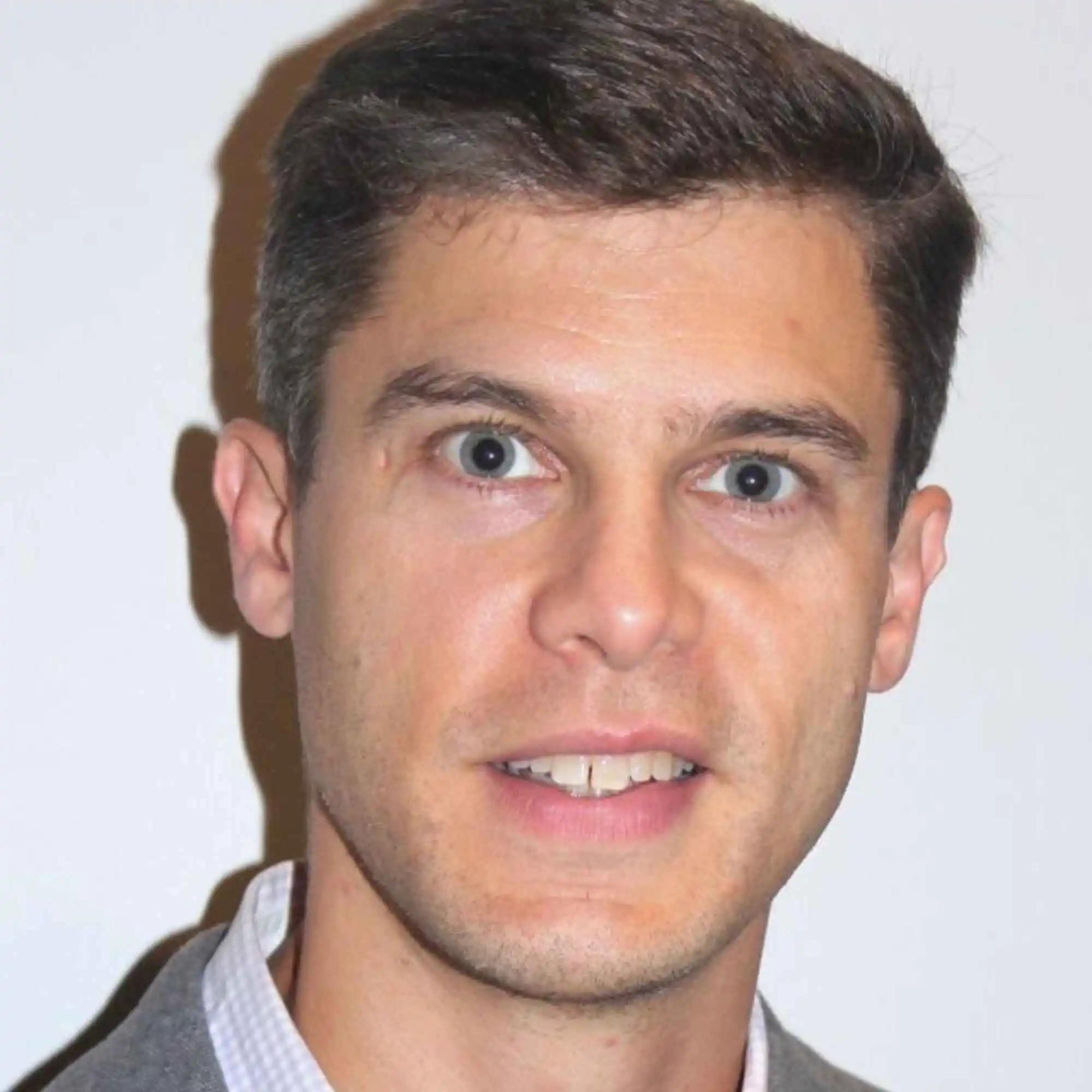 Bradley MacIntosh合作论文数Department of Medical Biophysics, Temerty Faculty of Medicine, University of Toronto7




