Background: CD4+ T-cells are essential in regulating dermal fibrosis and are a main source of IL-10. We have shown that IL-10 reduces dermal fibrosis by upregulating high molecular weight hyaluronan (HMW-HA). However, little of known the role of these cells in scarring. We hypothesize that IL-10 producing CD4+ cells attenuate dermal wound fibrosis via mediating inflammation and altering fibroblast extracellular matrix (ECM) production. Methods: C57BL/6J murine(6-10wk) splenocytes were enriched and sort into CD4+ IL-10 producing cells (Treg&Tr1). In vitro, fibroblasts were co-cultured with Treg or Tr1; fibrotic (Col1a1, aSMA) and ECM remodeling (HA synthases (HAS)1-3) markers were analyzed (qRT-PCR). In vivo, we performed bilateral 6mm dorsal full-thickness wounds on SCID mice (female,8-10wk), and adoptively transferred 106 CD4+, Treg, or Tr1 cells. Wounds were harvested at days 7/28 and analyzed for closure (H&E), fibrosis (trichrome), cell-cell spatial relationships (imaging mass cytometry) and inflammation panel (Luminex). Results: We confirmed the population (flow cytometry) and functionality (IL-10 ELISA) of Treg and Tr1. In the co-culture, Col1a1 in Tr1/Treg treated fibroblasts was reduced by 52.4% or 43.5%; and aSMA was reduced by 25.2% or 44.7%, respectively, compared to untreated fibroblasts. HAS2, a hyaluronan synthase which primarily produces HMW-HA, increased 3.11 or 2.95-fold with Tr1/Treg respectively. In vivo, Tr1 treated mice wound showed expedited wound closure at d7 and significantly greater aSMA (IHC), though both CD4+ and Tr1 treatments resulted in significantly reduced collagen content at d28 (trichrome). Reduced F4/80 were observed in all wounds treated with T cells (IHC). Luminex assay revealed wounds treated with Tr1 had decreased proinflammatory cytokines IL-6, MCP-1, and GM-CSF. Conclusions: IL-10 producing CD4+ cells regulate fibroblast ECM deposition and inflammatory cytokine balance to attenuate fibrosis. Promoting Tr1 recruitment, IL-10 production, and HMW-HA synthesis in wounds may a therapeutic target to improve wound healing.
You have accessJournal of UrologyInfections/Inflammation/Cystic Disease of the Genitourinary Tract: Kidney & Bladder I1 Apr 2017PD12-08 THE ROLE OF IL-10 IN UNILATERAL URETERAL OBSTRUCTION: REGULATION OF ANGIOGENESIS AND FIBROSIS Bethany Desroches, Xinyi Wang, Pu Duann, Meredith Rae, Swathi Balaji, and Sundeep Keswani Bethany DesrochesBethany Desroches More articles by this author , Xinyi WangXinyi Wang More articles by this author , Pu DuannPu Duann More articles by this author , Meredith RaeMeredith Rae More articles by this author , Swathi BalajiSwathi Balaji More articles by this author , and Sundeep KeswaniSundeep Keswani More articles by this author View All Author Informationhttps://doi.org/10.1016/j.juro.2017.02.683AboutPDF ToolsAdd to favoritesDownload CitationsTrack CitationsPermissionsReprints ShareFacebookTwitterLinked InEmail INTRODUCTION AND OBJECTIVES Clinical unilateral ureteral obstruction (UUO) results from pathologies like ureteral calculi, malignancy, trauma, and periureteral fibrosis. UUO requires prompt surgical intervention to prevent progressive inflammation, fibrosis, and kidney function impairment. Novel therapies are needed to recover renal function and architecture after obstruction. Previous reports show the anti-inflammatory cytokine interleukin-10 (IL-10) attenuates fibrosis in a murine model. We and others have shown IL-10 regulates angiogenesis and endothelial progenitor recruitment during dermal and ischemic cardiac tissue repair. We hypothesize that IL-10 can promote angiogenesis and prevent microvascular rarefaction in a murine UUO model. METHODS 8 week-old C57BL/6J (WT) mice and IL-10 null male mice were injected with lenti-IL-10/lenti-GFP (1x10¹⁰ IU) under the renal capsule. Three days after injection, UUO was performed. 14 days after UUO, UUO/sham kidneys and serum were collected for RNA, ELISA and immunohistochemical analysis of VEGF and TGFβ isoforms. Primary fibroblasts were isolated from 8-10 week-old male WT mice. IL-10 (50 ng/ml or 200 ng/ml) was added to cultures. VEGF and TGFβ-1 gene expressions were assessed by qPCR at 1, 2, 3 and 6h. Levels of TGFβ-1 and TGFβ-3 were determined at 48h by ELISA. Data presented as mean±SD, n=3/treatment group. P value by ANOVA. RESULTS IL-10 treatment, in vitro, increased VEGF expression (related to physiologic angiogenesis) and altered the expression of TGFβ isoforms (related to pathologic fibrosis). Lenti-Il10 treatment in mice with UUO reduced fibrotic changes between tubules (45±7%, p<0.05) and attenuated tubular dilatation (p<0.05, n=30/group). CD31, an endothelium marker essential to preserving tubular integrity, was normally expressed in healthy kidney parenchyma and decreased after UUO. IL-10 null mice showed a lower basal level of CD31 than WT mice. In both WT and IL-10 null mice, IL-10 treatment preserved CD31, suggesting a role in rescuing integral peritubular capillaries. CONCLUSIONS Ureteral obstruction can cause remodeled renal architecture and decrease in renal function. Our results in a murine UUO model show that IL-10 can effectively promote angiogenesis in vitro and prevent microvascular rarefaction in vivo. Mice treated with IL-10 show decreased fibrotic change and increased markers for tubular integrity. These results from the in vivo studies of UUO, and the in vitro studies of IL-10′s influence in angiogenesis and fibrosis of the kidney, may lead to novel treatments for the sequelae of urinary tract obstruction. © 2017FiguresReferencesRelatedDetails Volume 197Issue 4SApril 2017Page: e268 Advertisement Copyright & Permissions© 2017MetricsAuthor Information Bethany Desroches More articles by this author Xinyi Wang More articles by this author Pu Duann More articles by this author Meredith Rae More articles by this author Swathi Balaji More articles by this author Sundeep Keswani More articles by this author Expand All Advertisement Advertisement PDF downloadLoading ...
Lymphocytes have been shown to play a role in tissue fibrosis in several models, but their role in dermal wound repair is unclear. We previously demonstrated that hyaluronan (HA) can stimulate CD4+CD25+FoxP3+ T cells to produce an anti-inflammatory milieu, which is permissive of decreased fibrosis. We hypothesize that lymphocytes influence dermal healing and fibrosis. We make excisional 6mm stented wounds in SCID mice that lack of T and B cells, and compared to WT mice. Next, supple different lymphocytes subsets by adoptive transfer of 1)total lymphocytes; 2)CD4+CD25-T cells; 3)CD4+CD25+T cells. To investigate role of Tregs, wounds were created after selective depletion of CD4+CD25+FoxP3+Tregs in FoxP3DTR mice(FACS) analysis. Wounds harvested at day 7,14,30 and analyzed for wound closure(imaging), healing outcome(H&E), inflammation(CD45+ and F4/80+ cells/40X-HPF), and fibrosis(Trichrome; α-SMA). Data mean±SD, p values by ANOVA and t-test. SCID mice demonstrated increased dermal fibrosis (increased trichrome staining day14&30, α-SMA IHC day7) and an exaggerated inflammatory infiltrate compared to WT mice (CD45+,SCID34.50% vs WT11.00%;p < 0.05: Macrophages scores increased in SCID). Adoptive transfer of total lymphocytes (TL), CD4+CD25-Tcells and CD4+CD25+Tcells were successfully engrafted at day 7 by FACS; and by qPCR FoxP3 and CD25 locally. Transfer of all three cell types reduced inflammatory infiltrate (neutrophils: TL11.70%;CD4+CD25-Tcells 6.4%; CD4+CD25+T cells 20.10%; p < 0.05; macrophage scores decrease compared to control SCID). Treg depletion in FoxP3DTR mice significantly increased collagen content and fibrosis at D14(131077.2±12948.34 vs.96515.63±23828.77, p < 0.001), and increased CD45+ cells compared to the controls(37±18.1% vs.16.33±7.8%, p=0.017). Our data suggests that CD4+CD25+ T cells have a physiologic role in the regulation of wound fibrosis and repair, which is in part mediated by the attenuation of the inflammatory response and effects on the ECM. Understanding the individual contributions of lymphocyte subsets will permit an improved understanding of physiologic and pathologic wound repair.
Regenerative fetal dermal repair is characterized by a unique extracellular matrix (ECM) profile consisting of high levels of hyaluronan (HA) and elevated TGF-β3/TGF-β1 ratio. Interleukin-10 (IL-10) is essential for regenerative fetal dermal repair, and it recapitulates the fetal phenotype in adult tissue. The relationship between IL-10, HA, and TGF-β isoforms remains unclear. We hypothesize that IL-10 regulates HA and TGF-β isoforms expression, which is crucial to regenerative dermal repair. Primary fetal (E14.5) and adult (8wk) dermal fibroblasts (FBs) are isolated from C57BL/6J mice. FBs are treated with IL-10 +/- TGF-β Pan neutralizing Ab. Levels of HA, TGF-β1 and 3 are determined by ELISA. HAS synthases (HAS1-3) and hyaluronidases (HYAL 1-2, and KIAA 1199) are quantified by qPCR. Murine wounds (4mm) are treated with IL-10 or controls, and analyzed (qPCR, ELISA, H&E). Data present as mean +/- SD; p-values by ANOVA. Fetal FBs demonstrate increased HA levels (p<0.01) and an elevated TGF-β 3/1 ratio (p<0.05) compared to adult FBs at 48h. IL-10 treatment significantly increases HA levels (p<0.05) in adult FBs to the levels of the fetal FBs at 48h. IL-10 treatment increases HAS 1-2 gene expression and decreases HYAL1, 2 and KIAA 1199 at the first 3 hours (p<0.05) in adult FBs. IL-10 also results in a 5-fold increase of TGF-β3/ TGF-β1 ratio in adult FB. The neutralization of Pan TGF-β abrogated IL-10 effects on increasing HA. In vivo, overexpression of IL-10 increases HA level as well as TGF-β3/ TGF-β1 ratio in murine dermal wounds at day 3 (p<0.05), resulting in improved regenerative wound repair at day 30 compare to PBS or vector controls. This effect is abrogated in the presence of an HA inhibitor. Our data suggests that IL-10 regulates HA synthesis and degradation, and shifts TGF-β isoforms to a more fetal phenotype. Understanding the crosstalk of IL-10, HA and TGF-β will yield novel therapies that may result in regenerative wound healing.
Clinical studies suggest that patients with human immunodeficiency virus (HIV) infection have an increased risk for cardiovascular events including coronary artery disease, while there is little evidence about direct virus involvement in the vascular lesion formation. The objective of this study was to characterize vascular tissues obtained from HIV (+) and HIV (−) patients as well as from simian immunodeficiency virus (SIV)-infected and control Rhesus monkeys.
Purpose. Both aortic aneurysms and aortic dissections exhibit abnormal extracellular matrix properties. Connective tissue growth factor (CTGF) can induce connective tissue cell proliferation and extracellular matrix synthesis. The role of CTGF in thoracic aortic disease has never been investigated. We sought to compare the expression of CTGF in degenerative ascending aortic aneurysms and ascending aortic dissection. Methods. Intraoperative samples of ascending aorta were obtained from 47 patients: 16 patients had ascending aortic aneurysms with medial degeneration, 10 had acute aortic dissection, 9 had aneurysms due to chronic dissection. Control ascending aorta was obtained from organ donors and heart transplant recipients (n = 10). Patients with Marfan syndrome were excluded from this study. CTGF mRNA expression within aortic wall was semiquantitatively determined by real-time RT-PCR using GAPDH as the internal standard. Results. There was a significant increase in CTGF mRNA in degenerative aneurysms compared to control tissue (P = 0.04). Conversely, patients with acute dissection had decreased CTGF mRNA expression compared with nondissection aneurysms (P = 0.019) and controls (P = 0.06). The increase in CTGF expression in chronic dissections compared to acute dissections approached statistical significance (P = 0.075). Conclusions. The altered tissue levels of CTGF in aneurysms and dissections suggest possibly different molecular pathology in these aortic disorders. Further investigation regarding the role of CTGF in thoracic aortic disease is warranted.
 Sundeep Keswani合作论文数University of Cincinnati College of Medicine7
 S. Balaji合作论文数5M. Rae合作论文数Baylor Coll Med5H. Li合作论文数5Pu Duann合作论文数5
 Paul Bollyky合作论文数Medicine Department, Stanford University;Microbiology & Immunology Department, Stanford University4C.J. Chen合作论文数3
 Qizhi Cathy Yao合作论文数Division of Surgical Oncology, Baylor College of Medicine3Peter H Lin合作论文数3D. Nguyen合作论文数2W. Short合作论文数2Swathi Balaji合作论文数2A. Kagan合作论文数2L. Choi合作论文数2V. G. Sunkari合作论文数1S.A. LeMaire合作论文数1S.A. Carter合作论文数1Bethany Desroches合作论文数1L. Yu合作论文数1J. Cheng合作论文数1Jun Jiang合作论文数1
 Manish Butte合作论文数Department of Microbiology, Immunology, and Molecular Genetics, College of Life Sciences, University of California, Los Angeles1
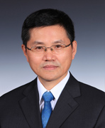 Yehua Gan (甘业华)合作论文数Hospital of Stomatology, Peking University1
 Katherine Y King合作论文数1
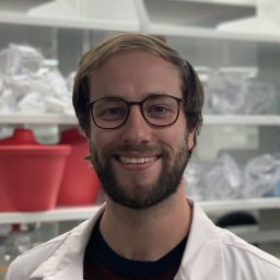 Moles Chad M合作论文数Cincinnati Children's Hospital Medical Center1
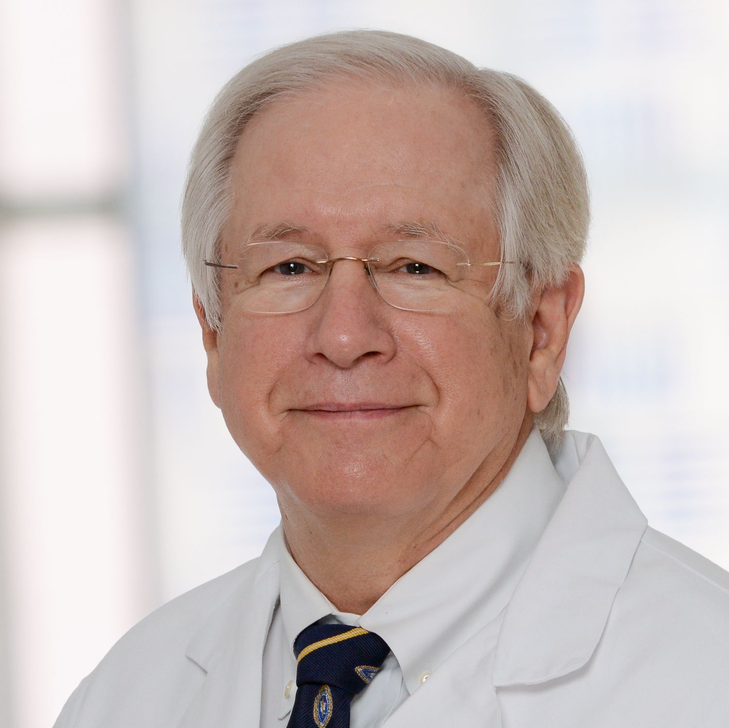 Joseph S. Coselli合作论文数Department of Surgery, Baylor College of Medicine1Qizhi Yao合作论文数Baylor College of Medicine1S. Wen合作论文数1
 Peter Lin合作论文数Baylor College of Medicine;Vascular Care Specialists of Los Angele1Hong Chai合作论文数1Alan B Lumsden合作论文数1
 Ryuji Ohashi合作论文数1
 Ying H Shen (沈英)合作论文数Division of Cardiothoracic Surgery, Department of Surgery, Baylor College of Medicine;Aortic Disease Research Laboratory, Department of Surgery, Baylor College of Medicine1Jonathan A. Wilks合作论文数1




