Optical coherence tomography (OCT) and its extension OCT angiography (OCTA) have become essential clinical imaging modalities due to their ability to provide depth-resolved angiographic and tissue structural information non-invasively and at high resolution. Within a field of view, the anatomic detail available is sufficient to identify several structural and vascular pathologies that are clinically relevant for multiple prevalent blinding diseases, including age-related macular degeneration (AMD), diabetic retinopathy (DR), and vein occlusions. The main limitation in contemporary OCT devices is that this field of view is limited due to a fundamental trade-off between system resolution/sensitivity, sampling density, and imaging window dimensions. Here, we describe a swept-source OCT device that can capture up to a 12 × 23-mm field of view in a single shot and show that it can identify conventional pathologic features such as non-perfusion areas outside of conventional fields of view. We also show that our approach maintains sensitivity sufficient to visualize novel features, including choriocapillaris morphology beneath the macula and macrophage-like cells at the inner limiting membrane, both of which may have implications for disease.
High-quality swept-source optical coherence tomography (SS-OCT) requires accurate k-sampling, which is equally vital for optical coherence tomography angiography (OCTA). Most SS-OCT systems are equipped with hardware-driven k-sampling. However, this conventional approach raises concerns over system cost, optical alignment, imaging depth, and stability in the clocking circuit. This work introduces an optimized numerical k-sampling method to replace the additional k-clock hardware. Using this method, we can realize high axial resolution (4.9-µm full-width-half-maximum, in air) and low roll-off (2.3 dB loss) over a 4-mm imaging depth. The high axial resolution and sensitivity achieved by this simple numerical method can reveal anatomic and microvascular structures with structural OCT and OCTA in both macular and deeper tissues, including the lamina cribrosa, suggesting its usefulness in imaging retinopathy and optic neuropathy.
Optical coherence tomography (OCT) is a well-established research tool for vision research in animal models capable of providing in vivo imaging of the retina. Structural OCT can be enhanced using OCT angiography (OCTA) processing in order to provide simultaneously acquired, automatically co-registered vascular information. Currently available OCT. Currently available OCTA lack either large field of view or high resolution. In this study we developed a wide-field (60-degree), high-resolution (10.5-µm optical transverse) and high-sensitivity (104-dB) OCTA-enabled system for non-human primate imaging and with it imaged multiple disease models, including models of age-related macular degeneration (AMD), Bardet-Biedl Syndrome (BBS), and the CLN7 variant of Batten disease. We demonstrate clear visualization of features including drusen, ellipsoid zone loss, vascular retinopathy, and retinal thinning in these eyes.
In this study, we present an optical coherence tomographic angiography (OCTA) prototype using a 500 kHz high-speed swept-source laser. This system can generate a 75-degree field of view with a 10.4 µm lateral resolution with a single acquisition. With this prototype we acquired detailed, wide-field, and plexus-specific images throughout the retina and choroid in eyes with diabetic retinopathy, detecting early retinal neovascularization and locating pathology within specific retinal slabs. Our device could also visualize choroidal flow and identify signs of key biomarkers in diabetic retinopathy.
In this study, we present a sensorless adaptive optics swept-source optical coherence tomographic angiography (sAO-SS-OCTA) imaging system for mice. Real-time graphics processing unit (GPU)-based OCTA image acquisition and processing software were applied to guide wavefront correction using a deformable mirror based on signal strength index (SSI) from both OCT and OCTA images. High-resolution OCTA images with aberrations corrected and contrast enhanced were successfully acquired. Fifty-degree field of view high-resolution montaged OCTA images were also acquired.
Optical coherence tomography (OCT) has changed the standard of care for diagnosis and management of macular diseases in adults. Current commercially available OCT systems, including handheld OCT for pediatric use, have a relatively narrow field of view (FOV), which has limited the potential application of OCT to retinal diseases with primarily peripheral pathology, including many of the most common pediatric retinal conditions. More broadly, diagnosis of all types of retinal detachment (exudative, tractional, and rhegmatogenous) may be improved with OCT-based assessment of retinal breaks, identification of proliferative vitreoretinopathy (PVR) membranes, and the pattern of subretinal fluid. Intraocular tumors both benign and malignant often occur outside of the central macula and may be associated with exudation, subretinal and intraretinal fluid, and vitreoretinal traction. The development of wider field OCT systems thus has the potential to improve the diagnosis and management of myriad diseases in both adult and pediatric retina. In this paper, we present a case series of pediatric patients with complex vitreoretinal pathology undergoing examinations under anesthesia (EUA) using a portable widefield (WF) swept-source (SS)-OCT device.
Recent advances in portable optical coherence tomography (OCT) and OCT angiography (OCTA) have resulted in wider fields of view (FOV) and shorter capture times, further expanding the potential clinical role of OCT technology in the diagnosis and management of retinopathy of prematurity (ROP). Using a prototype, handheld OCT device, retinal imaging was obtained in non-sedated infants in the neonatal intensive care unit (NICU) as well as sedated infants in the operating room of Oregon Health & Science University (OHSU) Hospital. In this observational study, we provide an overview of potential advantages of OCT-based disease assessment in ROP. We observed that next-generation OCT imaging (a) may be sufficient for objective diagnosis and zone/stage/plus disease categorization, (b) allows for minimally-invasive longitudinal monitoring of disease progression and post-treatment course, (c) provides three-dimensional mapping of the vitreoretinal interface, and (d) with OCTA, enables dye-free visualization of normal and pathologic vascular development.
In this study, we present the development of sensorless adaptive optics swept-source optical coherence tomographic angiography (sAO-SS-OCTA) imaging system for mice. GPU-based real-time OCTA image acquisition and processing software was applied to guide wavefront correction using a deformable mirror. High-resolution OCTA images with high capillary resolution and contrast have been successfully acquired. 45-degree field of view high-resolution montaged OCTA image was also acquired.
In this study, we developed a novel phase-stabilized complex-decorrelation (PSCD) optical coherence tomography (OCT) angiography (OCTA) method that can generate high quality OCTA images. This method has been validated using three different types of OCT systems and compared with conventional complex- and amplitude-based OCTA algorithms. Our results suggest that in combination with a pre-processing phase stabilization method, the PSCD method is insensitive to bulk motion phase shifts, less dependent on OCT reflectance than conventional complex methods and demonstrates extended dynamic range of flow signal, in contrast to other two methods.
The segmentation of en face retinal capillary angiograms from volumetric optical coherence tomographic angiography (OCTA) usually relies on retinal layer segmentation, which is time-consuming and error-prone. In this study, we developed a deep-learning-based method to segment vessels in the superficial vascular plexus (SVP), intermediate capillary plexus (ICP), and deep capillary plexus (DCP) directly from volumetric OCTA data. The method contains a three-dimensional convolutional neural network (CNN) for extracting distinct retinal layers, a custom projection module to generate three vascular plexuses from OCTA data, and three parallel CNNs to segment vasculature. Experimental results on OCTA data from rat eyes demonstrated the feasibility of the proposed method. This end-to-end network has the potential to simplify OCTA data processing on retinal vasculature segmentation. The main contribution of this study is that we propose a custom projection module to connect retinal layer segmentation and vasculature segmentation modules and automatically convert data from three to two dimensions, thus establishing an end-to-end method to segment three retinal capillary plexuses from volumetric OCTA without any human intervention.
Optical coherence tomography (OCT) and OCT angiography (OCTA) enable noninvasive structural and angiographic imaging of the eye. Portable handheld OCT/OCTA systems are required for imaging patients in the supine position. Examples include infants in the neonatal intensive care unit (NICU) and operating room (OR). The speed of image acquisition plays a pivotal role in acquiring high-quality OCT/OCTA images, particularly with the handheld system, since both the operator hand tremor and subject motion can cause significant motion artifacts. In addition, having a large field of view and the ability of real-time data visualization are critical elements in rapid disease screening, reducing imaging time, and detecting peripheral retinal pathologies. The arrangement of optical components is less flexible in the handheld system due to the limitation of size and weight. In this paper, we introduce a 400-kHz, 55-degree field of view handheld OCT/OCTA system that has overcome many technical challenges as a portable OCT system as well as a high-speed OCTA system. We demonstrate imaging premature infants with retinopathy of prematurity (ROP) in the NICU, a patient with incontinentia pigmenti (IP), and a patient with X-linked retinoschisis (XLRS) in the OR using our handheld OCT system. Our design may have the potential for improving the diagnosis of retinal diseases and help provide a practical guideline for designing a flexible and portable OCT system.
Wide-field OCT angiography (WF-OCTA) is challenging due to scanning speed limits and artifacts. A prototype with a graphic processing unit (GPU)-based self-tracking method was developed. High-resolution 75-degree WF-OCTA images were successfully acquired.
 Yali Jia合作论文数School of Medicine, Oregon Health & Science University;Center for Ophthalmic Optics and Lasers43
 Tristan T. Hormel合作论文数Casey Eye Institute, Oregon Health & Science University25
 David Huang (黄大衛)合作论文数Center for Ophthalmic Optics & Lasers, Casey Eye Institute, School of Medicine, Oregon Health & Science University;Department of Biomedical Engineering, School of Medicine, Oregon Health & Science University20
 Shaohua Pi合作论文数19
 Yukun Guo (郭玉坤)合作论文数School of Mathematics, Harbin Institute of Technology16
 William O Cepurna合作论文数13John C Morrison合作论文数12
 Acner Camino合作论文数Changchun University of Science and Technology, Changchun Univ. of Sci. and Technol.12
 Yifan Jian合作论文数School of Medicine, Oregon Health & Science University;Siloam Vision9
 Thomas Hwang合作论文数9Jie Wang合作论文数Casey Eye Institute, Oregon Health & Science University;Department of Biomedical Engineering, Oregon Health & Science University;Oregon Health & Science University, Oregon Health & Science University7
 John Peter Campbell合作论文数Oregon Health & Science University, Portland6
 Susan Ostmo合作论文数Oregon Health & Science University6
 Shuibin Ni合作论文数6
 Michael F. Chiang合作论文数Oregon Health and Science University5
 Guangru Liang合作论文数4Thanh-Tin Nguyen合作论文数4
 Shanjida Khan合作论文数Casey Eye Inst, Oregon Hlth & Sci Univ4
 Bingjie Wang合作论文数Casey Eye Institute, Oregon Health & Science University4Steven T. Bailey合作论文数Casey Eye Institute, Oregon Health &Science University;Casey Eye Institute, Oregon Health & Science University4John Morrison合作论文数3
 Min Gao合作论文数3
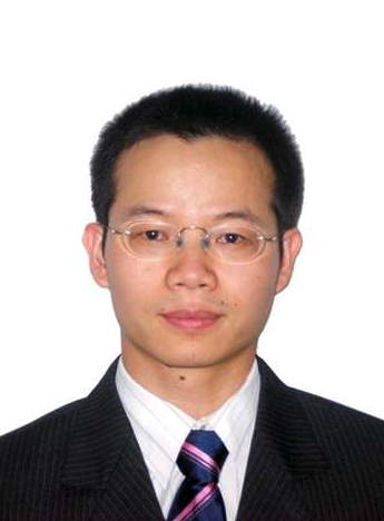 Qisheng You (游启生)合作论文数Beijing Tongren Hospital, Capital Medical University3Ringo Ng合作论文数2Naixing Huang合作论文数2
 Siyu Chen合作论文数2
 Martha Neuringer合作论文数Oregon National Primate Research Center2
 Miao Zhang合作论文数Genentech2Christina J. Flaxel合作论文数2Brigid Marshall合作论文数1Pengxiao Zang合作论文数Department of Biomedical Engineering, Oregon Health & Science University1
 Qi Sheng You合作论文数1
 Skalet Alison H合作论文数Francis I Proctor Foundation for Research in Opthalmology, University of California1
 Simonett Joseph M合作论文数Waisman Center.1Renner Lauren合作论文数1
 Liu Liang合作论文数1
 Xiaogang Wang合作论文数1Laurie Renner合作论文数1
 Phoebe Lin合作论文数Department of Ophthalmology and Visual Science, University of Illinois at Chicago1




