OBJECTIVES:Many studies have examined the general public's attitudes towards people with mental illness, but such studies are scarce in China. This study examined the perceptions of the Beijing population regarding their society's prevalent attitudes towards people with mental illness.METHODS:A total of 5000 individuals aged 18 or above living in Beijing were selected using a multistage, stratified, cluster and random sampling method. This was followed by a face-to-face interview which used a standardized questionnaire asking about societal attitudes towards individuals with mental illness.RESULTS:4602 out of 5000 eligible individuals met the inclusion criteria and participated in the interview. 4596 questionnaires were deemed valid and included in the analyses. A large proportion of respondents believed that most individuals within their society held negative attitudes and had a strong desire to distance themselves from people with mental illness. Respondents aged 60 or older, who lived farther to downtown Beijing, or with higher education tended to believe that most individuals have relatively positive and tolerant attitudes towards people with mental illness.CONCLUSIONS:Many people in Beijing perceive that most members of their society have negative beliefs towards people with mental illness. Further efforts are needed to determine if these perceptions are accurate and to reduce the stigma that is reinforced by these perceptions.
c-Src/Fyn, a non-receptor type tyrosine kinase, which is expressed by virtually all cells with very high expression levels found in neurons, suggesting a role in brain function. Lately, Src was found to be related to neuron-glia modulation, and the appreciable functions were found not only located at the plasma membrane but also in variety of intracellular locations. Besides that, Src could coordinate with other signal molecules and operated as molecular switches in synaptic cellular communication in response to brain injury. Src activation was also characterized to be required for conveying the recovery signals from immunosuppression, the regulatory networks involved the interaction between Src and mu opioid receptor, EGFR, IGF and IL-1 beta. Therefore, Src signaling was proposed to be the important therapeutic consideration regarding neuroimmune modulation in response to brain injury.
Compensatory adenylyl cyclase (AC) superactivation has been postulated to be responsible for the development of morphine tolerance and dependence, the underlying mechanism was demonstrated to comprise c-Src-dependent upregulation of AC5 within the lipid rafts. In the present study, we demonstrated that chronic morphine treatment sensitized EGFR signaling by augmenting EGFR phosphorylation and translocation into ER, which was essential for CRT-MOR tethering within the lipid rafts and AC5 superactivation. Intriguingly, synaptic clustering of CRT-MOR was dependent on EGFR phosphorylation and presumed to implicate in alignment and organization of synaptic compartments. Taken together, our data raised the possibility that an adaptive change in MOR and EGFR signal systems might establish CRT related subcellular communication, the signaling network within brain synaptic zone was proposed to implicate in morphine tolerance and dependence.
Age-dependent neuroimmune modulation following traumatic stress is accompanied by discordant upregulation of Fyn signaling in the frontal cortex, but the mechanistic details of the potential cellular behavior regarding IGF-1R/Fyn have not been established.
BACKGROUND:Neuroimmune modulation following traumatic stress is accompanied by cortical upregulation of c-Src expression, but the mechanistic details of the potential regulatory link between c-Src expression and immunosuppression have not been established.METHODS:We used a combination of techniques to measure temporal changes in: (i) the parallel expression of c-Src and microRNA222; (ii) levels of PAK1 (p21-activated kinase 1); and (iii) the association between PAK1 and interleukin 1β signaling, both in cortex of rats following traumatic stress and in primary cortical neurons. Techniques included real-time PCR, immunoprecipitation, western blotting and subcellular fractionation by discontinuous centrifugation. We also measured lymphocyte proliferation and natural killer (NK) cell activity.RESULTS:We confirm robust upregulation of c-Src expression following traumatic stress. c-Src upregulation was accompanied by marked increases in levels of miRNA222; other studied miRNAs were not affected by stress. We also established that PAK1 is a primary target for miRNA222, and that increased levels of miRNA222 following traumatic stress are accompanied by downregulation of PAK1 expression. PAK1 was shown to mediate the association of IL-1RI with lipid rafts and thereby enhance IL-1 signaling. Detailed analyses in cultured neurons and glial cells revealed that PAK1-mediated enhancement of IL-1RI activation is governed to a large extent by c-Src/miRNA222 signaling; this signaling played a central role in the modulation of lymphocyte proliferation and NK cell activity.CONCLUSIONS:Our results suggest that neuroimmune modulation following traumatic stress is mediated by a cascade that involves c-Src-mediated enhancement of miRNA222 expression and downregulation of PAK1, which in turn impairs signaling via IL-1β/IL1-RI, leading to immunosuppression. The regulatory networks involving c-Src/miRNA222 and PAK1/IL-1RI signaling have significant potential for the development of therapeutic approaches designed to promote recovery following traumatic injury.
To investigate the effect of electroacupuncture (EA) on the T-lymphocyte subsets and lymphocyte transformation in patients with gastrointestinal tumor during peri-operative and peri-chemotherapy periods.
This chapter will review the history of modern acupuncture research in China. The concept of “channels” and “collaterals” has been used for a long time in the traditional Chinese medicine (TCM). Clinical acupuncture yields therapeutic effects on many diseases, according to the general principles of “channels” and “collaterals”. In the past 5 decades, acupuncture research has been very popular in many major Chinese medical institutions, among which Shanghai Medical College of Fudan University (formerly Shanghai First Medical College, and then, Shanghai Medical University) has made substantial contribution to this field. Comprehensive scientific data obtained from normal subjects, patients, and animals have shown that needling of acupuncture points activates the afferent fibers of the peripheral nerves to elicit the De-Qi sensation, and subsequently, the nerve-mediated signals ascend to various levels of the central nervous system (CNS), producing analgesic effect. The pain relief is the most effective outcome of acupuncture treatment. As acupuncture enhances the analgesics’ effect of pain relief, the combination of acupuncture with small doses of analgesic drugs is now being adopted in the management of various types of pain and surgical anesthesia. In addition, clinical and bench studies on acupuncture therapy of other neurological diseases, such as epilepsy, cerebral ischemia, neuroimmune disorders, and woman’s reproductive disorders have also been successfully carried out in the recent years. This chapter will briefly summarize the research progress and present an overall picture of acupuncture research in China.
Acupuncture, one of the most prevalent methodologies of Traditional Chinese Medicine (TCM), has been used in Asian countries for curing numerous diseases for thousands of years. However, the true mechanisms underlying the effectiveness of acupuncture are still under debating. The meridian model based on TCM has been used so far, for guiding the practice of acupuncture. In this model, acupuncture is believed to treat the diseased organ of the patient by balancing the Yin and Yang conditions that are regulated by an energy substance (Qi) flowing constantly through the whole meridian, a network connecting all the organs of the body. Therefore, in the acupuncture treatment, it is crucial to select special acupoint(s) along the meridian that links the diseased organs, as well as to modulate the Qi flowing in the meridian through the induction of the needling sensation (De-Qi). On the other hand, a neurobiological model established in the recent decades, has supported the notion that an important mechanism of acupuncture in curing diseases is mediated by the nervous system. Stimulation by needles at acupoints is considered to initiate acupuncture signals through the nerve fibers (e.g., A(beta) and A(delta)) innervated at the deep tissue near the acupoints. The acupuncture signal is transmitted through the central nervous system, which activates and integrates with the neurons located in broad areas, such as those in the cortex, limbic system, brainstem, spinal cord, which in turn, regulate other systems. The nerve-mediated model provides us a better explanation regarding the biological mechanisms of acupuncture signal transmission in the body which has been broadly documented by both in vivo and in vitro studies under controlled conditions. In this chapter, we will review in particular, the research concerning the influence of acupuncture-elicited signals in the nervous system and how the neural pathways mediate the therapeutic effects of acupuncture.
We have previously reported that neuron and glia could collaboratively govern the immunomodulation in traumatic rats. Herein, we characterized the sequential involvement of cortical neuron, microglia, and astrocytes in the traumatic stress-mediated neuroimmune modulation. At day 1 of trauma, transient extracellular signal related kinase 1/2 (ERK1/2) activation was initiated in neuron and microglia, which was accompanied by RSK-1 expression in the cytosol. At day 3 of trauma, persistent ERK1/2 activation occurred in astrocytes, which were destined for the nucleus leading to Elk-1 expression. Furthermore, the functional overlap of ERK1/2 and neuroligin 1 in astrocytes was strengthened at day 3 of trauma and responsible for the recovery from the immnosuppression. These effects could be disrupted by beta-neurexin blockade. Altogether, we proposed the mechanism underlying the traumatic stress-induced immunosuppression, in which local activity ensured the initial establishment of neural circuitry in the frontal cortex. ERK1/2-signaling events are required for the temporal and spatial coordination between neuron and glial cells. Synapse 65: 433-440, 2011. (C)2010 Wiley-Liss, Inc.
Immunological changes initiated by major operative injury may result in inflammatory responses in both peripheral and central nervous system, which may lead to organ dysfunction. Recent studies indicate that β-adrenergic receptors (β-ARs) may mediate production of pro-inflammatory cytokines in the brain. In the present study propranolol (β-AR antagonist), but not prazosin (α1-AR antagonist), antagonized surgical trauma induced pro-inflammatory cytokine production in microglia cells isolated from rats. β-AR activation in the absence of pro-inflammatory stimuli increased IL-1β, TNF-α and IL-6 mRNA and protein expressions in the primary microglia cell culture. Isoproterenol (β-AR agonist) treatment induced a time- and concentration-dependent increase of IL-1β in cells. Both ERK1/2 and P38 MAPK inhibitor, but not PKA and JNK1/2 inhibitor abrogated isoproterenol-induced IL-1β and IL-6 production in microglia cells. In conclusion, the results suggest that β-ARs possess pro-inflammatory properties by modulating the functions of microglia cell.
In this chapter, the clinical and experimental studies on acupuncture-drug balanced anesthesia will be reviewed. The history of acupuncture anesthesia in China started in 1958. After two decades of clinical practice and experimental investigation, the combination of acupuncture (usually electro-acupuncture, EA) with drugs has been successfully used to improve anesthesia for surgical operations and pain treatment since the 1980s, and this technique is now known as the acupuncture-drug balanced anesthesia in China. It is more advantageous to use acupuncture combined with selected drugs to produce anesthesia, because acupuncture could regulate the functions of multiple organs of the body in addition to analgesia Moreover, the underlying mechanism has been elucidated by investigating the potentiation effects of some commonly used drugs on acupuncture analgesia, in various animal models. Substantial evidence has shown that the combination of acupuncture with these drugs promotes the release of endogenous opioid peptides (EOP). In addition, these drugs potentiate EA-induced expression of EOP gene. Furthermore, several lines of evidence show that EA induces an increase in both binding affinity and density of the opioid receptors, while the combined use of acupuncture with drugs can further enhance the EA effect. Currently, clinical and bench studies on acupuncture-drug balanced anesthesia are still in progress to improve the clinical efficacy and to better understand the working mechanisms. We expect that new discoveries through the translational research will bring more benefits to patients.
This chapter summarizes the clinical practice of acupuncture therapy for immune-mediated disorders and the mechanisms underlying the regulation of neuroimmune function by acupuncture. Numerous data indicate that acupuncture can regulate immune system, tune-up specific and nonspecific cellular and humoral immunity, and modulate leucocytosis, microbicidal activity, antibodies, globulin, complement, and interferon (IFN). The acupuncture-induced output signal has been observed to correct the dysfunction of immune system and induce a homeostatic effect on the body through the accommodation of nervous and immune systems. At the molecular level, the acupuncture-induced neuroimmune regulation is mediated through multiple pathways, and involves various bioactive molecules including steroids, neuropeptides, cytokines, and neurotransmitters, which form the basis for bidirectional-coordinated neuroimmune regulation, in response to homeostasis disturbances. An integrated investigation including the approaches of molecular biology, integrative physiology, and clinical research is considered to further improve the understanding of the acupuncture-mediated regulation of neuroimmune function, and eventually lead to better applications of acupuncture for the treatment of immune-related diseases.
The Purpose of this study was to investigate the possible needling effect on sympathetic activity by using functional MRI (fMRI). Twelve patients with left lower extremity pain were enrolled in our study. Each was given deep needling at left GB34 (yanglingquan) and GB39 (xuanzhong) points simultaneously. All patients got the strong DeQi sensation by manipulating the needles, and then electroacupucture (EA) was given and lasted for thirty minutes before fMRI scan. Then the fMRI scan was performed by scanning the whole brain with five blocks lasting 2 minutes each. The patients' palm skin temperatures were tested every five minutes as indication of the sympathetic activity from the beginning of EA to the end of our fMRI scan. Functional images were processed by using FEAT software at different levels including the simple single subject analysis, multi-subjects group mean analysis, and multi-subjects unpaired two difference analysis. Finally, 9 of 12 patients' palm temperatures increased gradually but the other three decreased. In comparison of two different palm skin temperature change groups, more significant activation over bilateral caudate head, right lentiform and periaqueductal gray (PAG) were found in the temperature-increased group, but palm temperature-decreased patients revealed more significant activation over bilateral anterior cingulated cortex (ACC), insula, primary somatosensory gyrus (SI), orbitofrontal cortex, occipital cortex, hippocampus and amygdala formation. Our study suggested that needling at analgesic points may modulate the sympathetic activity and such evident difference on brain responses may correlate with the clinical analgesia effects.
Regulation of neurite outgrowth is an important aspect not only for proper development of the nervous system but also for tissue regeneration after nerve injury and the treatment of neuropathological conditions. Here, we report that neurite outgrowth in cortical neuron and neuro 2A (N2A) cell was dependent on intact lipid rafts, as well as the enhanced localization of c-Src in the lipid rafts. Src inhibition or lipid rafts disruption could specifically block c-Src phosphorylation profile, pY416 Src increase and pY529 Src decrease, they also resulted in pY529 Src and c-terminal Src kinase (Csk) partition out of lipid rafts. Thus, we concluded that c-Src signal cascades within the lipid rafts is crucial for efficient neurite outgrowth.
Cumulative evidences suggest that electroacupuncture (EA) can modulate immune function, but the mechanism needs further study. In the present study, the contribution of EA on toll-like receptors 2 and 4 (TLR2/TLR4) and pro-inflammatory cytokine expression after surgical trauma stress were investigated. The mRNA level of both TLR2/4 and pro-inflammatory cytokine was measured by quantitative real-time PCR. ELISA and Western blot assay were chosen for TLR2/TLR4 protein expression and pro-inflammatory cytokine production, respectively. The results showed that surgical trauma stress increased TLR2 mRNA and TLR2/4 proteins in the spleen and augmented pro-inflammatory cytokines (e.g. IL-1beta) mRNA and protein expression in the spleen and plasma. These effects could be deteriorated by adrenalectomy (ADX). EA at "Zusanli" acupoint significantly inhibited surgical trauma-induced TLR2 mRNA and TLR2/4 protein expression in spleen and pro-inflammatory cytokine expression in the spleen and plasma. ADX, however, could not block the effect of EA. These results suggested that surgical trauma stress primes the innate immune system for enhanced TLR2 expression and pro-inflammatory cytokine production. EA inhibits TLR2/4 and pro-inflammatory cytokines to produce an anti-inflammatory effect in a surgical trauma stress model, without adrenal gland involvement.
Traumatic stress is well characterized to develop immuno-depression in our previous report. Here, we provide evidence that adult and aged rats showed similar decrease in lymphocyte proliferation and natural killer (NK) cell activity. However, compared with beginning recovering from traumatic stress after 3 day and fully recovered by 7 day in adult rats, aged rats begin the recovery phage later than 3 day and do not fully recovered by 7 day. In parallel, Fyn expression in cerebral cortex was augmented with the highest level at 3 day of trauma in both age groups of rats, although aged rats exhibited lower level than the younger cohorts. Immune consequences were consequently modified by intracerebroventricular (icv) injection of Fyn antibody or recombinant adenovirus expressing active Fyn. Finally, the increase in Fyn expression was converged on ERK1/2 (extracellular signal regulated kinase 1/2) activation. Taken together, the data indicated that immunological processes in response to traumatic stress was age dependent, Fyn-ERK1/2 signal pathway was required to convey the recovery signals.
Surgical trauma stress has been reported to induce immunosuppression. The mechanisms involved are still unclear. The present study was designed to assess the role of the sympathetic nervous system in regulating splenocyte apoptosis induced by surgical trauma stress. Our results showed that the rats that underwent surgical trauma stress exhibited a significant reduction in splenic cellularity, the loss of splenocytes was likely mediated by apoptosis, for a substantial increase in apoptosis was observed by using DNA gel electrophoresis and TUNEL assay. At the same time, an increase in Fas(CD95/Apo-1) protein expression in splenocytes was also observed. These effects were significantly abolished by either chemical sympathectomy or beta-adrenergic receptor antagonist propranolol. The data clearly revealed that the sympathetic nervous system especially beta-adrenergic receptors was involved in surgical trauma-induced immune alterations via a mechanism of apoptotic cell death.
 Gencheng Wu (吴根诚)合作论文数Department of Neurobiology, School of Basic Medical Sciences, Fudan University;Department of Integrative Medicine and Neurobiology, School of Basic Medical Sciences, Fudan University60
 Yanqing Wang (王彦青)合作论文数School of Basic Medical Sciences, Fudan University;Department of Integrative Medicine and Neurobiology, Fudan University14
 Hui Zhao (赵辉)合作论文数13Chong-Bin Zhu合作论文数12
 Jun Wang (汪军)合作论文数Department of Integrative Medicine and Neurobiology, School of Basic Medical Sciences, Fudan University11Jianwei Jiang合作论文数10LiNa Du合作论文数9Shao-Fen Xu合作论文数9He X P合作论文数7An-Zhong Zhang合作论文数4G-C Wu合作论文数4G Wu合作论文数4
 Jin Yu (俞进)合作论文数School of Physics and Astronomy, Shanghai Jiaotong University4
 Dafu Cui (崔大付)合作论文数Institute of Electrics, Chinese Academy of Sciences4
 Xiulin Zhang (张秀琳)合作论文数The Second Hospital, Cheeloo College of Medicine, Shandong University4Y Zhang合作论文数3XU Zhenfeng (许贞峰)合作论文数3Zhu J M合作论文数3Sheng Xiao合作论文数3
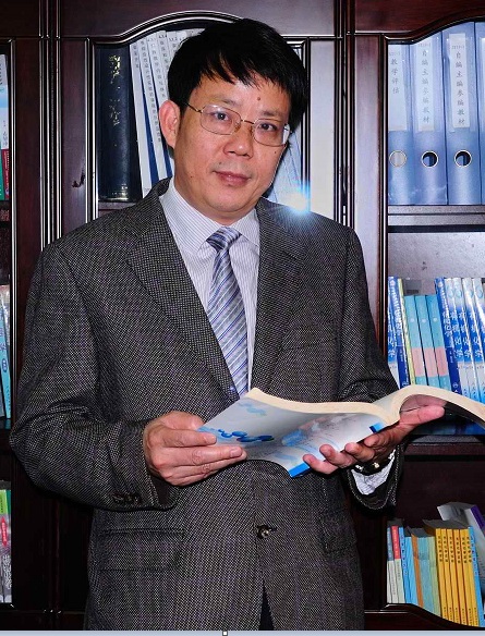 Changxi Yu (俞昌喜)合作论文数福建医科大学药学院3Lingyao Du合作论文数2yingsu huang (黄颖苏)合作论文数2Ji Li合作论文数2GS WU合作论文数2BY Chen合作论文数2HM Campiglia合作论文数2Xiaoyan Li.合作论文数2Xz Gu合作论文数2LF LI合作论文数2MZ LIAO合作论文数2
 Ying Xia (夏萤)合作论文数Shanghai Key Lab Acupuncture Mech & Acupoint Funct, Fudan Univ2JianWei Jiang (姜建伟)合作论文数2Law Ping-Yee合作论文数2Chengwu Chi合作论文数Institute of Biochemistry and Cell Biology2Tang Q M合作论文数2DaKuang Huang合作论文数2Xiaocong Zhao合作论文数2
 Horace H. Loh合作论文数Department of Pharmacology, Medical School, University of Minnesota2
 Xiaoyuan Feng (冯晓源)合作论文数Institute of Magnetic Resonance and Molecular Imaging in Medicine, East China Normal University2
 Mingya Li (李明亚)合作论文数School of Resources and Materials, Northeastern University at Qinhuangdao2Kuanyan Li合作论文数2Qiuzao He合作论文数2
 Yingqiliang Mao (毛应启梁)合作论文数Department of Integrative Medicine and Neurobiology, School of Basic Medical Sciences, Fudan University2
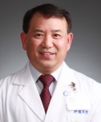 Sanjun Cai (蔡三军)合作论文数Fudan University Shanghai Cancer Center2Fei Ma合作论文数2Huiting Peng (彭惠婷)合作论文数2
 Yanqin Gao (高艳琴)合作论文数Institutes of Brain Science, Fudan University;State Key Laboratory of Medical Neurobiology, Fudan University2W Y Mo合作论文数1F. Yan合作论文数1




