AIM:The aim of the study is to compare the effect of extraperitoneal high uterosacral ligament suspension (EHUS) and sacrospinous ligament fixation on lower urinary tract symptoms (LUTS) and anatomical reduction in patients with pelvic organ prolapse (POP). METHODS:This retrospective cohort study concluded 91 patients with POP who underwent EHUS or sacrospinous ligament fixation (SSLF) for apical repair. The primary outcome was the effect of the two operations on anatomical reduction. The main secondary outcomes included improvement of questionnaire scores, satisfaction, and subjective prolapse rate. Categorical variables, Student's t test, and the Mann-Whitney U test were used for analysis. RESULTS:All patients' scores of POP-Q were significantly improved after surgery. The scores of urinary frequency and urgency in the SSLF group improved significantly from 6 months after surgery, with statistical significance (p < 0.05), while the scores of nocturia and urgency in the EHUS group improved from 1 month after surgery (p < 0.05). The scores of questionnaires in both groups were significantly lower than those before surgery, while the EHUS group showed more significant improvement (p < 0.05). CONCLUSION:EHUS is easy to operate, has a short operative time, fewer perioperative and postoperative complications, and the apical can be suspended well if the prolapse is not severe. It also has a certain degree of relief for LUTS, which is a new type of operation that is worthy of clinical promotion.
Background and Objectives: Portal hypertension (PH), as the main consequence of cirrhosis, leads to the development of gastroesophageal varices (GEVs). Variceal hemorrhage (VH) caused by the rupture of GEVs is a life-threatening emergency. Thus, the prediction of VH risk is considerably important. Our pilot study aimed to identify the risk factors of variceal hemorrhage (VH) in cirrhosis. Materials and Methods: Cirrhotic patients were prospectively included and divided into two groups according to the presence or absence of VH. Conventional ultrasound and shear wave dispersion (SWD) imaging were conducted to detect the portal vein diameter, spleen diameter, ascites, liver stiffness (LS) and shear wave dispersion slope (SWDS). The laboratory tests were recorded, including platelets (PLT), alanine transaminase (ALT), aspartate aminotransferase (AST), total bilirubin (TBIL) and albumin (ALB). The risk factors of VH were screened using univariate analyses and identified using multivariate logistic regression. The ROC curves were used to assess diagnostic accuracy. Comparisons between AUCs were performed using the Delong method. Results: Sixty-five patients with 22 VHs were finally included. The SWDS, spleen diameter and ascites were identified as independent risk factors for VH. The SWDS showed good performance for diagnosing VH (AUC = 0.768, 95% CI: 0.647–0.864), and sensitively identified 95.5% (95% CI: 77.2%–99.9%) of patients with VH. Including the three risk factors in multivariate logistic regression, we obtained a formula for diagnosing VH: −20.749 + 0.804 × SWDS + 0.449 × spleen diameter + 1.803 × ascites (no ascites = 0, ascites = 1). Comparison of AUCs revealed that the formula (AUC = 0.900, 95% CI: 0.800–0.961) performed better than LS, SWDS, and spleen diameter in diagnosing VH (p < 0.001; p < 0.05; p < 0.05). Conclusions: SWDS is a sensitive parameter for assessing the risk of VH. Combining the SWDS, spleen diameter and ascites resulted in good diagnostic accuracy.
肝脏本质为黏弹性体,兼具黏性和弹性力学特性.黏性反映肝组织抗形变能力,是表征组织病理变化的重要参数;但目前多数商用超声弹性成像忽略肝组织的黏性特征而将其假设为纯弹性体.超声剪切波频散(SWD)成像可同时获得反映肝脏黏性和弹性的相关参数,为评估肝脏弥漫性病变提供全新的视角.本文就超声SWD成像原理及其在肝脏弥漫性病变中的研究进展进行综述.
Cirrhosis is a chronic liver disease that seriously jeopardizes the life and health of patients. Currently, ultrasound (US) imaging is commonly used by the computer-aided diagnosis (CAD) system to diagnose cirrhosis. With the rapid development of artificial intelligence, deep learning methods for cirrhosis diagnosis using ultrasound image data have emerged. However, due to US images' complexity and variability, this input usually requires manual annotation. This study proposes LiverTL, an end-to-end deep learning approach for the automatic cirrhosis ultrasound image classification to overcome these limitations. LiverTL includes an automatic region of interest (ROI) detection module to support various ultrasound images' ROI extraction. Simultaneously, the classification module utilizes ROI areas and obtain the cirrhosis diagnosis results through the transfer learning network. We find that LiverTL achieves high classification accuracy on our evaluation data set. The cirrhosis data experiments suggest that a proper pre-training model for transfer learning is crucial for the classification results. These findings potentially pave the way to advance the diagnosis and therapy of cirrhosis.
目的 探讨分叶状毛细血管瘤的声像图表现.方法 回顾性分析11例经病理证实的分叶状毛细血管瘤患者的资料,总结其临床及超声特征.结果 病变均为单发,部分伴疼痛、出血,8例位于手部和颈面部,4例病变处曾有外伤史.7例累及皮肤层,4例仅位于皮下组织.病灶长径0.3~1.7 cm,平均(0.8±0.4) cm,呈低回声,内部可见小的低-无回声区.7例边界清晰.8例血流丰富,且其中5例可见基底部供血动脉.结论 分叶状毛细血管瘤的声像图具有一定特征,结合临床病史有助于同其他软组织肿物鉴别.
目的 探讨瞬时弹性成像系统FibroTouch脂肪衰减参数(FAP)诊断不同程度脂肪肝的意义,以及FAP的相关因素.方法 随机选取就诊于我院超声诊断科患者为被检测对象,行常规超声检查进行脂肪肝程度分级(S0、S1、S2、S3分别对应无、轻度、中度、重度);同时行FibroTouch检查,测定FAP,获得不同程度脂肪肝的最佳界值.同时分析FAP与BMI、SCD、LSM相关性.结果 共纳入受试者767例,其中S0 401例,S1 164例,S2 160例,S3 42例,ROC曲线分析得出,≥S1、≥S2、≥S3的FAP界值分别为243、268、285 dB/m,其对应的灵敏度、特异度分别为0.87%、0.80%、0.86%、0.84%、0.76%、0.95%.FAP值与BMI、SCD、LSM均呈正相关.结论 脂肪衰减参数可以定量评估肝脏脂肪化严重程度,对脂肪肝患者的随访及监测具有重要意义,并且与BMI、SCD、LSM具有相关性.
目的 探讨灰阶超声及弹性成像在诊断盆腔脂肪增多症中的应用价值.方法 前瞻性选取CT诊断为盆腔脂肪增多症的患者7例,平均年龄(40.4±6.4)岁.纳入健康受试者20例作为对照组,平均年龄(42±7)岁.使用声科SuperSonic Imagine Aixplorer超声诊断仪,线阵探头SL10-2测量皮下脂肪杨氏模量;凸阵探头SC6-1评估肾盂宽度,膀胱形态,膀胱周脂肪厚度及杨氏模量;腔内探头SE12-3评估直肠周围脂肪厚度及杨氏模量.结果 盆腔脂肪增多症患者均出现膀胱周围脂肪明显增多,使得膀胱受压呈泪滴形或梨形.弹性测值显示患者皮下脂肪、膀胱后脂肪及直肠周围脂肪硬度与健康受试者未见明显改变(P1=0.91、P2=0.52、P3=0.09).但是,前列腺前方脂肪硬度较健康对照组显著增加(P<0.01).结论 膀胱与前列腺间距增大合并膀胱形态改变为盆腔脂肪增多症的特征性表现.超声弹性成像为评估盆腔脂肪硬度提供了定量参数.
 Ligang Cui (崔立刚)合作论文数Peking University Third Hospital;Institute of Medical Technology, Health Science Center, Peking University6
 Ling Jiang (江凌)合作论文数Department of Ultrasound, Peking University Third Hospital2
 Weiwei Shen (沈伟伟)合作论文数1Fengwu Li (李凤舞)合作论文数1Shiyan Wang合作论文数1Shanshan Li (李姗姗)合作论文数1
 Yang Sun合作论文数Columbia University in the City of New York, Columbia University1Qiong He (何琼)合作论文数1Qiang Zhao (赵强)合作论文数1Wen Chen (陈文)合作论文数1
 Bingshan Li合作论文数1
 Li Zhang (张里)合作论文数School of Earth Sciences and Resources, China University of Geosciences, Beijing1
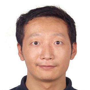 Hai Yang (杨海)合作论文数Department of Computer Science and Engineering, School of Information Science and Engineering, East China University of Science and Technology1
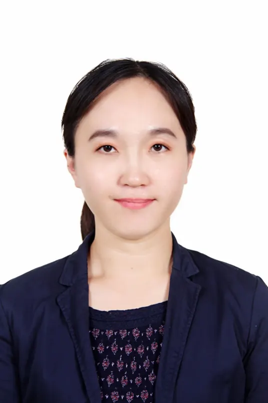 Jie Jiang (蒋洁)合作论文数Department of Ultrasound, Peking University Third Hospital1
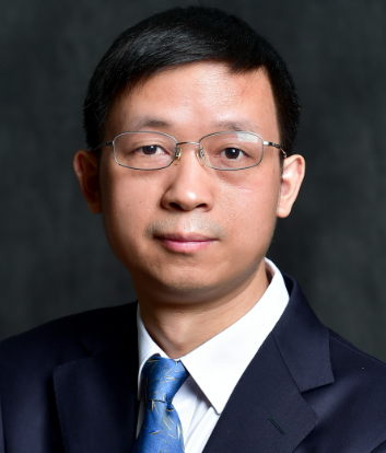 Jianwen Luo (罗建文)合作论文数School of Biomedical Engineering, Tsinghua University1
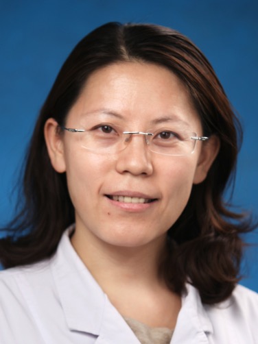 Xiuli Sun (孙秀丽)合作论文数Peking University People's Hospital1
 Jianying Liu (柳剑英)合作论文数Peking University Third Hospital;School of Basic Medical Sciences, Peking University1




