Schematic of the proposed mechanism by which NSD1 mutations enhance the malignant progression of EC.
BACKGROUND:Atrial fibrillation (AF) is the most common arrhythmia and is associated with high morbidity and mortality, particularly in the aging population. Current treatment and prevention strategies remain suboptimal, highlighting the urgent need to better understand the mechanisms underlying aging-associated AF. We recently reported a causal role of the stress-activated kinase JNK2 (c-Jun N-terminal kinase 2) in aging-associated AF pathogenesis, mediated by JNK2-driven sarcoplasmic reticulum Ca2+ dysfunction. However, the mechanisms by which cardiac JNK2 is activated during aging to promote AF remain unclear. Emerging evidence suggests that interorgan crosstalk contributes critically to the development of cardiovascular diseases. A hyperpermeable gastrointestinal epithelial barrier ("leaky gut"), commonly observed in aged individuals, is associated with elevated levels of proinflammatory cytokines and an increased risk of AF. Although proinflammatory cytokines have been proposed as predisposing factors for AF, clinical and experimental studies have yielded inconsistent results, underscoring the complexity of inflammation-associated AF pathogenesis. Here, we investigated whether cardiac JNK2 integrates diverse stress stimuli, including proinflammatory cytokines and lipopolysaccharide, to drive AF pathogenesis. METHODS:We used aged mice, intestinal epithelium-specific tight junction OD (occludin) knockdown (OD+/-) mice, and a well-established dextran sulfate sodium-induced leaky gut mouse model characterized by reduced gastrointestinal epithelial occludin expression. A series of physiological and molecular approaches was applied to assess cardiac and gastrointestinal responses. RESULTS:We found that leaky gut significantly activates atrial JNK2, which, in turn, drives Ca2+-triggered arrhythmic activity and increases AF inducibility in aged, dextran sulfate sodium-treated, and OD+/- mouse models. Restoration of gut barrier function in dextran sulfate sodium mice, a clinically relevant model, reduced AF susceptibility. Similarly, either JNK2 inhibition or TNF-α (tumor necrosis factor α) blockade abolished the increased AF risk associated with leaky gut. Furthermore, we demonstrate, for the first time, that leaky gut-associated proinflammatory cytokines, including TNF-α and IL-17A (interleukin-17A), together with lipopolysaccharide, activate cardiac JNK2. This activation promotes AF pathogenesis through JNK2-mediated arrhythmogenic mechanisms, including diastolic sarcoplasmic reticulum Ca2+ leak, Ca2+ waves, and delayed afterdepolarizations. CONCLUSIONS:Activated JNK2 functions as a pathological nodal integrator of leaky gut-associated stress signals, mediating gut-to-heart crosstalk and driving inflammation-induced AF pathogenesis. Targeting JNK2 may represent a novel therapeutic strategy for AF.
NSD1 mutation status and IHC scores in 99 cases of endometrial cancer specimens and the associated clinical information.
Catalytic activity of NSD1 is required to suppress proliferation, invasion, and glycolysis.
Restoration of PTEN expression and inhibition of AKT activation suppress glycolysis and malignant phenotypes in NSD1 KO cells.
BACKGROUND: Ovarian cancer (OC) is a fatal carcinoma for women. This study attempts to explore the role of vascular endothelial zinc finger 1 (VEZF1) in OC cell ferroptosis, thereby finding a new target for OC treatment. METHODS: Expressions of VEZF1, miR-545-3p and PLAG1 in HOSE cell line and OC cell lines were determined by RT-qPCR and western blot analysis. After VEZF1 was silenced in cells, cell proliferation was examined, contents of ROS, MDA, Fe2+, GSH were detected, and expressions of ACSL4 and GPX4 were tested. Afterwards, the binding relation between VEZF1 and miR-545-3p and between miR-545-3p and PLAG1 were verified. Functional rescue assays were formulated to validate the role of miR-545-3p knockdown or PLAG1 overexpression in OC cell ferroptosis. RESULTS: VEZF1 was overexpressed in OC, and VEZF1 silencing reduced cell proliferation, elevated levels of ROS, MDA, Fe2+, inactivated levels of GSH and GPX4, and enhanced ACSL4 expression. Functionally, VEZF1 transcriptionally inhibited miR-545-3p, and miR-545-3p targeted and inhibited PLAG1. miR-545-3p knockdown or PLAG1 overexpression could reverse the effect of VEZF1 silencing on OC cell ferroptosis. CONCLUSION: VEZF1 was overexpressed in OC and it limited OC cell ferroptosis by transcriptionally inhibiting miR-545-3p and elevating PLAG1 expression.
RNA-binding proteins (RBPs) are indispensable effectors of post-transcriptional gene regulation and are increasingly recognized as pivotal modulators of oncogenic signaling pathways. Cold shock domain-containing protein E1 (CSDE1) has been reported to be dysregulated or mutated in several malignancies; however, its functional significance and the mechanistic basis of its involvement in endometrial cancer (EC) progression remain poorly understood. Bioinformatic analysis of The Cancer Genome Atlas (TCGA) was employed to characterize CSDE1 expression and mutation profiles in EC. CRISPR/Cas9-mediated knockout (KO) was used to establish CSDE1-deficient EC cell lines. The impact on cell proliferation, migration, invasion, and epithelial–mesenchymal transition (EMT) was evaluated through standard functional assays. To identify downstream targets, transcriptomic profiling was integrated with RNA-protein interaction assay and mRNA stability analysis. Finally, functional rescue experiments were conducted using shRNA-mediated IL-6 silencing and pharmacological inhibition with the IL-6 antagonist Siltuximab. CSDE1 is frequently mutated and significantly downregulated in EC tissues compared to normal controls. Loss of CSDE1 markedly accelerated EC cell proliferation, migration, invasion, and the acquisition of EMT-associated phenotypes. Transcriptomic analysis identified IL-6 as a prominent downstream target that is significantly upregulated upon CSDE1 depletion. Mechanistically, CSDE1 directly binds to IL-6 mRNA and negatively regulates its stability, thereby restraining IL-6 expression. Notably, both genetic silencing of IL-6 and pharmacological neutralization of IL-6 signaling with Siltuximab effectively abrogated the aggressive malignant phenotypes induced by CSDE1 loss. These findings establish CSDE1 as a novel tumor suppressor in EC and elucidate a previously unrecognized post-transcriptional CSDE1–IL-6 regulatory axis. By linking RBP-mediated mRNA stability to oncogenic inflammatory signaling, our study provides critical insights into the molecular drivers of EC progression. Furthermore, we identify the IL-6 signaling pathway as a significant therapeutic vulnerability that could be exploited to treat CSDE1-deficient EC.
Stratified analysis of immunohistochemical correlations among NSD1, K98me, and PTEN expression according to NSD1 mutation type in endometrial carcinoma specimens.
MK-2206 suppresses enhanced uterine tumor growth driven by NSD1 mutations in an orthotopic patient-derived xenograft (PDX) model.
NSD1 loss enhances PTEN/AKT-mediated transcriptional activation of glycolytic genes.
 Isabelle Deschênes合作论文数59
 Xiaowei Xi (席晓薇)合作论文数Shanghai General Hospital40YiXia Yang (杨懿霞)合作论文数36
 Qin Yan (严沁)合作论文数School of Medicine, Tongji University;Obstetrics and Gynecology Hospital of Tongji University35
 Wen Lu (陆雯)合作论文数Obstetrics and Gynecology Hospital of Tongji University;School of Medicine, Tongji University31
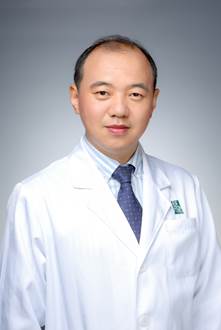 Shuangdi Li (李双弟)合作论文数School of Medicine, Tongji University;Shanghai First Maternity and Infant Hospital Affiliated to Tongji University28Laurita Kenneth R合作论文数25
 Yiran Li (李伊然)合作论文数School of Medicine, Tongji University;Obstetrics and Gynecology Hospital of Tongji University23
 Bilan Li合作论文数21
 Kai Wang (王凯)合作论文数School of Medicine, Tongji University;First Maternity and Infant Health Hospital Affiliated To Tongji University21
 Wei Bao合作论文数20
 Bin Cai (蔡斌)合作论文数Shanghai Red Cross Ninth People's Hospital20
 Yinyan He (贺银燕)合作论文数Shanghai General Hospital;Shanghai Jiao Tong University School of Medicine20Eckhard Ficker合作论文数19
 Huan Tong合作论文数Sichuan University, Sichuan University17Che Qi合作论文数17
 Jiarong Zhang (张佳荣)合作论文数The First People' s Hospital of Shanghai Affiliated to Shanghai Jiaotong University School of Medicine17
 Feizhou Jiang合作论文数16Haiyan Liu合作论文数16
 Dandan Yang合作论文数15XiaoMei Wu (吴晓梅)合作论文数15
 David S. Rosenbaum合作论文数School of Medicine, Case Western Reserve University15
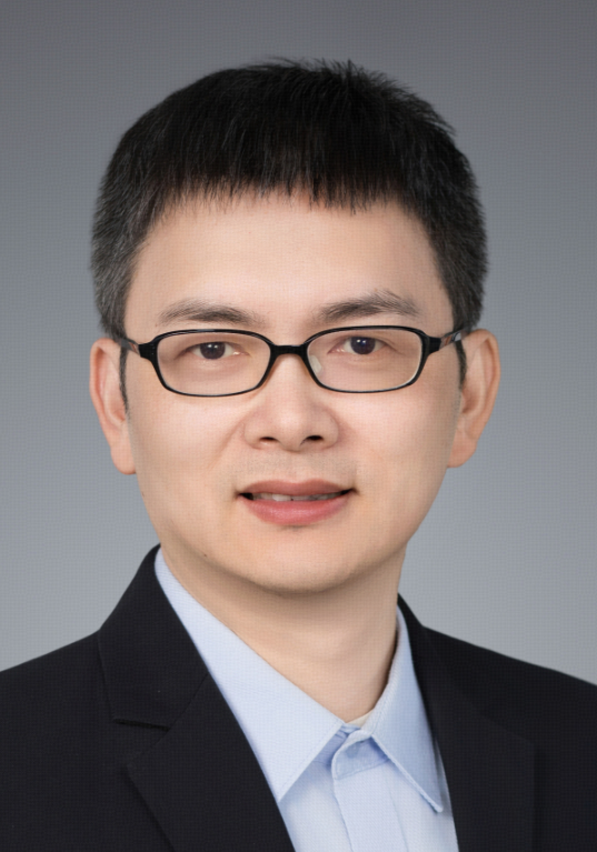 Zhiyong Mao (毛志勇)合作论文数School of Life Sciences and Technology, Tongji University15Ji-Dong Fu合作论文数15Xiaoying He合作论文数14
 Wang Fangyuan合作论文数Shanghai First People's Hospital, Shanghai Jiao Tong University14Xue-Lian Liu合作论文数13Xianyao Xu合作论文数13JianLi Chen (陈建利)合作论文数13
 Yun Liao (廖赟)合作论文数Tongren Hospital, Shanghai Jiao Tong University School of Medicine13Mengfei Wang合作论文数12Wang Huihui合作论文数12Drew Nassal合作论文数11Bailian Cai合作论文数11Yiding Bian合作论文数11Xiao-Yue Chen合作论文数11Zheng Chen合作论文数11Tingting Yang合作论文数11Wang Jingyun合作论文数11Thomas J. Hund合作论文数Washington University11
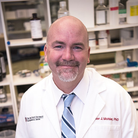 Peter Mohler合作论文数Departments of Physiology & Cell Biology & Internal Medicine, College of Medicine, The Ohio State University11Jieqi Ke合作论文数10Junjie Qu合作论文数10
 Ying Jiang (蒋颖)合作论文数School of Life Sciences and Technology, Tongji University10
 Qizhi He (贺其志)合作论文数School of Medicine, Tongji University10
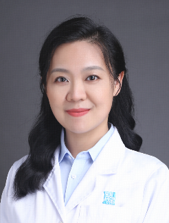 Kun Gao (高昆)合作论文数Obstetrics and Gynecology Hospital of Tongji University10A. Ramirez-Navarro合作论文数10Qiu Haifeng合作论文数9
 Binya Liu合作论文数9




