Background: Liver fibrosis (LF) is a common pathological process in the progression of multiple chronic liver diseases to cirrhosis, affecting millions of people worldwide annually. The incomplete understanding of its mechanisms has led to a lack of clinically effective therapeutic options. ErTao decoction (ETD, (sic)(sic)(sic)), a derivative combining the components of Erchen Decoction and Taohong Siwu Decoction, is rooted in the traditional Chinese medicine theory of "phlegm-dampness-blood stasis". However, the precise mechanism by which ETD exerts its therapeutic effects in LF remains unclear. Purpose: The purpose of study was to investigate the protective effect of ETD and elucidate its underlying molecular mechanism on LF. Methods: In this study, we employed a multifaceted approach to evaluate the effects of ETD on LF. We used H&E staining, Sirius red staining, immunofluorescence, immunohistochemical analysis, and Western blotting to assess the protective effects of ETD in a CCl4-induced fibrosis mouse model. In vitro validation was conducted using macrophages and hepatic stellate cells to further elucidate the mechanisms involved. STING-deficient mice were used to assess its regulatory effects on liver injury, inflammatory and activation through immunohistochemical staining and Western blotting. Furthermore, UHPLC-HRMS detection and computer-aided drug analysis were employed to identify and validate potential effective components of ETD for responsible for its therapeutic effects in treating LF. Results: In our in vivo and in vitro experiments, we found that ETD effectively reduced collagen fiber deposition and alleviated LF pathological changes by inhibiting macrophage inflammatory activation and suppressing NLRP3 and STING signaling. Notably, STING deficiency exhibited a protective effect against liver tissue injury and inhibited inflammatory activation of hepatic macrophages in LF model mice. Additionally, comprehensive analysis of the active ingredients in ETD strongly suggested that Naringin served as a pivotal bioactive constituent within ETD responsible for modulating STING signaling. Conclusions: Our study highlighted the protective effects of ETD on LF by inhibiting STING-mediated macrophage activation and NLRP3 inflammasome signaling. Notably, Naringin might serve as a promising novel STING
PurposeGastric cancer, characterised by a significant mortality rate, is one of the most prevalent malignant neoplasms globally. Exosomal non-coding RNAs play a key role in gastric carcinogenesis, metastasis and treatment resistance by regulating gene expression, remodelling the tumour microenvironment and mediating drug resistance. In the identification of early gastric cancer, these exosomal non-coding RNA molecules possess significant potential to be developed into biomarkers that do not entail invasive procedures.MethodsOur study undertook a comprehensive and profound literature examination in core databases like PubMed, Web of Science, ScienceDirect, Embase, Scopus, and Medline, with the aim of precisely evaluating the potential effectiveness of exosomal miRNAs, lncRNAs, and circRNAs in the diagnosis of gastric cancer.ResultsA sum of 52 studies were incorporated, comprising 164 studies. These studies identified a total of 59 miRNAs, 17 lncRNAs, and 16 circRNAs. For miRNAs, sensitivity was estimated at 0.72 (95% CI, 0.69 - 0.76) and specificity at 0.80 (95% CI, 0.77 - 0.83). For lncRNAs, the sensitivity was 0.86 (95% CI, 0.84 - 0.87) and the specificity was 0.82 (95% CI, 0.80 - 0.83). In the case of circRNAs, the sensitivity and specificity were 0.71 (95% CI, 0.63 - 0.78) and 0.88 (95% CI, 0.81 - 0.93) respectively. The AUC for miRNAs was calculated as 0.83. As for lncRNAs, its AUC was established to be 0.89. Regarding circRNAs, the determined AUC was 0.86.ConclusionThese results confirm the efficacy of exosomal ncRNAs as powerful biomarkers for the early diagnosis of gastric carcinomas, thereby laying a strong foundation for the advancement of novel diagnostic approaches.
Supplementary Table from Single-Hit Inactivation Drove Tumor Suppressor Genes Out of the X Chromosome during Evolution
Abstract Cancer-related genes are under intense evolutionary pressure. In this study, we conjecture that X-linked tumor suppressor genes (TSG) are not protected by the Knudson's two-hit mechanism and are therefore subject to negative selection. Accordingly, nearly all mammalian species exhibited lower TSG-to-noncancer gene ratios on their X chromosomes compared with nonmammalian species. Synteny analysis revealed that mammalian X-linked TSGs were depleted shortly after the emergence of the XY sex-determination system. A phylogeny-based model unveiled a higher X chromosome-to-autosome relocation flux for human TSGs. This was verified in other mammals by assessing the concordance/discordance of chromosomal locations of mammalian TSGs and their orthologs in Xenopus tropicalis. In humans, X-linked TSGs are younger or larger in size. Consistently, pan-cancer analysis revealed more frequent nonsynonymous somatic mutations of X-linked TSGs. These findings suggest that relocation of TSGs out of the X chromosome could confer a survival advantage by facilitating evasion of single-hit inactivation. Significance: This work unveils extensive trafficking of TSGs from the X chromosome to autosomes during evolution, thus identifying X-linked TSGs as a genetic Achilles' heel in tumor suppression.
Aim: To explore the expression and biological function of long intergenic non-protein coding RNA 1089 (LINC01089) in gastric cancer (GC) progression and its underlying mechanism. Methods: LINC01089 and microRNA-27a-3p (miR-27a-3p) expressions were detected with the quantitative real-time polymerase chain reaction (RT-qPCR). Cell proliferation, migration and invasion were evaluated by Cell Counting Kit-8 (CCK-8) and Transwell assay. Epithelial-mesenchymal transition (EMT)-related proteins were also measured by Western blot. The relationship between LINC01089 and miR-27a-3p was revealed by a bioinformatics analysis and dual-luciferase reporter assay. Results: LINC01089 was significantly down-regulated in GC tissues, as well as GC cell lines. GC patients with lower LINC01089 expression were more likely to have poor outcomes. Overexpression of LINC01089 significantly suppressed GC cells growth, migration and invasion and forbade the EMT process. LINC01089 was directly targeted at miR-27a-3p. The transfection of miR-27a-3p mimics reversed the inhibitory effects on proliferative and metastatic abilities of GC cells with LINC01089 overexpression. Conclusion: LINC01089 inhibits cell proliferation and metastasis in GC by targeting miR27a-3p/EMT axis, which should be considered as a promising therapeutic target.
Impaired autophagic degradation of intracellular lipids is causally linked to the development of non-alcoholic steatohepatitis (NASH). Pharmacological agents that can restore hepatic autophagic flux could therefore have therapeutic potentials for this increasingly prevalent disease. Herein, we investigated the effects of polydatin, a natural precursor of resveratrol, in a murine nutritional model of NASH and a cell line model of steatosis. Results showed that oral administration of polydatin protected against hepatic lipid accumulation and alleviated inflammation and hepatocyte damage in db/db mice fed methionine-choline deficient diet. Polydatin also alleviated palmitic acid-induced lipid accumulation in cultured hepatocytes. In both models, polydatin restored lysosomal function and autophagic flux that were impaired by NASH or steatosis. Mechanistically, polydatin inhibited mTOR signalling and up-regulated the expression and activity of TFEB, a known master regulator of lysosomal function. In conclusion, polydatin ameliorated NASH through restoring autophagic flux. The polydatin-regulated autophagy was associated with inhibition of mTOR pathway and restoration of lysosomal function by TFEB. Our study provided affirmative preclinical evidence to inform future clinical trials for examining the potential anti-NASH effect of polydatin in humans.
Treatment with cisplatin (DDP) is one of the standard therapies used to treat non-small-cell lung cancer (NSCLC) and fundamentally causes resistance in cancer cells, which eventually poses as an obstacle to the efficacy of chemotherapy in NSCLC. Efforts are on all over the world to explore a sensitizer of NSCLC to DDP. Here, we studied the effect of IL-7 on the resistance of NSCLC to chemotherapy. We observed that IL-7 treatment significantly enhanced DDP-induced effects in A549 and A549/DDP cells (DDP-resistant cells), including decreased cell viability and proliferation, as well as increased cell apoptosis and S arrest, indicating that IL-7 treatment resensitized DDP-resistant NSCLC cells to DDP. Subsequently, IL-7 enhanced the sensitivity of PI3K/AKT signaling and expressions of ABCG2 to DDP. By inhibiting IL-7 signaling via IL-7R knockdown or activating PI3K/AKT signaling via PI3K activation, the resensitization to DDP by IL-7 was abrogated, and the expression levels of ABCG2, p-PI3K, and p-AKT were found to be significantly higher. In vivo results also confirmed that IL-7 only in combination with DDP could remarkably induce tumor regression with reduced levels of ABCG2 in tumorous tissues. These findings indicate that IL-7, apart from its adjuvant effect, could overcome multidrug resistance of DDP to restore its chemotherapy sensitivity.
Autophagic impairment is implicated in nonalcoholic fatty liver disease (NAFLD), but the molecular mechanism is unclear. We found that autophagic flux was significantly inhibited in 3 murine models of NAFLD. Interestingly, the number of acidic organelles and the level of mature cathepsin D were reduced, suggesting defective lysosome acidification. Asparagine synthetase (ASNS) was induced by endoplasmic reticulum stress, leading to the generation of asparagine, which inhibited lysosome acidification. Both steatotic-and asparaginetreated hepatocytes showed reduced lysosomal acidity and retention of lysosomal calcium. Knockdown ofASNS in steatotic hepatocytes restored autophagic flux. As a potential biomarker, increased serum p62/sequestosome 1 (SQSTM1) level was an independent risk factor for patients with steatosis and lobular inflammation. Impaired autophagy inNAFLDis elicited by defective lysosome acidification, which is caused byASNS-induced asparagine synthesis under endoplasmic reticulum stress and subsequent retention of lysosomal calcium. p62/SQSTM1 could be used as a noninvasive biomarker in the diagnosis ofNAFLDpatients.
BACKGROUND:Cisplatin (DDP)-based chemotherapy is the common first-line therapy for lung cancer. However, their efficacy is often limited by primary drug resistance and/or acquired drug resistance. The aim of this study was to investigate the function of miRNA-146a (miR-146a) in DDP-resistant non-small cell lung cancer (NSCLC), as well as the underlying mechanisms.METHODS:The effect of overexpression of miR-146a and/or knockdown of cyclin J (CCNJ) in A549/DDP and SPC-A1/DDP cells were investigated as follows. The cellular sensitivity to DDP, cell apoptosis, cell cycle and cell mobility were detected by CCK-8, flow cytometry, hoechst staining and cell invasion/migration assay, respectively. The effects of miR-146a overexpression in NSCLC resistant cells were further analyzed in a nude mouse xenograft model.RESULTS:Overexpression of miR-146a and/or knockdown of CCNJ significantly increased the sensitivity to DDP in A549/DDP and SPC-A1/DDP cells compared to NC group via arresting cell cycle, enhancing cell apoptosis, inhibiting cell viability and motility in vitro and in vivo. Furthermore, miR-146a could specially degrade the mRNA of CCNJ, as examined by dual luciferase report assay.CONCLUSION:The study indicates a crucial role of miR-146a in the development of acquired drug resistance to DDP in NSCLC cells. Further understanding of miR-146a mediated crosstalk networks may promote the clinical use of miR-146a analogue in NSCLC therapy.
Intrinsic apoptosis eliminates cells with damaged DNA and cells with dysregulated expression of oncogene. PGAM5, a member of the phosphoglycerate mutase family, has two splicing variants: PGAM5L (the long form) and PGAM5S (the short form). It has been well established that PGAM5 is at the convergent point of multiple necrosis pathways. However, the role of PGAM5 in intrinsic apoptosis is still controversial. Here we report that the PGAM5L, but not PGAM5S is a prerequisite for the activation of Bax and dephosphorylation of Drp1 in arenobufagin and staurosporine induced intrinsic apoptosis. Knockdown of PGAM5L inhibits the translocation of Bax to the mitochondria and reduces mitochondrial fission. The interaction between PGAM5L and Drp1 was observed in both arenobufagin and staurosporine treated HCT116 cells, but not in HCT116 Bax(-/-) cells. Bax transfection rescues the formation of the triplex in both arenobufagin and staurosporine stimulated HCT116 Bax(-/-) cells. Arenobufagin shows remarkable anti-cancer effects both in orthotropic and heterotropic CRC models and demonstrates less toxic effects as compared with that of cisplatin. Bax-PGAM5L-Drp1 complex is detected in arenobufagin and staurosporine treated CRC cells in vitro and in arenobufagin and cisplatin treated tumor in vivo as well. In summary, our results demonstrate that Bax-PGAM5L-Drp1 complex is required for intrinsic apoptosis execution.
Cancer associated malnutrition(CAM) is the common cause of death in patients with cancer. Pathogenesis of CAM in traditional Chinese medicine( TCM) is deficiency of spleen qi or even asthenia of both the spleen and the kidney in the advanced stages. Tumor could induce a micro-environment that characterized by chronic inl ammation and immunosuppression. Tumor cells release cytokines and reactive oxygen species(ROS) into the micro-environment, resulting in the autophagy of skeletal muscles, adipocytes and interstitial cells. Cancer cells use glutamate, ketone and lactate released by the autophagy to fuel oxidative mitochondrial metabolism in mitochondrion of tumor cells. The reprogramming of metabolism between tumor and some normal tissues, such as skeletal muscles, directed by tumor is the internal essence of deficiency of spleen. The consumption of muscles and adipose tissues is the external manifestation. The metabolic coupling through autophagy is pivotal for the formation of cachexia characterized by deficiency of spleen qi or even asthenia of both the spleen and the kidney in the advanced stages. It is a novel idea on the theory of spleen governing muscles and limbs, as well as a new connotation on the therapy of invigorating the spleen.
 Jun Yu (于君)合作论文数Department of Medicine & Therapeutics, Faculty of Medicine, The Chinese University of Hong Kong4
 William K. K. Wu (胡嘉麒)合作论文数South Sichuan Institute of Translational Medicine;Department of Anaesthesia and Intensive Care, Faculty of Medicine, The Chinese University of Hong Kong;Peter Hung Pain Research Institute, The Chinese University of Hong Kong4
 Matthew T. V. Chan合作论文数Department of Anaesthesia and Intensive Care, Faculty of Medicine, The Chinese University of Hong Kong;Prince of Wales Hospital4
 Hung Chan合作论文数3
 Ka-fai To (杜家輝)合作论文数Department of Anatomical and Cellular Pathology, Faculty of Medicine, Chinese University of Hong Kong;State Key Laboratory in Oncology, Chinese University of Hong Kong3Xiaodong Liu合作论文数Chinese University of Hong Kong, The Chinese University of Hong Kong3
 Wei Kang (康巍)合作论文数School of Bioengineering, Dalian University of Technology3
 Xuegang Sun (孙学刚)合作论文数School of Traditional Chinese Medicine, Southern Medical University3
 Sunny Wong合作论文数Lee Kong Chian School of Medicine, Nanyang Technological University3
 Tony Gin合作论文数Department of Anaesthesia and Intensive Care, The Chinese University of Hong Kong3Ju-deng Zeng合作论文数2
 Jennifer X Zhang (張翔)合作论文数Department of Medicine & Therapeutics, Faculty of Medicine, The Chinese University of Hong Kong2Gang Wu合作论文数2
 Lin Shi合作论文数2Yuanyuan Huang合作论文数2
 Dan Huang (黄丹)合作论文数Department of Immunology and Microbiology, School of Life Sciences, Southern University of Science and Technology2Bin Ke (柯斌)合作论文数2Linlin Jing合作论文数2
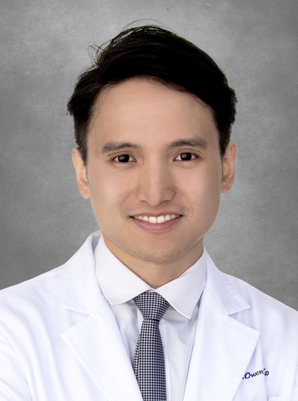 Owen H Ko (高浩)合作论文数Department of Medicine & Therapeutics, Faculty of Medicine, The Chinese University of Hong Kong2
 Sze Lok Alfred Cheng (鄭詩樂)合作论文数School of Biomedical Sciences, Faculty of Medicine, The Chinese University of Hong Kong2
 Maggie Haitian Wang (王海天)合作论文数The Jockey Club School of Public Health and Primary Care, Faculty of Medicine, The Chinese University of Hong Kong2Xie Chuan合作论文数2
 Danny Chan (陳振勝)合作论文数School of Biomedical Sciences, LKS Faculty of Medicine, The University of Hong Kong2
 Xiangchun Li (李祥春)合作论文数Tianjin Cancer Hospital, Tianjin Medical University2
 Lin Zhang合作论文数UPMC Hillman Cancer Center Shadyside2Huarong Chen合作论文数2Dariusz Plewczyński合作论文数Department of Information Processing Systems, Faculty of Mathematics and Information Science, Warsaw University of Technology2Chee Sam Chan合作论文数1Shanglong Kou合作论文数1Jeffery Ho合作论文数1Idy H T Ho合作论文数1Weiqiang Jiang合作论文数1Eagle S H Chu合作论文数1Ruiping Li合作论文数1Chaofeng Wu合作论文数1Yuxin Gong合作论文数1Junfang Liu合作论文数1Wenjuan Xu合作论文数1
 Xiansong Wang (王显嵩)合作论文数School of Pharmaceutical Sciences, Sun Yat-sen University1Xiyao Li合作论文数1Chung-chih Lin合作论文数Chang Gung University1Yongchang Cai合作论文数1Zhiliang Chen合作论文数1YuanLiang Liu (刘远亮)合作论文数1
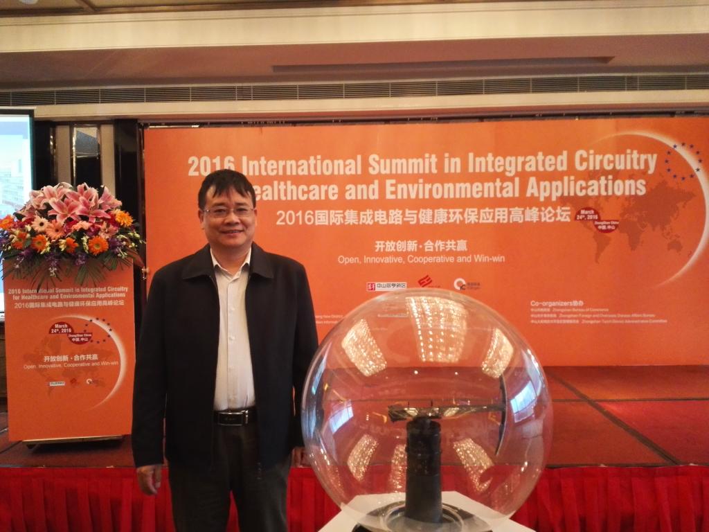 Feng Yang (杨丰)合作论文数南方医科大学生物医学工程学院1Deng Zhenwei合作论文数1Wu Feng合作论文数1
 Chang Liu (刘昌)合作论文数The Second Affiliated Hospital of Xi'an Jiaotong University1
 Yi-Jun Wang合作论文数College of Pharmacy and Health Sciences, St. John's University1




