BACKGROUND:Selenium has been shown to inhibit cancer development and growth through the mediation of selenium-binding proteins. Decreased expression of selenium-binding protein 1 has been reported in cancers of the prostate, stomach, colon, and lungs. No information, however, is available concerning the roles of selenium-binding protein 1 in uterine leiomyoma.METHODS:Using Western Blot analysis and immunohistochemistry, we examined the expression of selenium-binding protein 1 in uterine leiomyoma and normal myometrium in 20 patients who had undergone hysterectomy for uterine leiomyoma.RESULTS AND DISCUSSION:The patient age ranged from 34 to 58 years with a mean of 44.3 years. Proliferative endometrium was seen in 8 patients, secretory endometrium in 7 patients, and atrophic endometrium in 5 patients. Two patients showed solitary leiomyoma, and eighteen patients revealed 2 to 5 tumors. Tumor size ranged from 1 to 15.5 cm with a mean of 4.3 cm. Both Western blot analysis and immunohistochemistry showed a significant lower level of selenium-binding protein 1 in leiomyoma than in normal myometrium. Larger tumors had a tendency to show a lower level of selenium-binding protein 1 than smaller ones, but the difference did not reach a statistical significance. The expression of selenium-binding protein 1 was the same among patients with proliferative, secretory, and atrophic endometrium in either leiomyoma or normal myometrium. Also, we did not find a difference of selenium-binding protein 1 level between patients younger than 45 years and older patients in either leiomyoma or normal myometrium.CONCLUSIONS:Decreased expression of selenium-binding protein 1 in uterine leiomyoma may indicate a role of the protein in tumorigenesis. Our findings may provide a basis for future studies concerning the molecular mechanisms of selenium-binding protein 1 in tumorigenesis as well as the possible use of selenium in prevention and treatment of uterine leiomyoma.
Signal transducer and activator of transcription (STAT) proteins are both tyrosine- and serine-phosphorylated, mediating signal transduction and gene regulation. Following gene regulation, STAT activity in the nucleus is then terminated by a nuclear protein phosphatase(s), which remains unidentified. Using novel antibody arrays to screen the Stat1-specific protein phosphatase(s), we identified a SHP-2-Stat1 interaction in the A431 cell nucleus. SHP-2 and Stat1 nuclear localization and their association in response to either epidermal growth factor or interferon-gamma (IFN-gamma) were confirmed by immunofluorescent staining and affinity precipitation assays. The SHP-2 C-terminal region containing protein-tyrosine phosphatase activity interacted with the C-terminal SH2 transcriptional activation domain of Stat1. In SHP-2-/-mouse fibroblast cells, Stat1 phosphorylation at both the tyrosine residue Tyr(701) and the serine residue Ser(727) by IFNgamma was enhanced and prolonged. Consistently, purified GST-SHP-2 dephosphorylated Stat1 at both tyrosine and serine residues when immunoprecipitated phospho-Stat1 or a peptide corresponding to the sequence surrounding Tyr(p)(701) or Ser(p)(717) of Stat1 was used as the substrate. Overexpression of SHP-2 in 293T cells inhibited IFNgamma-dependent Stat1 phosphorylation and suppressed Stat1-dependent induction of luciferase activity. Our findings demonstrate that SHP-2 is a dual-specificity protein phosphatase involved in Stat1 dephosphorylation at both tyrosine and serine residues and plays an important role in modulating STAT function in gene regulation.
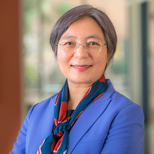 Cunxian Zhang合作论文数Department of Pathology and Laboratory Medicine, Brown University2
 Y Eugene Chin合作论文数1Am Dragoi合作论文数1W. Dwayne Lawrence合作论文数1
 M. Ruhul Quddus合作论文数Department of Pathology and Laboratory Medicine, Brown University1
 Andrew G Campbell合作论文数Division of Biology & Medicine, Department of Molecular Microbiology and Immunology, Brown University1Ling Mike Y合作论文数1
 Gen-Sheng Feng (冯根生)合作论文数Shenzhen Bay Laboratory1
 Peng Zhang (张鹏)合作论文数Shanghai Pulmonary Hospital;school of medicine, Tongji University1Fang Liu合作论文数1Zhiyong Han合作论文数1
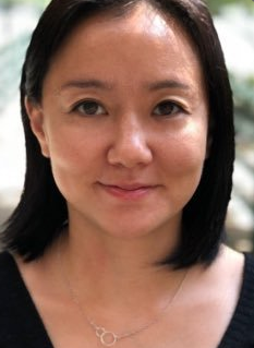 Y. Kate Hong合作论文数Neuroscience Institute, Carnegie Mellon University/Department of Neuroscience, University of Pittsburgh/Biological Sciences, Carnegie Mellon University/Department of Biological Sciences, Carnegie Mellon University1
 C James Sung合作论文数Department of Pathology and Laboratory Medicine, Brown University1Alicia Chung合作论文数1




