The gut microbiota regulates systemic metabolism, inflammation, and immunity, with evidence linking microbiota translocation through the gut-liver axis to hepatocellular carcinoma (HCC) progression. However, the specific bacteria and underlying mechanisms driving tumor progression remain unexplored. We identify that E. bolteae is enriched in the feces of HCC patients and is associated with poor prognosis. E. bolteae disrupts intestinal barrier integrity, translocates to the liver, and promotes tumor proliferation. Mechanistically, we show that E. bolteae's surface protein, penicillin-binding transpeptidase domain-containing protein (PbpT), directly interacts with the tumor cell receptor desmoglein 1 (DSG1), facilitating bacterial adhesion and attenuating DSG1's tumor-suppressive function. Additionally, this interaction activates the mitogen-activated protein kinase (MAPK) signaling pathway to accelerate HCC progression. Blockade of PbpT abrogates E. bolteae attachment and its role in promoting HCC progression. These findings identify the PbpT-DSG1-MAPK axis as a critical driver of HCC progression. Targeting PbpT may be a promising therapeutic strategy for HCC.
Abstract The tumor microenvironment is distinctive in primary and secondary liver cancer. B cells represent an important component of immune infiltrates. Here, we demonstrated that B cells are an important regulator in hepatocellular carcinoma (HCC) and colorectal cancer liver metastasis (CRLM) microenvironments. B cells displayed distinct developmental trajectories in HCC and CRLM. Single-cell analysis revealed that IgG+ plasma cells preferentially accumulated in HCC while IgA+ plasma cells were preferentially enriched in CRLM. Mechanistically, IgG+ plasma cells in HCC were recruited by tumor-associated macrophages via the CXCR3-CXCL10 axis, whereas IgA+ plasma cells in CRLM were recruited by metastatic tumor cells via CCR10-CCL28 signaling. Functionally, IgG+ plasma cells preferentially promoted pro-tumorigenic macrophages formation in HCC, and IgA+ plasma cells preferentially induced granulocytic myeloid-derived suppressor cells activation in CRLM. Clinically, increased infiltration of IgG+ plasma cells and macrophages in HCC was correlated to worse survival, while increased intratumoral IgA+ plasma cells and neutrophils in CRLM indicated poor prognosis. Taken together, this study demonstrated plasma and myeloid cell-mediated immunosuppression in HCC and CRLM, suggesting that selectively modulating primary or secondary tumor-related immunosuppressive regulatory networks might reprogram the microenvironment and provide an immunotherapeutic strategy for treating liver cancer.
The role of gut microbes in the pathogenesis of hepatocellular carcinoma (HCC) remains unclear. Here, we identified that Catenibacterium is enriched in both the feces and tumors of patients with HCC. C. mitsuokai accelerated HCC carcinogenesis in both conventional and germ-free mice. Furthermore, C. mitsuokai disrupted the gut barrier and translocated to the liver as live bacteria. Critically, the C. mitsuokai surface protein Gtr1/ RagA interacts with the gamma-catenin receptor on HCC cells, facilitating its attachment and colonization in the mouse liver. We further revealed that the pro-tumorigenic effect of C. mitsuokai depends on its secreted metabolite, quinolinic acid. Mechanistically, quinolinic acid binds to and activates the tyrosine kinase with immunoglobulin and epidermal growth factor homology domains 2 (TIE2) on HCC cells. Phosphorylated TIE2 subsequently activates the downstream oncogenic phosphatidylinositol 3-kinase/protein kinase B (PI3K/AKT) pathway, thereby promoting HCC progression. In summary, C. mitsuokai disrupts the gut barrier, colonizes HCC cells via Gtr1/RagA-gamma-catenin, and secretes quinolinic acid, which binds to TIE2 and drives the PI3K/ AKT pathway to promote HCC development.
The role of gut microbes in the pathogenesis of hepatocellular carcinoma (HCC) remains unclear. Here, we identified that Catenibacterium is enriched in both the feces and tumors of patients with HCC. C. mitsuokai accelerated HCC carcinogenesis in both conventional and germ-free mice. Furthermore, C. mitsuokai disrupted the gut barrier and translocated to the liver as live bacteria. Critically, the C. mitsuokai surface protein Gtr1/RagA interacts with the γ-catenin receptor on HCC cells, facilitating its attachment and colonization in the mouse liver. We further revealed that the pro-tumorigenic effect of C. mitsuokai depends on its secreted metabolite, quinolinic acid. Mechanistically, quinolinic acid binds to and activates the tyrosine kinase with immunoglobulin and epidermal growth factor homology domains 2 (TIE2) on HCC cells. Phosphorylated TIE2 subsequently activates the downstream oncogenic phosphatidylinositol 3-kinase/protein kinase B (PI3K/AKT) pathway, thereby promoting HCC progression. In summary, C. mitsuokai disrupts the gut barrier, colonizes HCC cells via Gtr1/RagA-γ-catenin, and secretes quinolinic acid, which binds to TIE2 and drives the PI3K/AKT pathway to promote HCC development.
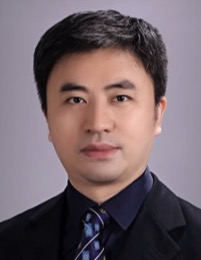 Ming Kuang (匡铭)合作论文数The First Affiliated Hospital, Sun Yat-sen University32
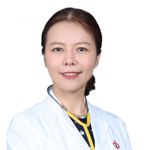 Sui Peng (彭穗)合作论文数The First Affiliated Hospital, Sun Yat-sen University29
 Lixia Xu (许丽霞)合作论文数The First Affiliated Hospital, Sun Yat-sen University27
 Zhihang Chen (陈志航)合作论文数26
 Shuling Chen (陈淑玲)合作论文数24Kaiyu Sun合作论文数22Zhang Guopei合作论文数22Zhijia Yao合作论文数21
 Qian Zhou合作论文数21
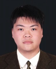 Dongming Kuang (邝栋明)合作论文数School of Life Sciences, Sun Yat-Sen University21
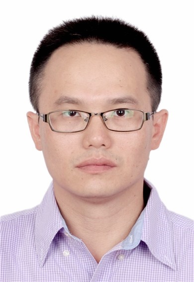 Shuibin Lin (林水宾)合作论文数The First Affiliated Hospital, Sun Yat-sen University6Xinyue Zhang合作论文数5Xuezhen Zeng合作论文数5
 Xiaoxing Li (李晓星)合作论文数Institute of Precision Medicine, The First Affiliated Hospital, Sun Yat-sen University;Cancer Center, Sun Yat-sen University4Shenghua Zhu合作论文数3
 Jennifer X Zhang (張翔)合作论文数Department of Medicine & Therapeutics, Faculty of Medicine, The Chinese University of Hong Kong3
 Weixin Liu合作论文数CUHK Shenzhen Research Institute, The Chinese University of Hong Kong3
 Liu Haining合作论文数3
 Qian Song合作论文数Shanghai Medical College, Fudan University3Ji Wang合作论文数3
 Chi-Chun Wong (黃子雋)合作论文数Department of Medicine & Therapeutics, Faculty of Medicine, The Chinese University of Hong Kong3Zhang Ying合作论文数3
 Ying Zhang合作论文数3
 Jun Yu (于君)合作论文数Department of Medicine & Therapeutics, Faculty of Medicine, The Chinese University of Hong Kong3Huayu Guan合作论文数2Gaomin Zheng合作论文数2Shangru Li合作论文数2Zi-Hao Dai合作论文数2Junbin Liao合作论文数2Qianwen Zeng合作论文数2
 Wanjie Zhu合作论文数2Xiaoxue Ren合作论文数2Yi Fang合作论文数1Xiaofei Liu合作论文数1Guanrui Liao合作论文数1Ju-deng Zeng合作论文数1Yingdong Hou合作论文数1
 Ruidong Xue (薛瑞栋)合作论文数Yunnan Baiyao International Medical Research Center, Peking University;Translational Cancer Research Center, Peking University First Hospital1Mengke Chen合作论文数1Juan Jiang合作论文数1Meinong Liang合作论文数1Xiao Zhao合作论文数1Siyi Zheng合作论文数1
 Shixian Hu合作论文数Univ Groningen, Dept Gastroenterol & Hepatol, Groningen, Netherlands1
 Tianhong Su合作论文数1Huadong Chen合作论文数1Qingxia Yang合作论文数1Qingfeng Zhang合作论文数State Key Laboratory of Oncology in South China, Collaborative Innovation Center for Cancer Medicine, Sun Yat-sen University Cancer Center, Guangzhou 510060, China.1Yu-Feng Zhou合作论文数1




