Background In-hospital neurologic deterioration (ND) in patients with acute ischemic stroke (AIS) is associated with worse prognosis, and endothelial dysfunction may be involved in this process. This study is aimed at evaluating the association between the endothelial activation and stress index (EASIX) and the risk of ND and short-term mortality in patients with AIS. Methods This retrospective cohort study included patients with AIS from the Medical Information Mart for Intensive Care IV (MIMIC-IV) database from 2008 to 2022. EASIX was calculated as (serum lactate dehydrogenase level [U/L] & times; creatinine level [mg/dL])/platelet count (10(9)/L). Multivariable logistic regression and Cox regression analyses were utilized to examine the associations of log-transformed EASIX (Ln (EASIX)) and ND and 28-day mortality, respectively. Propensity-score matching (PSM) and overlap weighting (OW) were performed to assess the robustness of the findings. Results A total of 1157 AIS patients were enrolled; 305 (26.36%) died within 28 days, and 673 (58.17%) experienced ND. Patients with Ln (EASIX) >= 0.40 had a higher risk of ND (OR = 1.56, 95% CI: 1.17-2.07) and 28-day mortality (HR = 1.47, 95% CI: 1.12-1.94) compared with those with Ln (EASIX) < 0.40. These associations remained significant after PSM and OW (all p < 0.05). Mediation analysis suggested that ND partially mediated the relationship between Ln (EASIX) and 28-day mortality risk, with a mediation proportion of 89.34% (beta = 0.35, 95% CI: 0.11-0.63). Conclusion Elevated EASIX was associated with an increased risk of ND and 28-day mortality in patients with AIS. ND may partially explain the EASIX-mortality association, but these hypothesis-generating findings warrant validation in future prospective studies.
Ischemic stroke (IS) is a severe condition regulated by complex molecular alterations. This study aimed to identify potential nicotinamide adenine dinucleotide (NAD+) metabolism-associated diagnostic markers of IS and explore their associations with immune dynamics. Weighted Gene Co-expression Network Analysis and single-sample gene set enrichment analysis (ssGSEA) were employed to identify key gene modules on the GEO dataset (GSE16561). LASSO regression was used to identify diagnostic genes. A diagnostic model was then developed using the training dataset, and its performance was assessed using a validation dataset (GSE22255 dataset). Associations between hub genes and immune cells, immune response genes, and human leukocyte antigen (HLA) genes were assessed by ssGSEA. A regulatory network was constructed using mirBase and TRRUST databases. A total of 20 NAD+ metabolic genes exhibited noteworthy expression variations. Within the module notably associated with NAD+ metabolism, 19 specific genes were included in the diagnostic model, which was validated on the GSE22255 dataset (AUC: 0.733). There were significant disparities in immune cell populations, immune response genes, and HLA gene expression, all of which were associated with the hub genes. A regulatory network composed of 153 edges and 103 nodes was constructed. This study advances our understanding of IS by providing insights into NAD+ metabolism and gene interactions, contributing to potential diagnostic innovations in IS.
MicroRNA (miRNA) dysfunction has been confirmed as a key event of ischemic stroke appearance. This study is aimed at revealing the role of miR-429 in the angiogenesis of HBMECs. The HBMECs were treated with oxygen and glucose deprivation (OGD) to establish the ischemic cell model. The qRT-PCR was used to measure the expression levels of the miR-429 in the serums of the patients or cells, and CCK-8, wound healing assay, and tube formation assay were used to observe the effects of miR-429 on the phenotype of HBMECs. Moreover, the Targetscan, dual-luciferase reporter assay, and Western blot were used to reveal the downstream target and regulation mechanism of miR-429 in OGD-induced HBMECs. The results showed that miR-429 was significantly upregulated in the serums of the patients, and overexpressed miR-429 could extremely inhibit the viability, migration, and tube formation of OGD-induced HBMECs. Furthermore, it was found that SNAI2 was a downstream factor of miR-429, and SNAI2 could rescue the effects of miR-429 on OGD-induced HBMECs. Besides, the Western blot showed that miR-429 could affect the activity of GSK-3β/β-catenin pathway via inhibiting the expression of SNAI2. In conclusion, this study suggests that miR-429 inhibits the angiogenesis of HBMECs through SNAI2-mediated GSK-3β/β-catenin pathway.
This study aimed to investigate whether ischemic postconditioning (IpostC) alleviates cerebral ischemia/reperfusion (I/R) injury involved in autophagy. Adult Sprague-Dawley rats were divided into five groups: sham (sham surgery), I/R (middle cerebral artery occlusion [MCAO] for 100 min, then reperfusion), IpostC (MCAO for 100 min, reperfusion for 10 min, MCAO for 10 min, then reperfusion), IpostC+3MA (3-methyladenine, an autophagy inhibitor, administered 30 min before first reperfusion), and IpostC+Veh (vehicle control for IpostC+3MA group). Infarct volume was measured using cresyl violet staining. Autophagy-related proteins were detected by western blot and immunohistochemistry. Autophagosomes, autophagolysosomes, and mitochondrial damage were identified by transmission electron microscopy. Cortical cell apoptosis was detected by the TUNEL assay. Neurologic function was assessed using the modified Neurologic Severity Score. IpostC improved neurological function and reduced infarct volume after I/R (P < 0.05). These effects of IpostC were inhibited by 3MA (P < 0.05). Autophagosome formation was increased in the I/R and IpostC+Veh groups (P < 0.05), but not in the IpostC+3MA group. The I/R group showed enhanced LC3-II/LC3-I ratio, p62, and Cathepsin B levels and decreased LAMP-2 level (all P < 0.05 vs. sham), indicating dysfunction of autophagic clearance. IpostC reduced p62 and Cathepsin B levels and increased the LC3-II/LC3-I ratio, and nuclear translocation of transcription factor EB (all P < 0.05); these effects of IpostC were reversed by 3MA, suggesting IpostC enhanced autophagic flux. Furthermore, IpostC attenuated I/R-induced mitochondrial translocation of Bax and mitochondrial cytochrome-c release (all P < 0.05); 3MA inhibited these effects of IpostC (P < 0.05). In conclusion, IpostC may alleviate cerebral I/R injury by activating autophagy during early reperfusion.
The present study assessed whether the protective effects of curcumin against cerebral ischemia injury were due to the suppression of overactivated autophagy. Curcumin is a well-known natural polyphenolic compound that effectively counteracts oxidation, inflammation, and various types of cancer. Several studies have demonstrated the protective effects of curcumin against ischemia-reperfusion injury in tissues from the lungs, cardiomyocytes, and liver. The present study employed brain injury models induced by middle cerebral artery occlusion (MCAO) in rats and PC12 oxygen-glucose-deprived (OGD) cells. Infarct area, neurological score, lactate dehydrogenase (LDH) activity, autophagy expression, cell apoptosis, and mRNA and protein expressions of caspase-3 were determined following curcumin supplementation. Compared to MCAO rats, curcumin-treated MCAO rats exhibited substantial reductions in neurological score, infarct area, and LDH activity. MCAO also increased LC3 II/I protein expression and decreased p62 protein expression, but curcumin supplementation significantly reversed these altered protein expressions. Caspase-3 protein expression increased by 46.2% in the MCAO group, but curcumin supplementation significantly reduced this expression. Similarly, apoptosis increased by 33.1% in OGD cells, but curcumin supplementation significantly reduced apoptosis to 21.6% and 9.3% at doses of 100 and 200 mg/kg, respectively. The mRNA and protein expressions of caspase-3 exhibited substantial increases in OGD cells but these expressions were significantly decreased following curcumin supplementation. Taken together, the present results indicate that curcumin represents a natural bioactive substance that can protect against cerebral ischemia via the suppression of overactivated autophagy.
Aim To investigate the effects of ischemic postconditioning (IpostC) in focal cerebral ischemic-refusion (I/R) injury in the rat transient middle cerebral artery occlusion (tMCAO) model. Methods Adult Sprague-Dawley rats were used to establish tMCAO models by Doppler detection of regional cerebral blood flow before and after surgery. Rats in the IpostC groups were treated with IpostC after 100 minutes of MCAO. Neurologic function was assessed using the modified Neurologic Severity Score and the rotarod test. The infarct volume was measured using TTC staining. Apoptosis related proteins (Bax, cytochrome C) were detected by Western blot, mitochondrial damage was identified by transmission electron microscopy, cortical cell apoptosis was detected by the TUNEL assay. Results IpostC can reduce infarct volume, improve neurologic function compared with the I/R-only group and attenuate the reduction of regional cerebral blood flow after I/R. Furthermore, IpostC can down-regulate cytochrome C release to the cytosol and Bax translocation to the mitochondria, and reduce TUNEL positive cells and mitochondrial damage. Conclusion This study showed that IpostC has a certain neuroprotective effect on cerebral ischemia-reperfusion injury, which is partly related to the antiapoptotic mechanisms and may become a new target in the clinical treatment of acute ischemic stroke in the future.
Ischemic stroke is the leading cause of disabilities worldwide. MicroRNA-377 (miR-377) plays important roles in ischemic injury. The present study focused on the mechanisms of miR-377 in protecting ischemic brain injury in rats. Cerebral ischemia was induced by middle cerebral artery occlusion (MCAO) in rats. Primary rat microglial cells and brain microvascular endothelial cells (BMECs) were exposed to oxygen-glucose deprivation (OGD). The concentrations of cytokines (TNF-, IL-1, IL-6, IFN-, TGF-, MMP2, COX2, and iNOS) in the culture medium were measured by specific ELISA. Tube formation assay was for the in vitro study of angiogenesis. Luciferase reporter assay was performed to confirm whether VEGF and EGR2 were direct targets of miR-377. The MCAO rats were intracerebroventricular (ICV) injection of miR-377 inhibitor to assess its protective effects in vivo. MiR-377 levels were decreased in the rat brain tissues at 1, 3, and 7d after MCAO. Both microglia cells and BMECs under OGD showed markedly lower expression levels of miR-377 while higher expression levels of EGR2 and VEGF compared to those under normoxia conditions. Knockdown of miR-377 inhibited microglial activation and the release of pro-inflammatory cytokines after OGD. Suppression of miR-377 promoted the capillary-like tube formation and cell proliferation and migration of BMECs. The anti-inflammation effect of EGR2 and the angiogenesis effect of VEGF were regulated by miR-377 after OGD. Inhibition of miR-377 decreased cerebral infarct volume and suppressed cerebral inflammation but promoted angiogenesis in MCAO rats. Knockdown of miR-377 lessened the ischemic brain injury through promoting angiogenesis and suppressing cerebral inflammation. J. Cell. Biochem. 119: 327-337, 2018. (c) 2017 Wiley Periodicals, Inc.
Our previous findings have demonstrated that autophagy regulation can alleviate the decline of learning and memory by eliminating deposition of extracellular beta-amyloid peptide (Aβ) in the brain after stroke, but the exact mechanism is unclear. It is presumed that the regulation of beta-site APP-cleaving enzyme 1 (BACE1), the rate-limiting enzyme in metabolism of Aβ, would be a key site. Neuro-2a/amyloid precursor protein 695 (APP695) cell models of cerebral ischemia were established by oxygen-glucose deprivation to investigate the effects of Rapamycin (an autophagy inducer) or 3-methyladenine (an autophagy inhibitor) on the expression of BACE1. Either oxygen-glucose deprivation or Rapamycin down-regulated the expression of BACE1 while 3-methyladenine up-regulated BACE1 expression. These results confirm that oxygen-glucose deprivation down-regulates BACE1 expression in Neuro-2a/APP695 cells through the introduction of autophagy.
Although ghrelin receptors have been demonstrated to be widely expressed in the central nervous system and peripheral tissues of mammals, it is still unknown whether ghrelin functions in cerebellar Purkinje neurons. In this study, we identified a novel functional role for ghrelin in modulating P-type Ca(2+) channel (P-type channel) currents (IBa) as well as action-potential firing in rat Purkinje neurons. Our results show that ghrelin at 0.1μM reversibly decreased IBa by ~32.3%. This effect was growth hormone secretagogue receptor 1a (GHS-R1a)-dependent and was associated with a hyperpolarizing shift in the voltage-dependence of inactivation. Intracellular application of GDP-β-S and pretreatment with pertussis toxin abolished the inhibitory effects of ghrelin. Dialysis of cells with the peptide QEHA (but not the scrambled peptide SKEE), and a selective antibody raised against the G-protein αo subunit both blocked the ghrelin-induced response. Ghrelin markedly increased protein kinase A (PKA) activity, and intracellular application of PKI 5-24 as well as pretreatment of the cells with the PKA inhibitor KT-5720 abolished ghrelin-induced IBa decrease, while inhibition of PKC had no such effects. At the cellular level, ghrelin induced a significant increase in action-potential firing, and blockade of GHS-R1a by BIM-28163 abolished the ghrelin-induced hyperexcitability. In summary, these results suggest that ghrelin markedly decreases IBa via the activation of GHS-R1a, which is coupled sequentially to the activities of Go-protein βγ subunits and the downstream PKA pathway. This could contribute to its physiological functions, including the spontaneous firing of action potentials in cerebellar Purkinje neurons.
 Kangyong Liu合作论文数5Nan Shi合作论文数4Zhang Yan合作论文数4
 Xiaojiang Sun (孙晓江)合作论文数Shanghai Ninth People's Hospital, School of Medicine, Shanghai Jiao Tong University4
 Jieqing Wan (万杰清)合作论文数Department of Neurosurgery, Renji Hospital, Shanghai Jiao Tong University School of Medicine3Shenghao Ding (丁圣豪)合作论文数3Fei Shen合作论文数2Jinfeng Li合作论文数2YaoHua Pan (潘耀华)合作论文数2Sun Yinyi合作论文数2Chen Wenqi合作论文数2
 Ting Zhang (张婷)合作论文数Shanghai Children's Hospital, School of Medicine, Shanghai Jiao Tong University2
 Bing Zhao (赵冰)合作论文数School of Basic Medical Sciences, Nanchang University;The First Affiliated Hospital of Nanchang University2Yiling Fan (樊翊凌)合作论文数2Wenhua Sun合作论文数1Yan ZHANG (张燕)合作论文数1Lei Jin合作论文数1Lei JIN (金磊)合作论文数1Qing Dong合作论文数1Yun Mo合作论文数1Jin-feng LI (李晋峰)合作论文数1Li Hua合作论文数1Rongfu Chen合作论文数1Ming Fang合作论文数1
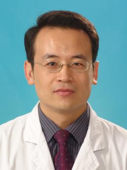 Jianliang Fu (付剑亮)合作论文数Shanghai Sixth People’s Hospital Affiliated to Shanghai Jiao Tong University1




