This study was performed to compare the phenotype and cytokines of γδT cells from mouse vagina and blood,for lay a foundation for further research of the immune function of vaginal γδT cells.The mononuclear cells from vagina and blood were isolated.Flow cytometry was used to detect the percentage of γδT cells,the expression of chemokine receptors,memory markers,cytotoxicity molecules and Erα;the expression of IL-17 and IFN-γ were detected after PMA+Ionomycin stimulation;the correlation between the amount of IL-17 produced byγδT cells and ERα density was analyzed.Data showed that the percentage of γδT cells in vaginal T cells was47.91%±3.12%,and γδT cells in vagina were mainly tissue resident memory γδT cells with high expression of CD44,CD69,CD103 and IL-17,but low expression of IFN-γ.In addition,the expression of IL-17 in spleen,peripheral blood,lung,bladder,uterus and vagina were positively correlated with ERα density.In conclusion,vaginal γδT cells were mainly tissue resident memory γδT cells which might play an important role in bacterial infection by producing IL-17.
Tissue-resident memory γδT cells at mucosal and epithelial sites play an important role for pathogen clearance, immunosurveillance, and participating in physiological processes. Different from other barrier sites, the immune cells in uterus face the protection against infections and tolerate an allogeneic fetus during a successful pregnancy. In the previous study, we found that tissue-resident memory γδT cells were enriched both in human and murine uterus and highly expressed IL-17 that promoted the invasion of trophocytes in vitro. In the current study, we found that γδT cells in uterus but not in blood or spleens expressed higher levels of estrogen receptors. The injection of estrogen into mice increased the proportion of γδT cells in uterus but not in spleens in vivo via CXCR3-CXCL10 chemokine axis. In addition, we found that estrogen enhanced the production of IL-17 but not IFN-γ in vivo and in vitro via interferon regulatory factor 4 but not RORγt and pSTAT3 at mRNA and protein levels. The analysis of cell transcriptome sequence further identified multiple differentially expressed genes between estrogen and control γδT cells. Our study demonstrated that estrogen directly act on γδT cells in uterus to enhance the production of IL-17 that might promote the invasion of trophocytes. Furthermore, our study might provide a new idea that estrogen increased the prevalence of autoimmune diseases in women by enhancing γδT cell-derived IL-17 production in uterus and uncover the critical pathological roles for estrogen in the development of autoimmune diseases.
Tissue-resident memory T (TRM) cells are well known to play critical roles in peripheral tissues during virus infection and tumor immunology. Our previous studies indicated that CD69+CD4+ and CD69+CD8+ T cells in tuberculous pleural effusion (TPE) were antigen-specific memory T cells. However, the phenotypical and functional characteristics of CD8+ TRM cells in tuberculosis remain unknown. We found that CD103+CD8+ T cells were the predominant subset of CD103+ lymphocytes in TPE; both CD103 and CD69 expressed on memory CD8+ T cells from TPE were significantly increased compared with those from paired peripheral blood. Phenotypically, CD103+CD69+ and CD103+CD69-CD8+ T cells expressed higher levels of CD45RO than CD103-CD69+CD8+ T cells did; CD103+CD69-CD8+ T cells highly expressed CD27, CD127, and CD62L and some chemokine receptors. We further compared the functional differences among the four distinct CD45RO+CD8+ T subsets identified by CD103 and CD69 expression. In consist with our published results, CD69+CD8+ T cells, but not CD103+CD8+, produced high levels of IFN-γ after treatment with BCG in the presence of BFA. Nevertheless, CD103-CD69+ and CD103+CD69+ memory CD8+ T cells expressed higher levels of Granzyme B, while CD103+CD69- memory CD8+ T cells were characterized as a possibly immunosuppressive subset by highly expressing CTLA-4, CD25, and FoxP3. Furthermore, TGF-β extremely increased CD103 expression but not CD69 in vitro. Together, CD103+CD8+ T cells form the predominant subset of CD103+ lymphocytes in TPE; CD103 and CD69 expression defines distinct CD8+ TRM-like subsets exhibiting phenotypical and functional heterogeneity. Our findings provide an important theoretical basis to optimize and evaluate new tuberculosis vaccines.
The field of tissue-resident B cells has received increasing attention, yet the feature of tissue B cells in respiratory system is unclear. Here, we first show that non-circulating B cells obtained from nasal, trachea and lung tissues are numerically and phenotypically distinct from their circulating counterparts. Analysis of single cell transcriptome sequence identified multiple differentially expressed genes between non-circulating B cells and circulating B cells, which illustrated their heterogeneity. Furthermore, we found high expression of CXCR3 on non-circulating B cells, and the chemokine CXCL11 was also up-regulated in the respiratory tissues, suggesting that CXCR3-CXCL11 axis might accelerate the local resident of non-circulating B cells in respiratory tract. Interestingly, intranasal immunization with BCG in mice elicited a sustained humoral immune response via induction of IgA and IgG Abs, which revealed the role of B cells. Meanwhile, tissue-resident B cells, IgA(+) and IgG(+) memory B cells (MBCs) in respiratory tissues, as well as plasma cells in bone marrow, were expanded and maintained, and these subsets probably developed into antibody-producing cells to participate in the local humoral immunity. Our data illustrate the phenotype and function of tissue B cells in the upper and lower airways, provide references for the prospective development of vaccines.
目的:探讨小鼠膀胱组织中T细胞的表型和细胞因子表达.方法:获取小鼠膀胱单个核细胞,流式细胞术检测小鼠膀胱组织中T细胞的比例、趋化因子受体和细胞因子的表达.HE染色观察膀胱结构,免疫组化和免疫荧光观察正常小鼠与MB49原位肿瘤小鼠膀胱组织中CD3+T和γδT细胞的分布.结果:正常小鼠膀胱中含有组织驻留记忆T细胞,表达记忆分子CD44和组织驻留标志分子CD69和CD103,其中γδT细胞的比例显著高于外周血并高表达趋化因子受体CXCR3和细胞因子IL-17、TNF-α.与正常膀胱组织相比,MB49原位肿瘤小鼠膀胱组织中CD4+T细胞显著降低,CD8+T和γδT细胞比例显著升高,并高表达IFN-γ.此外,γδT细胞还显著高表达IL-17和TNF-α.结论:小鼠膀胱组织中具有组织驻留记忆αβT和γδT细胞,且表达细胞因子IFN-γ,IL-17和TNF-α,为进一步深入研究膀胱癌奠定基础.
γδT cells are non-conventional T cells and serve as the bridge for connecting the innate and adaptive immune systems. γδT cells form a substantial population at barrier sites and play an important role in the development of physiology, inflammation, autoimmune diseases and tumors. γδT cells not only distribute in the maternal-fetal interface during pregnancy but also in non-pregnant uterus. However, the phenotypes and functions of γδT cells in uterus were not clear. In the current study, we found that the percentages of γδT cells were significantly higher in uterus than peripheral blood and most of γδT cells in uterus were distributed in endometrium. Further studies indicated that the majority of γδT cells in uterus were memory cells with higher expression of CD44 and CD27 but lower expression of CD62L and CCR7 compared to those in blood. In addition, we found that γδT cells in uterus were tissue resident memory γδT cells expressing CD69, expressed high levels of CCR6, GranzymeB and CD107a. Moreover, γδT cells in uterus were activated and fully expressed transcription factor RORγt. After short time of activation, γδT cells in uterus significantly expressed high levels of IL-17 but not IFN-γ, which promotes the invasion of murine trophocytes. Taken together, our study will lay the foundation for future research on uterine γδT cells in pregnancy and autoimmune disease.
Tissue-resident memory T cells (TRM) are different from effector memory T cells (TEM) and central memory T cells (TCM) and contribute to the protective immunity against local challenges. Currently, we found that CD4+ and CD8+ TRM cells in the nasal mucosa, trachea, lungs, and lavage fluids were heterogeneous on the expression of CD69 and CD103 as well as the production of cytokines including IFN-γ, IL-2, and TNF-α. After intranasal vaccination of mice with BCG, respiratory tissues expressed higher levels of the chemokine CXCL16 and TRM cells expressed CXCR6 to CXCL16. In addition, antigen-specific CD4+ and CD8+ TRM cells expressed cytokines following the stimulation with BCG and persisted in the nasal mucosa, trachea, and lungs for more than a hundred days. At the same time, mice were infected intranasally with live BCG and the results showed that vaccinated mice cleared up live BCG faster than nonvaccinated mice in the respiratory system. Taken together, our data demonstrated that intranasal vaccination of mice with BCG could induce antigen-specific CD4+ and CD8+ TRM cells in the respiratory system and have the ability to provide protection against pulmonary reinfection.
OBJECTIVE: To investigate the effect of bone marrow mesenchymal stem cells (BMSCs) on repairing brachial plexus injury in rabbits and their influence on expression of the extracellular signal-regulated kinase (ERK) pathway. MATERIALS AND METHODS: With big-ear rabbits as the objects, the BMSCs were first isolated, and the cluster of differentiation (CD)45- and CD90+ BMSCs were sorted out via flow cytometry. BMSCs were transfected with red fluorescent protein (RFP), and the transfection effect was detected. Then, the big-ear rabbits were subjected to brachial plexus root avulsion injury (BPAI) to establish injury Model group and sham-operation group (Sham group). Later, the BMSCs were transfected with RFP to construct RFP-BMSCs. The RFP-BMSCs (5×10⁶, Treat group) and normal saline (Model group) were intraperitoneally injected, and the recovery rate of wet weight of the upper limb muscle was measured by weighing. The injured nerve tissues were embedded for hematoxylin and eosin (HE) staining and observation of pathological changes. The electrophysiological measurement of the compound muscle action potential (CMAP) on the injured side was conducted for the rabbits to be sacrificed immediately using an electromyogram instrument, and the CMAP amplitude and latency were applied to evaluate the recovery of upper limb muscle. Finally, the location of RFP-BMSCs in the nerve tissues was traced by a fluorescence microscope, and the protein expression levels of phosphorylated ERK (p-ERK) and phosphorylated mitogen-activated protein kinase (p-MAPK) in the injured nerve tissues were determined by means of Western blotting. RESULTS: Persistently expressed red fluorescence was observed in CD45- and CD90+ BMSCs sorted via flow cytometry under the fluorescence microscope, indicating that the RFP-BMSCs were constructed successfully. Compared with Sham group, Model group had a remarkably decreased recovery rate of wet muscle weight (p<0.05), while Treat group exhibited a notably increased recovery rate of wet muscle weight in comparison with Model group. The CMAP amplitude was reduced markedly (p<0.05), while the CMAP latency was prolonged significantly (p<0.05) in Model group compared with those in Sham group. Moreover, Treat group had distinctly higher CMAP amplitude and evidently shorter CMAP latency than Model group (p<0.05). It was discovered under the fluorescence microscope that RFP-BMSCs were visibly arranged on both sides of nerve fibers in Treat group. The expressions of p-MAPK and p-ERK were raised prominently in Model group in comparison with those in Sham group (p<0.05), and they were lowered apparently in Treat group compared with those in Model group (p<0.05). CONCLUSIONS: BMSCs can repair the impaired brachial plexus neurons and restore their physiological functions, and the protective effect of the BMSCs on the neurons is associated with the mediated MAPK/ERK pathway.
目的:观察索曲妥林(sotrastaurin或AEB071)对人外周血中单个核细胞(PBMCs)、纯化CD4+T细胞和CD8+T细胞分泌细胞因子的抑制作用及其机制.方法:采集健康志愿者的静脉血,肝素抗凝,密度梯度离心法分离PBMCs后纯化CD4+T和CD8+T细胞,应用抗CD3抗体联合CD28抗体进行刺激,加或不加入AEB071培养后,检测细胞因子的产生、转录因子的表达和细胞的活化与增殖.结果:索曲妥林处理不促进外周血T细胞的凋亡,而能抑制T细胞分泌细胞因子IFN-γ、TNF-α和IL-2,且呈时间和剂量依赖性;进一步研究发现,索曲妥林通过下调STAT-1和STAT-4的磷酸化以及T-bet的表达抑制细胞因子的产生.同时经研究发现,索曲妥林处理后抑制T细胞的活化和增殖.结论:索曲妥林抑制T细胞转录因子的表达与磷酸化而抑制细胞因子的产生,从而抑制人体免疫功能.为此,进一步研究索曲妥林免疫抑制的机制,对索曲妥林在器官移植、银屑病和溃疡性结肠炎的治疗方面的应用,具有参考价值.
Tonsils are important lymphoid organs in which B cells and T cells complete their maturation and identify cells that are infected by pathogens. However, the functions of T cells in human tonsils remain unclear, especially the characteristics of polyfunctional CD4+ T helper cells. In this study, we used multi-color flow cytometry to analyze the expression or co-expression of effector cytokines in CD4+ T cells from tonsillar tissues. We have demonstrated that tonsillar CD4+ T cell can express various Th effector cytokines after short-term polyclonal stimulation, and that cytokine-producing CD4+ T cells were CD45RO+ T cells. In addition, we analyzed the co-expression of two or more kinds of cytokines at the level of a single cell. The results showed that tonsillar CD4+ T cells exhibited polyfunctionality by co-expressing two to five kinds of cytokines in the same time. These data furnished a basic theory for further understanding the differentiation of polyfunctional Th cells in human tonsils and their functions in resisting invasive microorganisms.
Omenn syndrome is a rare autosomal recessive disorder characterized by severe, combined immunodeficiency and autoimmune features. In this case study, we found Omenn syndrome in a 3-month-old boy with recurrent infection, erythroderma, axillary lymphadenopathy, and hepatosplenomegaly. The numbers of eosinophile granulocytes and the levels of immunoglobulin E in his blood were distinctly elevated. Circulating B cells were absent, and the numbers of activated T lymphocytes were present in his peripheral blood. The production of T cell cytokines was significantly higher in the patient compared to the control samples except for interferon gamma. Whole exome sequencing revealed that the patient carried compound heterozygous mutations in the RAG1 gene, which included a previously undescribed frameshift mutation (exon 2, 2491_2497del, p. K830fsX4) and a missense mutation (exon 2, 2923 C > T, p.R975W).
目的 研究一氧化氮(nitric oxide,NO)对T细胞和单核细胞分泌细胞因子的作用及机制.方法 用抗人CD3抗体或抗人CD3抗体联合抗人CD28抗体或LPS刺激培养外周血单个核细胞(PBMCs)、纯化T细胞或纯化单核细胞,加或不加一氧化氮培养,ELISA检测培养细胞上清液中细胞因子的含量;流式细胞术检测T细胞表面活化分子CD69和CD25的表达、T-box转录因子(T-bet)的表达;STAT1和STAT4的磷酸化程度以及T细胞的分裂情况.结果 一氧化氮呈时间和剂量依赖方式抑制T细胞产生IFN-γ、TNF-α和IL-2,但促进单核细胞IL-1β、IL-6和TNF-α的分泌,且不诱导细胞的凋亡.进一步研究发现其抑制细胞晚期活化分子的表达和细胞的分裂,以及抑制T细胞相关转录因子T-bet的表达,但不抑制STAT1和STAT4的磷酸化程度.结论 一氧化氮通过非细胞毒性作用抑制活化的T细胞产生细胞因子,但促进活化的单核细胞的细胞因子的分泌,其机制是通过抑制转录因子T-bet信号通路而抑制T细胞细胞因子的分泌,该研究将为临床应用提供理论依据.
BACKGROUND:Bisphenol A is widely used in the manufacture of polycarbonate plastics and has caused increasing concern over its potential adverse impacts on spermatogenesis. However, the effect of bisphenol A on spermiogenesis is yet to be explored.OBJECTIVES:To evaluate whether bisphenol A has adverse effects on DNA integrity and protamination of spermatogenic cell.MATERIALS AND METHODS:Newborn male mice were subcutaneously injected with bisphenol A (0.1, 5 mg/kg body weight, n = 15) or coin oil (control group, n = 20) daily from post-natal day 1 until 35. At post-natal day 70, epididymis caudal spermatozoa and testes were collected. Sperm count, sperm motility, and sperm morphology were analyzed. The sperm chromatin structure assay was performed to examine the sperm DNA fragmentation. Terminal deoxynucleotidyl transferase dUTP nick end labeling (TUNEL) method was used to assess apoptosis of spermatogenic cells. The ultrastructural features of testicular sections were examined under a transmission electron microscope. Western blot and RT-PCR were used to detect the expression levels of transition protein (Tnp) 1 and Tnp2, protamine (Prm) 1 and Prm2 protein, and mRNA in mice testes.RESULTS:Bisphenol A significantly reduced sperm counts, impaired sperm motility, and increased the percentage of malformed spermatozoa. Poor sperm chromatin integrity and increased TUNEL-positive spermatogenic cells were also observed in mice exposed to bisphenol A. Ultrastructural analysis of testes showed that bisphenol A exposure caused incomplete chromatin condensation, retention of residual cytoplasm, and abnormal acrosome formation. In addition, the relative expression levels of Tnp2 and Prm2 in mice testes decreased significantly in bisphenol A groups.DISCUSSION AND CONCLUSION:Our findings identified that neonatal bisphenol A exposure may negatively contribute to the sperm quality in adult mice. Mechanistically, we showed that bisphenol A reduced sperm chromatin integrity along with increased DNA damage, which may be due to poor protamination of spermatozoa.
The study is aimed to investigate the differences between Vδ1+,Vδ2+ and Vδ1-Vδ2-cells in frequencies,subpopulations,phenotypes and biological functions.We isolated peripheral blood mononuclear cells from healthy donors and detected the surface makers (TCRγδ,Vδ1,Vδ2,CD4,CD8,CD45RO,CD62L),intracellular cytokines (IFN-γ IL-17A),transcription factors (T-bet,RoRγt),and Granzyme A by flow cytometry directly or after stimulation with PMA plus Ionomycin.The cytokines produced by purified cells stimulated with PMA plus Ionomycin were detected by ELISA.According to the expression of TCRγδ,γδT cells could be divided into three groups,and the level of TCRγδ expression was related to the expression of Vδ1 and Vδ2.About 20%-40% of Vδ1+ and Vδ2+ cells express CD8 molecules,but not express CD4 molecules,whereas 60% of Vδ1-Vδ 2-γδT cells express CD8 and 20% of them express CD4 molecule.Approximately 6% of Vδ1 + and 80% of Vδ2+ cells express CD45RO,whereas about 30% of Vδ1-Vδ2-γδT cells express CD45RO.After stimulation,all γδT cells express high levels of IFN-γand IL-17,and the Vδ2+ T cells expressed the highest levels of ctokines (P< 0.01),compared with other subsets,which was consistent with the results of purified cells detected by ELISA.Further studies had shown that Granzyme A and transcription factors T-bet and RoRγt were highly expressed in Vδ 1+,Vδ2+ and Vδ1-Vδ2-cells.In conclusion,although Vδ1+,Vδ2+ and Vδ1-Vδ2-γδT cells has similarity in high secretion of cytokines (IFN-γ),there are significantly differences in their frequencies,phenotypes and productions of cytokines.These results will be a good foundation for further study in γδT cells.
To investigate the recovery effect and mechanism of IL-12 on the immune suppression induced by chemotherapeutic medicine, human PBMCs were stimulated with or without anti-CD3 plus anti-CD28 in the presence or absent of chemotherapeutics and IL-12. The production of m RNA and expression of IFN-γ and TNF-α were measured by different methods; the expression of IFN-γ and transcription factors including T-bet, p-STAT-1 and p-STAT-4 were analyzed by fluorescence activated cell sorter (FACS) or Western blotting. PBMCs from PPD+ individuals were stimulated with BCG for the production of cytokines in the presence or absence of chemotherapeutics and IL-12. We found that chemotherapeutics had significant suppressive effects on the production of IFN-γ and TNF-α in a dose-dependent manner, and IL-12, in a dose-dependent manner, relieved the inhibitory effect of chemotherapeutics on the cytokine production. In addition, chemotherapeutics also inhibited BCG-induced antigen-specific production of cytokines, which could be recovered by IL-12. Further study revealed that chemotherapeutics down-regulated the phosphorylation of STAT4 and STAT1 and the expression of T-bet. However, IL-12 could only recover the phosphorylation of STAT4. All the results indicated that IL-12 might play an important role in the reconstruction of immune function against the infections and cell mutations in cancer patients with chemotherapy.
Recent studies indicated that CXCR5+CD8+ T cells in lymph nodes could eradicate virus-infected target cells. However, in the current study we found that a subset of CXCR5+CD8+ T cells in the germinal centers from human tonsils or lymph nodes are predominately memory cells that express CD45RO and CD27. The involvement of CXCR5+CD8+ T cells in humoral immune responses is suggested by their localization in B cell follicles and by the concomitant expression of costimulatory molecules, including CD40L and ICOS after activation. In addition, CXCR5+CD8+ memory T cells produced significantly higher levels of IL-21, IFN-γ, and IL-4 at mRNA and protein levels compared to CXCR5-CD8+ memory T cells, but IL-21-expressing CXCR5+CD8+ T cells did not express Granzyme B and perforin. When cocultured with sorted B cells, sorted CXCR5+CD8+ T cells promoted the production of antibodies compared to sorted CXCR5-CD8+ T cells. However, fixed CD8+ T cells failed to help B cells and the neutralyzing antibodies against IL-21 or CD40L inhibited the promoting effects of sorted CXCR5+CD8+ T cells on B cells for the production of antibodies. Finally, we found that in the germinal centers of lymph nodes from HIV-infected patients contained more CXCR5+CD8+ T cells compared to normal lymph nodes. Due to their versatile functional capacities, CXCR5+CD8+ T cells are promising candidate cells for immune therapies, particularly when CD4+ T cell help are limited.
 Changyou Wu (吴长有)合作论文数Clifford Hospital75
 Sifei Yu合作论文数23Yannan Zhang (张燕楠)合作论文数18Suihua Lao (劳穗华)合作论文数16Qiongli Wu合作论文数11Zitao Li (李子涛)合作论文数11Juan Shen (沈娟)合作论文数9Jiang-Jun Ma合作论文数7Shuangpeng Kang合作论文数7
 Hui Wang (王慧)合作论文数Zhongshan School of Medicine, Sun Yat-sen University7
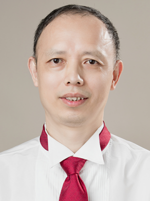 Huabin Li (李华斌)合作论文数Department of Nutrition, School of Public Health, Sun Yat-sen University6Xiaoying Fu合作论文数6Lei Jia合作论文数5Jianping Zhang合作论文数5Li Xiao合作论文数4Baiqing Li合作论文数4Yan-ying FAN (范彦英)合作论文数4
 Xianlan Zhang (张贤兰)合作论文数Guangzhou Chest Hospital;Guangzhou Medical College4Fengyu Huang合作论文数4Li Li合作论文数3
 Jun Huang (黄俊)合作论文数Pritzker School of Molecular Engineering, The University of Chicago3Yun Liu合作论文数3Peidong Chi (池沛冬)合作论文数3
 Junqi Huang (黄俊琪)合作论文数3L. Yi合作论文数2Yanquan Xu合作论文数2Qin Li合作论文数2Yuxia Jiang合作论文数2HUANG He (黄河)合作论文数2N Miao合作论文数2D. Wu合作论文数2Shuhua XIE (谢淑华)合作论文数2Shuiping JIN (金水平)合作论文数2Junfang He合作论文数2Rui MENG (孟锐)合作论文数2Jun ZHAO (赵珺)合作论文数2ZhaoLing Zhu (朱兆玲)合作论文数2
 Erxia Shen (沈二霞)合作论文数School of Basic Medical Sciences, Guangzhou Medical University;Sino-French Hoffmann Institute2Wu Xingmei合作论文数2Tang Zeli合作论文数2
 Xinchun Chen (陈心春)合作论文数Department of Pathologen Biology, School of Basic Medical Sciences, Shenzhen University Medical School;Gannan Medical University2Maohua Zhou合作论文数2
 Lin Xu (徐霖)合作论文数Zhongshan School of Medicine, Sun Yat-Sen University2
 Hui Chen (陈慧)合作论文数Boya (Liberal Arts) College, Sun Yat-sen University2
 Kai Zhao (赵凯)合作论文数Heilongjiang University2




