Boundary lubrication under harsh working conditions results in severe wear of water-lubricated bearing materials, e.g., a tail bearing in a ship. Inspired by cartilage lubrication, we prepare a smart hydrogel with balanced hydration and load-bearing properties through the construction of polyvinyl alcohol (PVA)-chitosan/sodium alginate (CS/SA) double networks and the introduction of aramid nanofibers. The hydrogels are blended with ultrahigh molecular weight polyethylene (UHMWPE) particles into new bionic biphasic hydrogel-containing composites. Thorough assessments (chemical, thermal, surface, and bulk mechanical properties) of the hydrogels and composites revealed that the high hydrophilicity of the hydrogel particles encapsulated in bulk UHMWPE facilitates water absorption, leading to improved friction performance under the boundary lubrication mode, e.g., at startup. The stripped hydrogel pits and induced microtextures between the friction interfaces as the hydration layer play a role in separating the friction interface, effectively reducing the friction contact. Under a 40 N load, the friction coefficient and wear rate of one composite are 28.7% and 14% lower than those of the plain UHMWPE composite, respectively. After soaking in seawater for 28 days and holding at 50 degrees C for 1 h, the mechanical properties of the composite material are still better than those of plain UHMWPE. Taken together, the smart biphasic hydrogel-containing composites were able to improve the lubrication state according to the operating conditions.
Traditional lubrication methods face challenges related to spatial constraints and suboptimal tribological performance in high-speed small-module gears. To address these limitations, this study prepared supramolecular gel lubricants using three gelators-12-hydroxystearic acid (12-HSA), glycerol monostearate (GMS) and salicylic acid (SA), reacting with two base oils (PAO10 and 500SN). The gelation mechanisms, thermodynamic stability, rheological behaviour, and frictional properties were elucidated by integrating molecular dynamics (MD) simulations, Fourier transform infrared spectroscopy (FT-IR), differential scanning calorimetry (DSC), rheological measurements and four-ball tribological tests. The results indicated that 12-HSA exhibited the strongest gelation ability. The hydroxyl group (-OH) and carboxyl group (-COOH) in its molecule can form primary coordination clusters through 'O-H & ctdot;O' oriented hydrogen bonds, thereby constructing a three-dimensional crosslinked network to achieve the curing of the low-polarity, low-molecular-weight PAO10 matrix. The stable gelation was achieved at 1 wt.% and demonstrated an ultralow critical gelation concentration. With the increase of 12-HSA, the gel melting peak temperature shifted from 120 degrees C to 135 degrees C, and creep-recovery tests showed a marked increase in shear recovery rate, indicating that strengthened intermolecular interactions were the key drivers for enhanced network crosslinking strength and thermal stability. The tribological performance of the supramolecular gel was optimal at 5 wt.% 12-HSA, in which the friction coefficient decreased by 22.0%, and wear scar diameter and wear scar volume were reduced by 11.8% and 11.6%, respectively, owing to the formation of a stable boundary film through synergistic lubrication. This study provides an efficient adaptive lubrication solution for high-speed small-module gears and offers valuable insights into the design of supramolecular gel lubricants.
Objective:Mechanical thrombectomy alters cerebrovascular hemodynamics in ways that remain poorly characterized. This in vitro study investigated the pressure restoration rate (PRR), a quantitative measure of how quickly pressure returns to baseline in an occluded vessel, during thrombectomy using aspiration and stent-retriever devices in different cerebrovascular anatomical configurations. Methods:A cerebrovascular glass model with pulsatile flow was constructed to simulate good (symmetrically connected) and poor (unilateral) collateral anatomy. Pressure sensors were integrated into the system to record hemodynamic responses during thrombectomy procedures using SOFIA 5F and 6F aspiration catheters (Terumo Neuro, Aliso Viejo, CA, USA) and a Solitaire 6 × 30-mm stent retriever (Medtronic, Irvine, CA, USA). PRR was calculated in the middle cerebral artery at a 5-ms resolution and compared across devices and anatomical variants using Kruskal-Wallis tests. Results:Aspiration thrombectomy exhibited significantly higher PRRs than the stent-retriever thrombectomy across both good and poor collateral models (p <0.05). Median PRRs were 17.9 μmmHg/5 ms (SOFIA 5F), 13.5-14.3 μmmHg/5 ms (SOFIA 6F), and 0.8-1.3 μmmHg/5 ms (stent retriever). Aspiration was associated with a rapid, near-instantaneous return to baseline pressure, whereas stent-retriever thrombectomy produced a more gradual restoration with a plateau phase followed by a lower magnitude instantaneous return. Momentary reciprocal pressure fluctuations were also observed in neighboring vessels during clot removal. Conclusion:This study demonstrates that thrombectomy induces different hemodynamic flow return patterns in the targeted vessel, with technique-specific PRR values. High PRRs, particularly with aspiration, may subject compromised cerebral vessels to potentially injurious pressure surges during reperfusion. These findings suggest a possible mechanistic link between device-technique modality and complications such as hemorrhagic transformation, warranting further investigation in clinical settings.
Objective:Embolization of thrombus to a new vascular territory (ENT) is a feared and important complication of cerebral thrombectomy in acute ischemic stroke. This study explores the utilization of 2 simultaneous stent retrievers to prevent ENT, as well as its alternative setups. Methods:An in vitro glass model with a unilateral anterior cerebral vasculature was used to recreate a challenging scenario that intrinsically facilitates ENT. Four different thrombectomy techniques were tested and compared across a total of 50 in vitro procedures: Technique [1]-single stent retriever; Techniques [2] and [3]-dual stent retrievers with asymmetric and symmetric stent retrieval, respectively; and Technique [4]-dual stent retrievers combined with distal aspiration. The success rate and time to perform the procedures were compared using the Fisher's exact test and Mann-Whitney U test, respectively. Results:The collective performance of dual-stent techniques [2 + 3 + 4] yielded successful recanalization on 1st pass without ENT in 24/30 cases (80%), which was statistically significantly better than the 8/20 (40%) achieved with the single-stent technique [1] (p = 0.006). The dual stent retrievers combined with an aspiration catheter [4] performed best, yielding successful recanalization on 1st pass without ENT in 10/10 cases (100%), which was also statistically significantly better compared to the single-stent technique [1] (p = 0.002). All other possible comparisons across the different techniques did not yield any statistically significant differences. Conclusion:Overall, the results suggest that applying dual stent retrievers performs better than the single-stent-retrieval technique in preventing ENT and achieving a single-pass procedure. Among all techniques, combining dual stent retrievers with aspiration through a distal catheter performed best.
The study of in vivo thrombus formation has principally been performed using intravital microscopy in mice and other species. These have allowed us to visualise the molecular and cellular processes that regulate thrombus formation inside the body. However current in vivo arterial thrombosis models are difficult to standardise between labs and frequently produce results that do not reliably translate successfully in human clinical trials. Here we provide a step-by-step description with accompanying video tutorials to demonstrate how to produce a 3D humanised thrombus-on-a-chip model, which uses perfusion of fluorescently-labelled human blood over a mechanically-injured human tissue engineered arterial construct (TEAC) within a 3D printed microfluidic flow chamber to replicate thrombus formation within a healthy artery. We also provide a written methodology on how to use 3D printing to produce a mechanical injury press that can reproducibly damage the TEAC as a stimulus for thrombus formation as part of a mechanical injury model. Perfusion of the uninjured TEAC with whole human blood containing DiOC6-labelled platelets without initiating notable thrombus formation. The mechanical injury press was shown to induce a reproducible puncture wound in the TEAC. Fluorescence microscopy was used to demonstrate that thrombus formation could be observed reproducibly around sites of injury. This humanised thrombosis-on-a-chip model can replace the use of animals in in vivo thrombosis models for preclinical assessment of anti-thrombotic therapies. This method also offers multiple scientific advantages: allowing new drugs to be directly tested on human blood from a diverse array of donors, facilitating use of a realistic and reproducible injury modality as well as removing the potential confounding effects of general anaesthetics in animal studies. The use of human thrombus-on-a-chip models combining TEACs offers a new methodology to reduce animal use whilst improving the predictive capabilities of preclinical trials of anti-thrombotic therapies.
The study of in vivo thrombus formation has principally been performed using intravital microscopy in mice and other species. These have allowed us to visualise the molecular and cellular processes that regulate thrombus formation inside the body. However current in vivo arterial thrombosis models are difficult to standardise between labs and frequently produce results that do not reliably translate successfully in human clinical trials. Here we provide a step-by-step description with accompanying video tutorials to demonstrate how to produce a 3D humanised thrombus-on-a-chip model, which uses perfusion of fluorescently-labelled human blood over a mechanically-injured human tissue engineered arterial construct (TEAC) within a 3D printed microfluidic flow chamber to replicate thrombus formation within a healthy artery. We also provide a written methodology on how to use 3D printing to produce a mechanical injury press that can reproducibly damage the TEAC as a stimulus for thrombus formation as part of a mechanical injury model. Perfusion of the uninjured TEAC with whole human blood containing DiOC6-labelled platelets without initiating notable thrombus formation. The mechanical injury press was shown to induce a reproducible puncture wound in the TEAC. Fluorescence microscopy was used to demonstrate that thrombus formation could be observed reproducibly around sites of injury. This humanised thrombosis-on-a-chip model can replace the use of animals in in vivo thrombosis models for preclinical assessment of anti-thrombotic therapies. This method also offers multiple scientific advantages: allowing new drugs to be directly tested on human blood from a diverse array of donors, facilitating use of a realistic and reproducible injury modality as well as removing the potential confounding effects of general anaesthetics in animal studies. The use of human thrombus-on-a-chip models combining TEACs offers a new methodology to reduce animal use whilst improving the predictive capabilities of preclinical trials of anti-thrombotic therapies.
Cryogels were fabricated by combining polyvinyl alcohol (PVA) and chitosan of varying molecular weights (Mw). In this study, the effects of chitosan Mw, types of boron-containing molecules on network formation, and boron release rate in resulted cryogels were investigated. The PVA/chitosan blend maintained a constant 4.5% (w/v) polymer content. PVA to chitosan weight ratio of 6:1 was maintained and fixed. Five percent w/w boric acid and borax (sodium tetraborate) crosslinkers were added in PVA and chitosan mixtures to construct cryogels. The freeze-dried specimens underwent crosslinking evaluation, chemical composition analysis by FTIR, and boron release studies by ICP-MS. The pore morphology and the swelling capacity of the cryogel have been assessed by SEM and incubation in water, respectively. Mechanical test was also used to evaluate the effect of borax and Mw of chitosan on cryogels' mechanical properties. It was demonstrated that the types of boron supply had a significant role on the cryogelation capability. For different chitosan Mw, the cryogels made using borax showed stable cryogels. In contrast, even after altering the chitosan Mw, the formula with boric acid was unable to create stable cryogels. In addition, boron release assay showed that the quantity of free boron in the incubation solutions decreased as the Mw of the chitosan component of the cryogel was reduced. Cell culture studies with MC3T3-E1 pre-osteoblast cells in the cryogels indicated that borax-crosslinked samples exhibited sustained cell viability. Alizarin red staining assay was used to study mineralization capacity of boron-containing hydrogels, which confirmed increase of mineralization in low molecular weight chitosan groups.
The limbus, located at the cornea-conjunctiva junction, houses limbal epithelial stem cells that maintain the corneal epithelium and respond to tissue damage. The limbal epithelial thickness is an indicator of corneal structural or functional alterations. This study proposes the use of optical attenuation coefficient (OAC) for automatic segmentation of limbal epithelial layer and examines age-related differences in limbal epithelial thickness and OAC values. We scanned 11 cadaveric limbal tissues from donors aged 4-96, utilizing a lab-built optical coherence tomography (OCT) system. An optimized depth-resolved estimation method was employed to estimate OAC. The limbal epithelial boundaries were detected based on OAC values to generate epithelial thickness map and en face OAC projected map. The average thickness for donors <= 65 years (Group A) was 73.9 +/- 12.7 mu m, thicker than the 66.0 +/- 16.8 mu m observed in donors >65 years (Group B). Group A's average OAC (0.80 +/- 0.10 mm-1) was significantly higher than Group B's (0.39 +/- 0.09 mm-1)(P<0.001). Our findings demonstrate that OAC has potential as an indicator for non-invasively screening abnormal limbal conditions in clinical practice.
The limbal niche is an adult source of epithelial stem cells which regenerate the cornea epithelium. The architecture and biomechanical properties of the limbus have previously been demonstrated to change due to aging and disease. This study aims to non-destructively and simultaneously quantify these limbal niche properties, along with their age-related changes. A lab-built vibrational optical coherence elastography (OCE) device consisting of a phase-sensitive optical coherence tomography (OCT) with a vibrational stimulator has been used to collect structural images and the depth-resolved elasticity of human corneoscleral tissues (aged 4-96 years old). The limbal palisades of Vogt (POV) were delineated well in OCT images which were validated by histology. The POVs have been spatially mapped with simultaneous elasticity measurements in cross-sections, showing tissue stiffness distributions across the undulations. A significant influence of age on the dimensions of the POVs was explored. The elastic modulus within the limbal niches for the ≥65-year-old group was significantly higher than that of the <65-year-old group.
Acute cardiovascular events result from clots caused by the rupture and erosion of atherosclerotic plaques. This paper aimed to produce a functional biomimetic hydrogel of the neointimal layer of the atherosclerotic plaque that can support thrombogenesis upon exposure to human blood. A biomimetic hydrogel of the neointima was produced by culturing THP-1-derived foam cells within 3D collagen hydrogels in the presence or absence of atorvastatin. Prothrombin time and platelet aggregation onset were measured after exposure of the neointimal models to platelet-poor plasma and washed platelet suspensions prepared from blood of healthy, medication-free volunteers. Activity of the extrinsic coagulation pathway was measured using the fluorogenic substrate SN-17. Foam cell formation was observed following preincubation of the neointimal biomimetic hydrogels with oxidized LDL, and this was inhibited by pretreatment with atorvastatin. The neointimal biomimetic hydrogel was able to trigger platelet aggregation and blood coagulation upon exposure to human blood products. Atorvastatin pretreatment of the neointimal biomimetic layer significantly reduced its pro-aggregatory and pro-coagulant properties. In the future, this 3D neointimal biomimetic hydrogel can be incorporated as an additional layer within our current thrombus-on-a-chip model to permit the study of atherosclerosis development and the screening of anti-thrombotic drugs as an alternative to current animal models.
As the deadliest type of skin cancer, melanoma has a high mortality rate and takes away thousands of lives in the UK every year. However, if detected at earlier stage, the survival rate largely increases. With the development of machine learning, many well-known pre-trained models were used to detect melanoma accurately through imaging analysis. The overall performance is far beyond skillful human experts. This paper examined the performance of a pre-trained modelVisual Geometry Group network (VGG) on International Skin Imaging Collaboration (ISIC) 2019 challenge dataset in automatically classifying melanoma and non-melanoma diseases. The highest accuracy achieved was 0.9067 with AU ROC over 0.93. Ablation studies illustrated potential factors that could affect model performance, including training data size, frozen layers, classifier nodes and data augmentation methods.
Raman spectroscopy provides spectral information related to the specific molecular structures of substances and has been well established as a powerful tool for studying biological tissues and diagnosing diseases. This article reviews recent advances in Raman spectroscopy and its applications in diagnosing various critical diseases, including cancers, infections, and neurodegenerative diseases, and in predicting surgical outcomes. These advances are explored through discussion of state-of-the-art forms of Raman spectroscopy, such as surface-enhanced Raman spectroscopy, resonance Raman spectroscopy, and tip-enhanced Raman spectroscopy employed in biomedical sciences. We discuss biomedical applications, including various aspects and methods of ex vivo and in vivo medical diagnosis, sample collection, data processing, and achievements in realizing the correlation between Raman spectra and biochemical information in certain diseases. Finally, we present the limitations of the current study and provide perspectives for future research.
Cryogel formation is an effective approach to produce porous scaffolds for tissue engineering. In this study, cryogelation was performed to produce boron-containing scaffolds for bone tissue engineering. A combination of the synthetic polymer, poly(vinyl alcohol) (PVA), and the natural polymers, chitosan and starch, was used to formulate the cryogels. Boron was used with a dual purpose: as an additive to alter gelation properties, and to exploit its bioactive effect since boron has been found to be involved in several metabolic pathways, including the promotion of bone growth. This project designs a fabrication protocol enabling the competition of both physical and chemical cross-linking reactions in the cryogels using different molecular weight PVA and borax content (boron source). Using a high ratio of high-molecular-weight PVA resulted in the cryogels exhibiting greater mechanical properties, a lower degradation rate (0.6–1.7% vs. 18–20%) and a higher borax content release (4.98 vs. 1.85, 1.08 nanomole) in contrast to their counterparts with low-molecular-weight PVA. The bioactive impacts of the released borax on cellular behaviour were investigated using MG63 cells seeded into the cryogel scaffolds. It was revealed that the borax-containing scaffolds and their extracts induced MG63 cell migration and the formation of nodule-like aggregates, whilst cryogel scaffolds without borax did not. Moreover, the degradation products of the scaffolds were analysed through the quantification of boron release by the curcumin assay. The impact on cellular response in a scratch assay confirmed that borax released by the scaffold into media (~0.4 mg/mL) induced bone cell migration, proliferation and aggregation. This study demonstrated that boron-containing three-dimensional PVA/starch–chitosan scaffolds can potentially be used within bone tissue engineering applications.
Current in vitro thrombosis models utilise simplistic 2D surfaces coated with purified components of the subendothelial matrix. The lack of a realistic humanised model has led to greater study of thrombus formation in in vivo tests in animals. Here we aimed to develop 3D hydrogel-based replicas of the medial and adventitial layers of the human artery to produce a surface that can optimally support thrombus formation under physiological flow conditions. These tissue-engineered medial- (TEML) and adventitial-layer (TEAL) hydrogels were developed by culturing human coronary artery smooth muscle cells and human aortic adventitial fibroblasts within collagen hydrogels, both individually and in co-culture. Platelet aggregation upon these hydrogels was studied using a custom-made parallel flow chamber. When cultured in the presence of ascorbic acid, the medial-layer hydrogels were able to produce sufficient neo-collagen to support effective platelet aggregation under arterial flow conditions. Both TEML and TEAL hydrogels possessed measurable tissue factor activity and could trigger coagulation of platelet-poor plasma in a factor VII-dependent manner. Biomimetic hydrogel replicas of the subendothelial layers of the human artery are effective substrates for a humanised in vitro thrombosis model that could reduce animal experimentation by replacing current in vivo models.
The role of the mechanical environment in defining tissue function, development and growth has been shown to be fundamental. Assessment of the changes in stiffness of tissue matrices at multiple scales has relied mostly on invasive and often specialist equipment such as AFM or mechanical testing devices poorly suited to the cell culture workflow.In this paper, we have developed a unbiased passive optical coherence elastography method, exploiting ambient vibrations in the sample that enables real-time noninvasive quantitative profiling of cells and tissues. We demonstrate a robust method that decouples optical scattering and mechanical properties by actively compensating for scattering associated noise bias and reducing variance. The efficiency for the method to retrieve ground truth is validated in silico and in vitro, and exemplified for key applications such as time course mechanical profiling of bone and cartilage spheroids, tissue engineering cancer models, tissue repair models and single cell. Our method is readily implementable with any commercial optical coherence tomography system without any hardware modifications, and thus offers a breakthrough in on-line tissue mechanical assessment of spatial mechanical properties for organoids, soft tissues and tissue engineering.
In the biomedical field, there is a demand for the development of novel approaches for the investigation of optical epithelial anatomical features with biomimetic materials. These materials are not only required to replicate structures but also enable dynamic modelling for disease states such as limbal stem cell deficiency and ageing. In the present study, the effective generation of reversible wrinkled polydimethylsiloxane (PDMS) substrates was undertaken to mimic the undulating anatomy of the limbal epithelial stem cell niche. This undulating surface pattern was formed through a dual treatment with acid oxidation and plasma using an innovatively designed stretching frame. This system enabled the PDMS substrate to undergo deformation and relaxation, creating a reversible and tuneable wrinkle pattern with cell culture applications. The crypt-like pattern exhibited a width of 70–130 µm and a depth of 17–40 µm, resembling the topography of a limbal epithelial stem cell niche, which is characterised by an undulating anatomy. The cytocompatibility of the patterned substrate was markedly improved using a gelatin methacrylate polymer (GelMa) coating. It was also observed that these wrinkled PDMS surfaces were able to dictate cell growth patterns, showing alignment in motile cells and colony segregation in colony-forming cells when using human and porcine limbal cells, respectively.
Thrombus formation is highly dependent upon the physico-chemical environment in which it is triggered. Our ability to understand how thrombus formation is initiated, regulated, and resolved in the human body is dependent upon our ability to replicate the mechanical and biological properties of the arterial wall. Current in vitro thrombosis models principally use reductionist approaches to model the complex biochemical and cellular milieu present in the arterial wall, and so researcher have favored the use of in vivo models. The field of vascular tissue engineering has developed a range of techniques for culturing artificial human arteries for use as vascular grafts. These techniques therefore provide a basis for developing more sophisticated 3D replicas of the arterial wall that can be used in in vitro thrombosis models. In this review, we consider how tissue engineering approaches can be used to generate 3D models of the arterial wall that improve upon current in vivo and in vitro approaches. We consider the current benefits and limitations of reported 3D tissue engineered models and consider what additional evidence is required to validate them as alternatives to current in vivo models.
The micrometer scale sac-like alveoli are the most important and essential unit for gas exchange in the lung. Thus, design and fabrication of scaffolds for alveoli regeneration by tissue engineering approach should meet a few topography and functional requests such as large surface area, flexibility, and high gas permeability to their native counterpart. Testing the gas permeability of scaffolds through a fast and simple technique is also highly demanded to assist new scaffold development. This study fabricated alveolus-like scaffolds with regular pore shape, high pore connectivity, and high porosity produced by inverse opal technique alongside randomly distrusted porous scaffolds by salt leaching technique from two different materials (polyurethane and poly(L-lactic acid)). The scaffold surface was modified by immobilization of VEGF. A facile and new technique based on the bubble meter principle enabling to measure the gas permeability of porous scaffolds conveniently has been developed specifically. The cellular response of the scaffolds was assessed by culturing with bone marrow mesenchymal stem cells and coculturing with lung epithelial NL20 and endothelial HUVECs. Our results showed that the newly designed gas permeability device provided rapid, nondestructive, reproducible, and accurate assessment of gas permeability of different scaffolds. The porous polyurethane scaffolds made by inverse opal method had much better gas permeability than other scaffolds used in this study. The cellular work indicated that with VEGF surface modification, polyurethane inverse opal scaffolds induced alveolus-like tissues and have promising application in lung tissue engineering.
Cardiovascular diseases are still among the leading causes of mortality and morbidity worldwide. The build-up of fatty plaques in the arteries, leading to atherosclerosis, is the most common cause of cardiovascular diseases. The central player in atherosclerotic plaque formation is the foam cell. Foam cells are formed when monocytes infiltrate from the blood stream into the sub-endothelial space, differentiating into macrophages. With the subsequent uptake and storage of lipoprotein, especially low-density lipoprotein (LDL), they change their phenotype to lipid laden cells. Lowering circulating LDL levels, or initiating cholesterol efflux/reverse cholesterol transport in foam cells, is one of the current clinical therapies. Prescription of the pleiotropic drugs, statins, is the most successful therapy for the treatment and prevention of atherosclerosis. In this study, we used a foam cell model from the macrophage cell line, RAW 246.7, and applied the label-free Fourier Transform Infrared Spectroscopy (FTIR) method, i.e. synchrotron-based microFTIR spectroscopy, to study the lipid efflux process initiated by statins in a dose and time dependent manner. We used glass coverslips as substrates for IR analysis. The optical images (visible and fluorescent light) clearly identify the localization and lipid distribution within the foam cells, and the associated changes before and after culturing them with atorvastatin at concentrations of 0.6, 6 and 60 μg mL-1, for a culture duration between 24 to 72 hours. MicroFTIR spectroscopic spectra uniquely displayed the reduction of lipid content, with higher lipid efflux observed at higher doses of, and longer incubation time with, atorvastatin. Principal Component Analysis (PCA) and t-distributed Stochastic Neighbor Embedding (t-SNE) analysis demonstrated defined cluster separation at both lipid (3000-2800 cm-1) and fingerprint (1800-1350 cm-1) regions, with more profound discrimination for the atorvastatin dose treatment than time treatment. The data indicate that combining synchrotron-based microFTIR spectroscopy and using glass substrates for foam cells can offer an alternative tool in atherosclerosis investigation at a molecular level, and through cell morphology.
 Alicia El Haj合作论文数School of Chemical Engineering, College of Engineering and Physical Sciences, University of Birmingham;Healthcare Technologies Institute, Institute of Translational Medicine, University of Birmingham86
 Pierre Bagnaninchi合作论文数Centre for Regenerative Medicine, Edinburgh Medical School,The University of Edinburgh25
 Mark Ahearne合作论文数School of Medicine, Keele University22Josep Sule-Suso合作论文数13
 Halil Murat Aydin合作论文数Bioengineering Division and Center of Bioengineering, Hacettepe University11
 Ricky Wang合作论文数College of Engineering, University of Washington10
 Yvonne Reinwald合作论文数Department of Biomedical Sciences, National Institute of Health10Ganesh D Sockalingum合作论文数CNRS, Université de Reims Champagne-Ardenne10
 Erhan Bişkin合作论文数Kimya Mühendisliği Bölümü, Mühendislik Fakültesi, Hacettepe Üniversitesi9
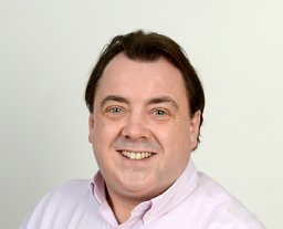 Nicholas R. Forsyth合作论文数Department of Cell Biology, University of Texas Southwestern Medical Center;Fax: +1 214 648 8694, Tel: +1 214 648 3282;Department of Cell Biology, University of Texas Southwestern Medical Center9
 Cassilda Cunha-Reis合作论文数CBQF–Centro de Biotecnologia e Química Fina-Laboratório Associado, Universidade Católica Portuguesa8
 Samantha Louise Wilson合作论文数Division of Clinical Neuroscience, University of Nottingham8Alan G.S. Harper合作论文数8
 Nureddin Ashammakhi合作论文数Department of Biomedical Engineering, Henry Samueli School of Engineering, University of California, Los Angeles;Department of Radiological Sciences, David Geffen School of Medicine, University of California, Los Angeles;Center for Minimally Invasive Therapeutics, California NanoSystems Institute, University of California, Los Angeles7
 Isaac Liu合作论文数School of Engineering, Faculty of Science, Engineering and Medicine, The University of Warwick7I Wimpenny合作论文数7
 Zhihong Huang合作论文数6Kong Y Then合作论文数6
 Anthony J Deegan合作论文数Dept Bioengn, Univ Washington6
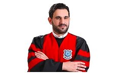 Hamza Abu Owida合作论文数Med Engn Dept, Al Ahliyya Amman Univ6
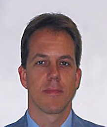 Cinque Gianfelice合作论文数Diamond Light Source6Ian Wimpenny合作论文数6
 Nicola Maffulli合作论文数Queen Mary University of London6
 Katie Bardsley合作论文数5
 Jonathan Mason合作论文数Univ Edinburgh5bin hu合作论文数5M.A. Wood合作论文数5
 Saaeha Rauz合作论文数Institute of Inflammation and Ageing, College of Medical and Dental Sciences, University of Birmingham5
 Sarah Waters合作论文数Mathematical Institute, University of Oxford;St Anne's College, University of Oxford5Jan Herman Kuiper合作论文数Department of Postgraduate Medicine, Keele University5
 Paul Dumas合作论文数SOLEIL Synchrotron5Rkk Wang合作论文数4E BAAS合作论文数4
 Ryan Dimmock合作论文数4Wanjiku Njoroge合作论文数4
 Chengqing Yuan (袁成清)合作论文数School of Transportation and Logistics Engineering, Wuhan University of Technology;School of Energy and Power Engineering, Wuhan University of Technology4Bölgen Nimet合作论文数Engineering Faculty, Mersin University4Ling Yuting合作论文数4
 Abigail Rutter合作论文数4
 Isaac Kuo-Kang Liu合作论文数4
 Paul Roach合作论文数School of Environmental & Life Sciences, University of Newcastle4
 Wen-Wu Li合作论文数4
 Petek Korkusuz合作论文数Faculty of Medicine, Hacettepe University4Jacek Pijanka合作论文数4Homayemem Weli合作论文数3
 Xie Bowen合作论文数3
 Yilong Zhang合作论文数Sch Sci & Engn, Univ Dundee3Lu Luo合作论文数3
 Christophe Sandt合作论文数3




