Biodiversity loss in the present era requires new tools for studying nonmodel organisms. Elephants are both an endangered species and excellent models for studying complex phenotypes including size, social behavior and longevity. Here we report the first derivation of elephant (Elephas maximus) induced pluripotent stem (emiPS) cells. We achieved emiPS cells using two approaches: (1) a two-step process of chemical media induction and colony selection followed by over-expression of elephant transcription factors; and (2) a one-step process with transcription factors and HRAS mutant, HRASG12V. For both protocols, we inhibited TP53 retrogenes, which are hypothesized to confer unique cancer resistance in elephants. To confirm their reprogrammed state, we generated a functional omics catalog of emiPS cells. While these emiPS cells remain transgene-dependent, we inactivated the transgenes and differentiated emiPS cells into all three germ layers via tri-lineage differentiation, embryoid body generation and direct differentiation into putative cell types from all three layers. These methods will open new frontiers for cellular models of nonmodel organisms, including for genetic rescue and conservation.
We previously reported that WAVE1, a major activator of Arp2/3 complex-mediated actin polymerization, is downregulated in postmortem brains of Alzheimer's disease (AD) and that WAVE1 regulates amyloid precursor protein trafficking and amyloid-β production. However, its role in tau pathology remains unknown. Here, we demonstrate that WAVE1 activity is suppressed in P301S tau mice through elevated inhibitory phosphorylation. Strikingly, WAVE1 gene ( Wasf1 ) knockout in P301S tau mice significantly reduces tau hyperphosphorylation and improves cognition, suggesting a compensatory role for WAVE1 suppression in tau pathogenesis. Single-nucleus RNA sequencing reveals that Wasf1 deletion in P301S tau mice reverses microglial state transitions, with minimal impact on other brain cell types. Wasf1 mRNA is highly translated in microglia in non-Tg mice, while its expression is downregulated in P301S tau mice. Wasf1 knockdown in BV2 microglia cells enhances the degradation of engulfed tau fibrils, indicating WAVE1 as an endogenous regulator of microglial function. Additionally, CellChat analysis indicates that Wasf1 deletion alters microglial autocrine signaling and their interactions with other cell types in P301S tau mice. These findings, taken together, suggest that Wasf1 deletion restores homeostatic microglial function, mitigates tau pathology, and alleviates cognitive deficits, highlighting WAVE1 as a potential therapeutic target for tauopathy-related dementias.
Rationale:Direct reprogramming of fibroblasts into endothelial cells (rECs) using ETV2 shows promise for vascular regeneration. However, current approaches using integrating viral vectors pose clinical translation barriers, and poor long-term cell survival limits therapeutic efficacy. Objective:To develop a clinically compatible method for generating rECs using non-integrating adenoviral ETV2 (Ad-ETV2) and enhance their engraftment and therapeutic efficacy through peptide amphiphile (PA) nanomatrix encapsulation. Methods and Results:Human dermal fibroblasts were reprogrammed using Ad-ETV2 and characterized by flow cytometry, RNA sequencing, and functional assays. Therapeutic efficacy was evaluated in murine hindlimb ischemia with or without PA-RGDS encapsulation over 12 months. Ad-ETV2 induced robust endothelial gene expression (CDH5, KDR, PECAM1) within 6 days, with 40-50% reprogramming efficiency. KDR+ Ad-rECs demonstrated functional endothelial properties including Ac-LDL uptake, tube formation, and exceptional proangiogenic factor secretion (200-fold higher HGF than HUVECs). RNA sequencing revealed rapid transcriptional reprogramming with fibroblast gene suppression and endothelial/angiogenic gene activation. In hindlimb ischemia, Ad-rECs significantly enhanced blood flow recovery and capillary density versus controls. Long-term analysis revealed sustained vascular contribution through three mechanisms: direct incorporation, perivascular support, and vessel guidance, persisting throughout 12 months-the longest reported follow-up for reprogrammed cells. PA-RGDS encapsulation markedly improved cell retention; while 75% of cells were lost by 3 months, retention stabilized thereafter with minimal additional loss through 12 months. Conclusions:Adenoviral ETV2 delivery enables efficient generation of clinically compatible rECs without genomic integration. These cells demonstrate potent and sustained therapeutic efficacy through multiple vascular regeneration mechanisms. PA-RGDS encapsulation significantly enhances long-term engraftment, establishing this combined approach as a promising platform for treating ischemic cardiovascular diseases.
Chimeric Antigen Receptor (CAR)-T cell therapy is a novel personalized treatment that engineers patient immune cells to fight against cancer cells. Recently, CAR-T cell therapy has demonstrated remarkable success in the treatment of hematopoietic cancers, whereas the treatment of solid tumors is more challenging, likely in part due to their severely immunosuppressive tumor microenvironment. To address distinct molecular responses of CAR-T cells between blood and solid tumors, we performed synthetic analysis of single-cell transcriptomics of CAR-T cells and identified unique immunosuppressive subpopulations and aberrant signalling inductions in CD4+ and CD8+ CAR-T cells in the context of solid tumors. Furthermore, we also found that PD-1-independent exhaustion-like CD8+ CAR-T cells, characterized by high expression of TNFRSF9 and CCL3 , were preferentially generated under solid tumor stimulation. Collectively, our comprehensive analyses provide essential molecular insights into solid tumor-stimulated CAR-T cells and assists in overcoming the limited efficacy of CAR-T cell therapy against solid tumors. ### Competing Interest Statement Y.T. works as a consultant in Colossal Biosciences. The remaining authors have no conflict of interest to declare.
Identifying cell-type-specific enhancers is critical for developing genetic tools to study the mammalian brain. We organized the "Brain Initiative Cell Census Network (BICCN) Challenge: Predicting Functional Cell Type-Specific Enhancers from Cross-Species Multi-Omics" to evaluate machine learning and feature-based methods for nominating enhancer sequences targeting mouse cortical cell types. Methods were assessed using in vivo data from hundreds of adeno-associated virus (AAV)-packaged, retro-orbitally delivered enhancers. Open chromatin was the strongest predictor of functional enhancers, while sequence models improved prediction of non-functional enhancers and identified cell-type-specific transcription factor codes to inform in silico enhancer design. This challenge establishes a benchmark for enhancer prioritization and highlights computational and molecular features critical for identifying functional cortical enhancers, advancing efforts to map and manipulate gene regulation in the mammalian cortex.
Alcohol dependence (AD) is one of the most prevalent neuropsychiatric disorders. Multiple polymorphisms in the Fyn tyrosine kinase gene (FYN) were found to be associated with AD. The function of AD-associated FYN variants remains largely unknown due to the absence of an appropriate model for studying them. Here, we generated human embryonic stem cell lines homozygous/heterozygous for rs706895 C/T alleles in 5' untranslated region (5' UTR) of FYN by CRISPR-Cas9 editing to explore the AD association. Transcriptome and reporter gene analyses demonstrated that induced neurons with the rs706895 C allele showed a significantly higher expression level of FYN under ethanol treatment. Our results suggest that FYN 5' UTR variant rs706895 may influence an individual's vulnerability to AD by altering FYN expression. Targeting AD-associated variants may provide a better understanding of disease mechanisms and a reliable basis for the personalized AD treatment.
The human brain represents one of the most complex organs in our body, with development regulated by an intricate genetic program. Recently, non-genetic factors, such as prenatal stress, infection, and diet, have been shown to influence brain development. Radiofrequency radiation (RF; 800-2,400 MHz), emitted by natural and artificial sources such as microwaves and cell phones, represents a non-invasive environmental factor. Using human cortical organoids (hCOs) derived from human embryonic stem cells (hESCs), we investigate RF's effects on corticogenesis. We find that RF exposure regulates the differentiation of human and non-human primate radial glia progenitors, maintaining stem cell identity and delaying differentiation. Neurons differentiated under RF treatment show induction of expression of human endogenous retroviruses. Importantly, inhibitors for the BET (bromodomain and extraterminal) protein rescue RF-induced developmental defects in hCOs. Our findings reveal a mechanism by which RF modulates early brain development, offering a non-biological approach to regulate neural stem cell self-renewal.
The E2F transcription factors constitute a core transcriptional network that governs cell division and oncogenesis in multi-cellular organisms, although their molecular mechanisms remain incompletely understood. Here, we show that elevated expression of the transcription factor FOXK1 promotes transcription of E2F target genes and cellular transformation. High expression of FOXK1 in patient tumors is also strongly correlated with E2F gene expression. Mechanistically, we demonstrate that FOXK1 is O-GlcNAcylated, and loss of this modification impairs FOXK1 ability to promote cell proliferation and tumor growth. We also show that expression of FOXK1 O-GlcNAcylation-defective mutants results in reduced recruitment of the H2AK119 deubiquitinase and tumor suppressor BAP1 to E2F target genes. This event is associated with a transcriptional repressive chromatin environment and reduced cell proliferation. Our results define an essential role of FOXK1 O-GlcNAcylation in co-opting the tumor suppressor BAP1 to promote cancer cell progression through orchestration of the E2F pathway.
Application of single-cell/nucleus genomic sequencing to patient-derived tissues offers potential solutions to delineate disease mechanisms in humans. However, individual cells in patient-derived tissues are in different pathological stages, and hence, such cellular variability impedes subsequent differential gene expression analyses. To overcome such a heterogeneity issue, we present a novel deep learning approach, scIDST, that infers disease progression levels of individual cells with weak supervision framework. The disease progression-inferred cells display significant differential expression of disease-relevant genes, which cannot be detected by comparative analysis between patients and healthy donors. In addition, we demonstrate that pretrained models by scIDST are applicable to multiple independent data resources and are advantageous to infer cells related to certain disease risks and comorbidities. Taken together, scIDST offers a new strategy of single-cell sequencing analysis to identify bona fide disease-associated molecular features.
Integrative analysis of publicly available scRNA-seq data facilitates deeper understanding of biological phenomena with strong statistical power and high resolution. A new study in this issue of PLOS Biology examined the fidelity of various brain organoid protocols in reference to human primary developing brain by gene co-expression relationships with million-scale collection of public scRNA-seq data sets.
Gene transcription is a highly regulated process, and deregulation of transcription factors activity underlies numerous pathologies including cancer. Albeit near four decades of studies have established that the E2F pathway is a core transcriptional network that govern cell division in multi-cellular organisms1,2, the molecular mechanisms that underlie the functions of E2F transcription factors remain incompletely understood. FOXK1 and FOXK2 transcription factors have recently emerged as important regulators of cell metabolism, autophagy and cell differentiation3-6. While both FOXK1 and FOXK2 interact with the histone H2AK119ub deubiquitinase BAP1 and possess many overlapping functions in normal biology, their specific functions as well as deregulation of their transcriptional activity in cancer is less clear and sometimes contradictory7-13. Here, we show that elevated expression of FOXK1, but not FOXK2, in primary normal cells promotes transcription of E2F target genes associated with increased proliferation and delayed entry into cellular senescence. FOXK1 expressing cells are highly prone to cellular transformation revealing important oncogenic properties of FOXK1 in tumor initiation. High expression of FOXK1 in patient tumors is also highly correlated with E2F gene expression. Mechanistically, we demonstrate that FOXK1, but not FOXK2, is specifically modified by O-GlcNAcylation. FOXK1 O-GlcNAcylation is modulated during the cell cycle with the highest levels occurring during the time of E2F pathway activation at G1/S. Moreover, loss of FOXK1 O-GlcNAcylation impairs FOXK1 ability to promote cell proliferation, cellular transformation and tumor growth. Mechanistically, expression of FOXK1 O-GlcNAcylation-defective mutants results in reduced recruitment of BAP1 to gene regulatory regions. This event is associated with a concomitant increase in the levels of histone H2AK119ub and a decrease in the levels of H3K4me1, resulting in a transcriptional repressive chromatin environment. Our results define an essential role of O-GlcNAcylation in modulating the functions of FOXK1 in controlling the cell cycle of normal and cancer cells through orchestration of the E2F pathway.
The transmembrane death receptor Fas transduces apoptotic signals upon binding its ligand, FasL. Although Fas is highly expressed in cancer cells, insufficient cell surface Fas expression desensitizes cancer cells to Fas-induced apoptosis. Here, we show that the increase in Fas microaggregate formation on the plasma membrane in response to the inhibition of endocytosis sensitizes cancer cells to Fas-induced apoptosis. We used a clinically accessible Rho-kinase inhibitor, fasudil, that reduces endocytosis dynamics by increasing plasma membrane tension. In combination with exogenous soluble FasL (sFasL), fasudil promoted cancer cell apoptosis, but this collaborative effect was substantially weaker in nonmalignant cells. The combination of sFasL and fasudil prevented glioblastoma cell growth in embryonic stem cell-derived brain organoids and induced tumor regression in a xenograft mouse model. Our results demonstrate that sFasL has strong potential for apoptosis-directed cancer therapy when Fas microaggregate formation is augmented by mechano-inhibition of endocytosis.
Age is the primary risk factor for Parkinson’s disease (PD), but how aging changes the expression and regulatory landscape of the brain remains unclear. Here we present a single-nuclei multiomic study profiling shared gene expression and chromatin accessibility of young, aged and PD postmortem midbrain samples. Combined multiomic analysis along a pseudopathogenesis trajectory reveals that all glial cell types are affected by age, but microglia and oligodendrocytes are further altered in PD. We present evidence for a disease-associated oligodendrocyte subtype and identify genes lost over the aging and disease process, including CARNS1, that may predispose healthy cells to develop a disease-associated phenotype. Surprisingly, we found that chromatin accessibility changed little over aging or PD within the same cell types. Peak–gene association patterns, however, are substantially altered during aging and PD, identifying cell-type-specific chromosomal loci that contain PD-associated single-nucleotide polymorphisms. Our study suggests a previously undescribed role for oligodendrocytes in aging and PD. Aging is a risk factor of Parkinson’s disease (PD). Adams, Song et al. present a multiomics analysis of the human midbrain showing age-induced changes in genes associated with glial function, with further alterations of oligodendrocytes in PD.
Forkhead box protein A1 (FOXA1), a pioneering transcriptional factor known for its critical roles in prostate and ERα−positive breast cancer, is also expressed in human epidermal growth factor receptor-2 (HER2/ErbB2)-positive breast cancers. However, its role in HER2-pos tumors is less well understood. Here we investigate the function of FOXA1 in HER2/ErbB2- positive breast cancers. The loss of FOXA1 was associated with a marked decrease in the viability of HER2-positive and HER2 amplified cell lines, suggesting a pivotal involvement of FOXA1 in these breast cancers. Employing patient-derived single-cell RNA sequencing and spatial transcriptomics, we demonstrate that FOXA1 is co-expressed with ErbB2 in HER2- positive breast cancers. Suppression of FOXA1 expression led to the reduction of HER2 expression and signaling. Chromatin Immunoprecipitation Sequencing (ChIP-seq) and Assay for Transposase-Accessible Chromatin using sequencing (ATAC-seq) identified FOXA1 binding motifs in the ErbB2 promoter and regulatory element regions, which controlled ErbB2 gene expression. Notably, FOXA1 knockdown was observed to enhance Epithelial-Mesenchymal Transition (EMT) signaling and impede luminal tumor differentiation. Furthermore, we find that FOXA1 and TRPS1 combine to regulate TEAD/YAP-TAZ activity. Taken together, these findings highlight the essential role of FOXA1 in maintaining HER2 expression and a luminal cell phenotype in HER2-positive breast cancers.### Competing Interest StatementThe authors have declared no competing interest.
ABSTRACTThe crisis of biodiversity loss in the anthropogenic era requires new tools for studying non-model organisms. Elephants, for example, are both an endangered species and excellent models studying complex phenotypes like size, social behavior, and longevity, but they remain severely understudied. Here we report the first derivation of elephant (Elephas maximus) induced pluripotent stem cells (emiPSCs) achieved via a two-step process of chemical-media induction and colony selection, followed by overexpression of elephant transcription factorsOCT4, SOX2, KLF4, MYC±NANOGandLIN28A, and modulation of theTP53pathway. Since the seminal discovery of reprogramming by Shinya Yamanaka, iPSCs from many species including the functionally extinct northern white rhinocerous have been reported, but emiPSCs have remained elusive. While for multiple species the reprogramming protocol was adopted with little changes compared to model organisms like mouse and human, our emiPSC protocol requires a longer timeline and inhibition ofTP53expansion genes that are hypothesized to confer unique cancer resistance in elephants. iPSCs unlock tremendous potential to explore cell fate determination, cell and tissue development, cell therapies, drug screening, disease modeling, cancer development, gametogenesis and beyond to further our understanding of this iconic megafauna. This study opens new frontiers in advanced non-model organism cellular models for genetic rescue and conservation.
The lack of physiological parity between 2D cell culture and in vivo culture has led to the development of more organotypic models, such as organoids. Organoid models have been developed for a number of tissues, including the liver. Current organoid protocols are characterized by a reliance on extracellular matrices (ECMs), patterning in 2D culture, costly growth factors and a lack of cellular diversity, structure, and organization. Current hepatic organoid models are generally simplistic and composed of hepatocytes or cholangiocytes, rendering them less physiologically relevant compared to native tissue. We have developed an approach that does not require 2D patterning, is ECM independent, and employs small molecules to mimic embryonic liver development that produces large quantities of liver-like organoids. Using single-cell RNA sequencing and immunofluorescence, we demonstrate a liver-like cellular repertoire, a higher order cellular complexity, presenting with vascular luminal structures, and a population of resident macrophages: Kupffer cells. The organoids exhibit key liver functions, including drug metabolism, serum protein production, urea synthesis and coagulation factor production, with preserved post-translational modifications such as N-glycosylation and functionality. The organoids can be transplanted and maintained long term in mice producing human albumin. The organoids exhibit a complex cellular repertoire reflective of the organ and have de novo vascularization and liver-like function. These characteristics are a prerequisite for many applications from cellular therapy, tissue engineering, drug toxicity assessment, and disease modeling to basic developmental biology.
Three-dimensional (3D) genomics shows immense promise for studying X chromosome inactivation (XCI) by interrogating changes to the X chromosomes' 3D states. Here, we sought to characterize the 3D state of the X chromosome in naïve and primed human pluripotent stem cells (hPSCs). Using chromatin tracing, we analyzed X chromosome folding conformations in these cells with megabase genomic resolution. X chromosomes in female naïve hPSCs exhibit folding conformations similar to the active X chromosome (Xa) and the inactive X chromosome (Xi) in somatic cells. However, naïve X chromosomes do not exhibit the chromatin compaction typically associated with these somatic X chromosome states. In H7 naïve human embryonic stem cells, XIST accumulation observed on damaged X chromosomes demonstrates the potential for naïve hPSCs to activate XCI-related mechanisms. Overall, our findings provide insight into the X chromosome status of naïve hPSCs with a single-chromosome resolution and are critical in understanding the unique epigenetic regulation in early embryonic cells.
Human brain organoids provide unique platforms for modeling several aspects of human brain development and pathology. However, current brain organoid systems mostly lack the resolution to recapitulate the development of finer brain structures with subregional identity, including functionally distinct nuclei in the thalamus. Here, we report a method for converting human embryonic stem cells (hESCs) into ventral thalamic organoids (vThOs) with transcriptionally diverse nuclei identities. Notably, single-cell RNA sequencing revealed previously unachieved thalamic patterning with a thalamic reticular nucleus (TRN) signature, a GABAergic nucleus located in the ventral thalamus. Using vThOs, we explored the functions of TRN-specific, disease-associated genes patched domain containing 1 (PTCHD1) and receptor tyrosine-protein kinase (ERBB4) during human thalamic development. Perturbations in PTCHD1 or ERBB4 impaired neuronal functions in vThOs, albeit not affecting the overall thalamic lineage development. Together, vThOs present an experimental model for understanding nuclei-specific development and pathology in the thalamus of the human brain.
Although mitochondrial activity is critical for angiogenesis, its mechanism is not entirely clear. Here we show that mice with endothelial deficiency of any one of the three nuclear genes encoding for mitochondrial proteins, transcriptional factor (TFAM), respiratory complex IV component (COX10), or redox protein thioredoxin 2 (TRX2), exhibit retarded retinal vessel growth and arteriovenous malformations (AVM). Single-cell RNA-seq analyses indicate that retinal ECs from the three mutant mice have increased TGFβ signaling and altered gene expressions associated with vascular maturation and extracellular matrix, correlating with vascular malformation and increased basement membrane thickening in microvesels of mutant retinas. Mechanistic studies suggest that mitochondrial dysfunction from Tfam , Cox10 , or Trx2 depletion induces a mitochondrial localization and MAPKs-mediated phosphorylation of SMAD2, leading to enhanced ALK5-SMAD2 signaling. Importantly, pharmacological blockade of ALK5 signaling or genetic deficiency of SMAD2 prevented retinal vessel growth retardation and AVM in all three mutant mice. Our studies uncover a novel mechanism whereby mitochondrial dysfunction via the ALK5-SMAD2 signaling induces retinal vascular malformations, and have therapeutic values for the alleviation of angiogenesis-associated human retinal diseases.
Microglia play a role in the emergence and preservation of a healthy brain microenvironment. Dysfunction of microglia has been associated with neurodevelopmental and neurodegenerative disorders. Investigating the function of human microglia in health and disease has been challenging due to the limited models of the human brain available. Here, we develop a method to generate functional microglia in human cortical organoids (hCOs) from human embryonic stem cells (hESCs). We apply this system to study the role of microglia during inflammation induced by amyloid-β (Aβ). The overexpression of the myeloid-specific transcription factor PU.1 generates microglia-like cells in hCOs, producing mhCOs (microglia-containing hCOs), that we engraft in the mouse brain. Single-cell transcriptomics reveals that mhCOs acquire a microglia cell cluster with an intact complement and chemokine system. Functionally, microglia in mhCOs protect parenchyma from cellular and molecular damage caused by Aβ. Furthermore, in mhCOs, we observed reduced expression of Aβ-induced expression of genes associated with apoptosis, ferroptosis, and Alzheimer's disease (AD) stage III. Finally, we assess the function of AD-associated genes highly expressed in microglia in response to Aβ using pooled CRISPRi coupled with single-cell RNA sequencing in mhCOs. In summary, we provide a protocol to generate mhCOs that can be used in fundamental and translational studies as a model to investigate the role of microglia in neurodevelopmental and neurodegenerative disorders.
 In-Hyun Park合作论文数Neuroscience, Biological & Biomedical Sciences, Yale University39
 Yangfei Xiang (向阳飞)合作论文数School of Life Science and Technology, ShanghaiTech University17
 Kim Kun-Yong合作论文数16Benjamin Patterson合作论文数Department of Genetics, Yale Stem Cell Center, Yale School of Medicine, New Haven, CT 06520, USA14
 Bilal cakır合作论文数Sabri Ulker R&D Ctr Bldg, IZU Food & Agr Res Ctr GTAUM14
 Xin Wang合作论文数12Hysolli Eriona合作论文数10
 Sugang Xu合作论文数9
 Sherman Weissman合作论文数Yale School of Medicine9Tithra Chap合作论文数8
 Xinghua Pan合作论文数6
 Young-Jin Kang合作论文数PTC Therapeutics, Inc.6
 Filippos Balasis合作论文数5Jung Yong Wook合作论文数5
 Gareth j Sullivan合作论文数5
 Ferdi Ridvan Kiral合作论文数Division of Neurobiology, Freie Universität Berlin, Königin Luise Straße 1-3, 14195 Berlin, Germany4
 Young-sup Yoon合作论文数4
 Patra Prabir K合作论文数Department of Mechanical Engineering, University of Bridgeport;Department of Biomedical Engineering, University of Bridgeport;University of Bridgeport, University of Bridgeport4
 Jong Hoon Kim合作论文数Division of Biotechnology, College of Life Sciences and Biotechnology, Korea University4
 Sangho Lee合作论文数Department of Medicine, Emory University School of Medicine3
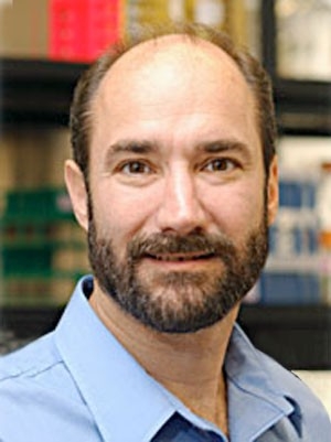 Michael Snyder合作论文数Department of Genetics, School of Medicine, Stanford University;Center for Genomics and Personalized Medicine;Karolinska Institute3
 Huiping Zhang (张会平)合作论文数School of Social Research, Renmin University of China3Siller Richard合作论文数Institute of Basic Medical Sciences, University of Oslo3Pingnan Sun (孙平楠)合作论文数3
 Mehmet Hamdi Kural合作论文数Department of Biomedical Engineering, Worcester Polytechnic Institute3
 Laura Niklason合作论文数Department of Anesthesiology, School of Medicine, Yale University3
 Yoon Seong Kim合作论文数Weill Medical College, Cornell University3Ghia Euskirchen合作论文数3Akihiro Sakaguchi合作论文数2Woo Sub Yang合作论文数2yee sook cho合作论文数2Kazunari Nakaue合作论文数2
 Levi Adams合作论文数College of Medicine, University of Central Florida2Masatomo Yoshida合作论文数2Xinran Liu (欣然劉)合作论文数2
 Louis Masclef合作论文数Univ Montreal, Maisonneuve Rosemont Hosp, Res Ctr, Montreal, PQ H3C 3J7, Canada2
 Haifeng Zhang合作论文数Pathology Faculty, Yale School of Medicine, Yale University2
 Wang Min合作论文数Department of Pathology and Vascular Biology, Yale School of Medicine, Yale University2
 Mallette Frédérick A合作论文数Dept Biochim, Univ Montreal2Laura Hulea合作论文数Goodman Cancer Centre, McGill University2
 Ji Woong Han合作论文数2
 Lindsay A. Farrer合作论文数Schools of Medicine and Public Health, Boston University2Schcolnik Cabrera Alejandro合作论文数Facultad de Medicina, Universidad Nacional Autónoma de México2Kim SangSung合作论文数2
 Appleton Evan合作论文数Graduate Program in Bioinformatics, Boston University2
 Joel Gelernter合作论文数Department of Neuroscience, School of Medicine, Yale University;Division of Human Genetics, School of Medicine, Yale University2
 Hwang Sungmin合作论文数Department of Microbiology and Cell Science, University of Florida2




