The chlamydial Major Outer Membrane Protein (MOMP) is a promising subunit vaccine candidate due to its abundance in the outer membrane (OM), the presence of four surface-exposed variable domains (VDs) containing neutralizing and serotyping epitopes, and confirmed T-cell epitopes located in its constant domains (CDs). However, recombinant and denatured forms of MOMP have failed to elicit immune responses comparable to native preparations, indicating that conformation is critical for immunogenicity. Here, we present two cryo-EM structures of native Chlamydia muridarum MOMP, isolated from infectious elementary bodies (EBs). EB MOMP forms a distinct trimer with a stem of three narrow, non-permeable β-barrels and an extracellular, folded antigenic cap that displays VDs. In complex with a neutralizing Fab fragment of a conformational antibody, the cap undergoes structural reorganization that shows how epitope presentation is modulated by immune engagement. These structures reveal the molecular basis of MOMP's serovar specificity and provide a template for structure-based vaccine design.
Type IV pili (T4P) mediate surface motility, host interactions, and DNA uptake through cycles of extension and retraction. While the primary retraction ATPase PilT has been extensively characterized, its homolog PilU remains less well understood despite being demonstrated as a PilT-dependent retraction ATPase. Here, we determined six PilU structures by cryo-electron microscopy and x-ray crystallography. The structures reveal a homohexameric assembly stabilized by interactions between the C-terminal catalytic domain of one subunit and the N-terminal PAS-like domain of a neighboring subunit. PilU adopts multiple conformational states, exhibiting different combinations of open and closed interfaces even in the absence of nucleotide. Comparison with PilT highlights structural features that likely underlie PilU's weak ATPase activity and its dependence on PilT for function. Together, these findings provide a structural framework for understanding PilU's role within the T4P retraction machinery.
The correct description of quantum scattering places the observed scattering contributions on the Ewald's sphere and its Friedel mate copy. In electron microscopy, due to the large radius of the Ewald's sphere, these scattering contributions are typically merged during data analysis. We present an approach that separates and factorizes those contributions into real and imaginary components of the image. When an inverted solution is calculated, the map derived from the real component of the image generates an inverted solution, while the map derived from the imaginary component of the image generates an inverted and sign-flipped solution. Therefore, the sign of correlation between reconstructions derived from the real and imaginary components provides the automatic determination of handedness and additional validation for the quality of 3D reconstructions. The factorization and its implementation are robust enough to be routinely used in single-particle reconstructions, even at resolutions below the limit where the curvature of the Ewald's sphere affects the overall signal-to-noise ratio.
To understand immunogenicity of chlamydial Major Outer Membrane Protein (MOMP), we determined the structures of MOMP from Chlamydia muridarum elementary bodies and its complex with a Fab fragment of a neutralizing monoclonal antibody.Natively folded MOMP forms a trimer of 10-stranded β-barrel protomers, with their extracellular regions folded into a single, compact magnesium- binding cap that contains immunogenic variable domains (VDs) from all three protomers. Sphingolipid molecules bound at the barrel interfaces further stabilize the trimer. The extracellular cap features positively charged cavities that may bind chlamydial and host molecules. The periplasmic region contains conserved cysteine residues, consistent with MOMP’s role in stabilizing the chlamydial outer membrane complex (COMC) through intramolecular disulfide bonds with other MOMP molecules and COMC proteins.The β-barrels’ channels are too narrow to transfer previously characterized substrates, and are blocked by the extracellular cap and their N- terminal segments on the periplasmic side. Thus, native EB MOMP cannot function as a porin, suggesting that alternative MOMP folds may exist during intermediate and reticulate body stages, or that other proteins may perform this role.In the MOMP-Fab complex (3:3 stoichiometry), each Fab binds two MOMP protomers, inducing structural changes in VD1 and VD4, which partially opens the antigenic cap without exposing the species-specific motif TTWNPTISG (TTLNPTIAG in C. trachomatis).These findings challenge long-standing assumptions about MOMP’s architecture and open new avenues for research into MOMP’s role in chlamydial pathogenesis and development of therapeutics.
In cryoEM 3D reconstruction, noise is spread over the entire 3D space, while the signal is present only within the molecular mask. The signal-to-noise ratio (SNR) within the molecular mask is the SNR of interest in data analysis. Any method that does not apply the molecular mask to noise underestimates the SNR. For instance, Fourier Shell Correlation (FSC) curves recalculated between two half-maps after the data are deposited in the PDB do not apply the molecular mask to noise, consequently providing a misleading impression of resolution. These approaches, relying on calculations without mask constraints on noise, try to avoid introducing biases within the mask area that could potentially arise at low resolution. Here, we present a method of FSC calculations in which masking does not introduce spurious correlation between two maps. In this approach, we split the particle image signal in real space into two components: 1) the real part, which is the signal used in traditional calculations; and 2) the imaginary part, which is the signal present only when Ewald's sphere calculation is considered. If the two components are used in reconstruction separately, they are independent of each other. Then, the FSC is calculated between two maps derived from the real part of all particles for one map and from the imaginary part of all particles for the second map. Typically, reconstruction from the real component is used to refine particle orientations, introducing bias into subsequent reconstruction based on the real component. However, reconstruction based on the imaginary component is not biased by the refinement using only the real component. This way, reconstruction from the imaginary component can be used as an unbiased reference for validating reconstruction from the real component. This validation works even at relatively low resolution (10 Å data if the number of particles is large), despite relying on Ewald's sphere curvature. The byproduct of this validation is the determination of the structure's handedness, which only requires medium resolution data. In cryoEM 3D reconstruction, noise is spread over the entire 3D space, while the signal is present only within the molecular mask. The signal-to-noise ratio (SNR) within the molecular mask is the SNR of interest in data analysis. Any method that does not apply the molecular mask to noise underestimates the SNR. For instance, Fourier Shell Correlation (FSC) curves recalculated between two half-maps after the data are deposited in the PDB do not apply the molecular mask to noise, consequently providing a misleading impression of resolution. These approaches, relying on calculations without mask constraints on noise, try to avoid introducing biases within the mask area that could potentially arise at low resolution. Here, we present a method of FSC calculations in which masking does not introduce spurious correlation between two maps. In this approach, we split the particle image signal in real space into two components: 1) the real part, which is the signal used in traditional calculations; and 2) the imaginary part, which is the signal present only when Ewald's sphere calculation is considered. If the two components are used in reconstruction separately, they are independent of each other. Then, the FSC is calculated between two maps derived from the real part of all particles for one map and from the imaginary part of all particles for the second map. Typically, reconstruction from the real component is used to refine particle orientations, introducing bias into subsequent reconstruction based on the real component. However, reconstruction based on the imaginary component is not biased by the refinement using only the real component. This way, reconstruction from the imaginary component can be used as an unbiased reference for validating reconstruction from the real component. This validation works even at relatively low resolution (10 Å data if the number of particles is large), despite relying on Ewald's sphere curvature. The byproduct of this validation is the determination of the structure's handedness, which only requires medium resolution data.
GPUs offer significant potential for accelerating diffraction data processing leading to potential speed-ups of up to 10,000-fold. However, achieving this potential is substantially limited by the extensive effort required to redesign algorithms so that they are optimized for GPU architectures. Diffraction data analysis inherently lends itself to vectorization, yet existing data structures frequently introduce serial dependencies incompatible with the parallel architecture of GPUs, severely diminishing computational efficiency. Historically, diffraction data algorithms development was heavily influenced by memory constraints, as memory-efficient algorithms often performed better and were faster on CPUs. Transitioning to GPUs introduces distinct challenges, notably designing computations around two- or three-dimensional grids, wherein each grid point's calculation is handled independently by dedicated GPU threads. This grid-based parallelism enables GPUs to manage hundreds of thousands of simultaneous threads effectively.I will describe these challenges and opportunities with specific examples of both legacy and newly developed GPU-oriented algorithms. These include (a) transforming detector-coordinate data into a reciprocal lattice aligned with crystallographic axes, analogous to the traditional XDS algorithm, and (b) using libraries that streamline computational code development, by integration of GPU-based FFTs with intuitive coordinate system representations. Such approaches will allow us to fully leverage GPU capabilities in diffraction data analysis.
ABSTRACT During infection, bacterial pathogens rely on secreted virulence factors to manipulate the host cell. However, in gram-positive bacteria, the molecular mechanisms underlying the folding and activity of these virulence factors after membrane translocation are not clear. Here, we solved the protein structures of two secreted parvulin and two secreted cyclophilin-like peptidyl-prolyl isomerase (PPIase) ATP-independent chaperones found in gram-positive streptococcal species. The extracellular parvulin-type PPIase, PrsA in Streptococcus pneumoniae and Streptococcus mutans maintain dimeric crystal structures reminiscent of folding catalysts that consist of two domains, a PPIase and foldase domain. Structural comparison of the two cyclophilin-like extracellular chaperones from S. pneumoniae and Streptococcus pyogenes with other cyclophilins demonstrates that this group of cyclophilin-like chaperones has novel structural appendages formed by 9- and 24-residue insertions. Furthermore, we demonstrate that deletion of prsA and slrA genes impairs the secretion of the cholesterol-dependent pore-forming toxin, pneumolysin in S. pneumoniae. Using protein pull-down and biophysical assays, we demonstrate a direct interaction between PrsA and SlrA with Ply. Then, we developed chaperone-assisted folding assays that show that the S. pneumoniae PrsA and SlrA extracellular chaperones accelerate pneumolysin folding. In addition, we demonstrate that SlrA and, for the first time, S. pyogenes PpiA exhibit PPIase activity and can bind the immunosuppressive drug, cyclosporine A. Altogether, these findings suggest a mechanistic role for streptococcal PPIase chaperones in the activity and folding of secreted virulence factors such as pneumolysin.IMPORTANCEStreptococcal species are a leading cause of lower respiratory infections that annually affect millions of people worldwide. During infection, streptococcal species secrete a medley of virulence factors that allow the bacteria to colonize and translocate to deeper tissues. In many gram-positive bacteria, virulence factors are secreted from the cytosol across the bacterial membrane in an unfolded state. The bacterial membrane-cell wall interface is exposed to the potentially harsh extracellular environment, making it difficult for native virulence factors to fold before being released into the host. ATP-independent PPIase-type chaperones, PrsA and SlrA, are thought to facilitate folding and stabilization of several unfolded proteins to promote the colonization and spread of streptococci. Here, we present crystal structures of the molecular chaperones of PrsA and SlrA homologs from streptococcal species. We provide evidence that the Streptococcus pyogenes SlrA homolog, PpiA, has PPIase activity and binds to cyclosporine A. In addition, we show that Streptococcus pneumoniae PrsA and SlrA directly interact and fold the cholesterol-dependent pore-forming toxin and critical virulence determinant, pneumolysin.
Current guidelines for depositing cryogenic electron microscopy single particle reconstruction (cryo-EM SPR) data require submission of unfiltered, unmasked, and unsharpened raw half-maps. The Fourier Shell Correlation (FSC) between the half-maps is then used as a proxy for the signal-to-noise ratio (SNR) to estimate the reconstruction's resolution. This policy was introduced to enable independent validation of reported resolutions. Although developed to safeguard data integrity and minimize bias, these guidelines do not account for specific features of modern cryo-EM processing software, in particular weighting schemes that are not retained in half-map depositions and yet in general are necessary to recapitulate resolution estimates. As a results, resolution estimates and other validation statistics based on half-maps FSC may be under- or overestimated. Here, we describe the limitations of the current deposition guidelines and propose an alternative: depositing cryo-EM results in Fourier (reciprocal) space together with the mandatory deposit of molecular masks or their descriptors. This approach addresses the current limitations, preserves critical information from the reconstruction process, and better supports downstream analyses.
Ultra-high-resolution crystal structures of proteins provide critical insights into protein structure, dynamics, hydrogen bonding, and solvent networks. Crambin, a small hydrophobic storage protein consisting of 46 residues (4.7 kDa), is found in the embryonic tissue of seeds from Crambe abyssinica. This protein is renowned for its ability to crystallize readily, forming some of the best-ordered macromolecular crystals known, which diffract X-rays to the highest sub-atomic resolution recorded for any protein to date.We have previously reported the room temperature structure of crambin, refined to an exceptional resolution of 0.70 Å using SHELXL. That analysis revealed intricate details of the dynamic solvent network, characterized by alternative side chain conformations and shifts in water molecule positions. In this work, we extend our investigation by presenting new structural data collected at cryogenic temperatures: 15K using liquid helium and 100K using liquid nitrogen cooling.We will report the ultra-high-resolution structures at 15K and 100K, providing a comparative analysis of the solvent networks across these different temperature datasets. This comparison aims to deepen our understanding of the solvent and protein dynamics, offering valuable insights into the protein interactions within solvent environments. Our findings underscore the significance of ultrahigh-resolution crystallography in elucidating the complex interplay between proteins and their solvent environments, with potential implications for the broader field of structural biology.
Gram-negative bacteria harness the proton motive force (PMF) within their inner membrane (IM) to uphold cell envelope integrity, an indispensable aspect for both division and survival. The IM TolQ-TolR complex is the essential part of the Tol-Pal system, serving as a conduit for PMF energy transfer to the outer membrane. Here we present cryo-electron microscopy reconstructions of Acinetobacter baumannii TolQ in apo and TolR-bound forms at atomic resolution. The apo TolQ configuration manifests as a symmetric pentameric pore, featuring a transmembrane funnel leading toward a cytoplasmic chamber. In contrast, the TolQ-TolR complex assumes a proton nonpermeable stance, characterized by the TolQ pentamer's flexure to accommodate the TolR dimer, where two protomers undergo a translation-based relationship. Our structure-guided analysis and simulations support the rotor-stator mechanism of action, wherein the rotation of the TolQ pentamer harmonizes with the TolR protomers' interplay. These findings broaden our mechanistic comprehension of molecular stator units empowering critical functions within the Gram-negative bacterial cell envelope.
Ultrahigh-resolution structures provide unprecedented details about protein dynamics, hydrogen bonding and solvent networks. The reported 0.70 Å, room-temperature crystal structure of crambin is the highest-resolution ambient-temperature structure of a protein achieved to date. Sufficient data were collected to enable unrestrained refinement of the protein and associated solvent networks using SHELXL. Dynamic solvent networks resulting from alternative side-chain conformations and shifts in water positions are revealed, demonstrating that polypeptide flexibility and formation of clathrate-type structures at hydrophobic surfaces are the key features endowing crambin crystals with extraordinary diffraction power.
TolQ-TolR-TolA is an energy-transducing inner-membrane sub-complex conserved across Gram-negative bacteria. It maintains connection with periplasmic TolB-Pal sub-complex in an energy-dependent manner through TolA protein spanning the periplasm. Utilizing proton motive force (PMF), Tol-Pal complex contributes to the integrity of outer membrane and the whole cell envelope. TolQ remains the only part of Tol-Pal complex with unknown structure up to now. To address this, we determined 3.02 and 3.34 Å cryo-electron microscopy (cryo-EM) structures of the integral inner-membrane protein TolQ and in complex with TolR, respectively from Acinetobacter baumannii.
Single particle reconstruction (SPR) in cryoEM is an image processing task with an elaborate hierarchy that starts with many very noisy multi-frame images. Efficient representation of the intermediary image structures is critical for keeping the calculations manageable. One such intermediary structure is called a particle stack and contains cut-out images of particles in square boxes of predefined size. The micrograph that is the source of the boxed images is usually corrected for motion between frames prior to particle stack creation. However, the contrast transfer function (CTF) or its Fourier Transform point spread function (PSF) are not considered at this step. Historically, the particle stack was intended for large particles and for a tighter PSF, which is characteristic of lower resolution data. The field now performs analyses of smaller particles and to higher resolution, and these conditions result in a broader PSF that requires larger padding and slower calculations to integrate information for each particle. Consequently, the approach to handling structures such as the particle stack should be reexamined to optimize data processing. Here we propose to use as a source image for the particle stack a complex-valued image, in which CTF correction is implicitly applied as a real component of the image. We can achieve it by applying an initial CTF correction to the entire micrograph first and perform box cutouts as a subsequent step. The final CTF correction that we refine and apply later has a very narrow PSF, and so cutting out particles from micrographs that were approximately corrected for CTF does not require extended buffering, i.e. the boxes during the analysis only have to be large enough to encompass the particle. The Fourier Transform of an exit-wave reconstruction creates an image that has complex values. This is a complex value image considered in real space, opposed to standard SPR data processing where complex numbers appear only in Fourier space. This extension of the micrograph concept provides multiple advantages because the particle box size can be small and calculations crucial for high resolution reconstruction such as Ewald sphere correction, aberration refinement, and particle-specific defocus refinement can be performed on the small box data.
The Escherichia coli cytochrome bo3 ubiquinol oxidase is a four-subunit heme-copper oxidase that serves as a proton pump in the E. coli aerobic respiratory chain. Despite many mechanistic studies, it is unclear whether this ubiquinol oxidase functions as a monomer, or as a dimer in a manner similar to its eukaryotic counterparts-the mitochondrial electron transport complexes. In this study, we determined the monomeric and dimeric structures of the E. coli cytochrome bo3 ubiquinol oxidase reconstituted in amphipol by cryogenic electron microscopy single particle reconstruction (cryo-EM SPR) to a resolution of 3.15 and 3.46 Å, respectively. We have discovered that the protein can form a dimer with C2 symmetry, with the dimerization interface maintained by interactions between the subunit II of one monomer and the subunit IV of the other monomer. Moreover, the dimerization does not induce significant structural changes in the monomers, except the movement of a loop in subunit IV (residues 67-74).
AbstractTheE. colicytochromebo3ubiquinol oxidase is a four-subunit heme-copper oxidase that serves as a proton pump in theE. coliaerobic respiratory chain. Despite many mechanistic studies on this protein, it is unclear whether this ubiquinol oxidase functions as a monomer, or as a dimer in a manner similar to its eukaryotic counterparts – the mitochondrial electron transport complexes. In this study, we determined the monomeric and dimeric structures of theE. colicytochromebo3ubiquinol oxidase reconstituted in amphipol by cryogenic electron microscopy single particle reconstruction (cryo-EM SPR) to a resolution of 3.15 Å and 3.46 Å, respectively. We have discovered that the protein can form a dimer in C2 symmetry, with the dimerization interface maintained by interactions between the subunit II of one monomer and the subunit IV of the other monomer. Moreover, the dimerization does not induce significant structural changes in each monomer, except the movement of a loop in subunit IV (residues 67–74).
The His-tag is a widely used affinity tag that facilitates purification by means of affinity chromatography of recombinant proteins for functional and structural studies. We show here that His-tag presence affects how coproheme decarboxylase interacts with the air-water interface during grid preparation for cryoEM. Depending on His-tag presence or absence, we observe significant changes in patterns of preferred orientation. Our analysis of particle orientations suggests that His-tag presence can mask the hydrophobic and hydrophilic patches on a protein’s surface that mediate the interactions with the air-water interface, while the hydrophobic linker between a His-tag and the coding sequence of the protein may enhance other interactions with the air-water interface. Our observations suggest that tagging, including rational design of the linkers between an affinity tag and a protein of interest, offer a promising approach to modulating interactions with the air-water interface.
In electron microscopy, the resolution of results has a softer character compared to X-ray crystallography.The core procedure involved in estimating resolution is comparing two reconstructions derived from two halves of the data.The 3D reconstruction process uses image data and the end results are real-space maps.However, intermediate calculations are performed mainly in reciprocal space, and due to the low signal-to-noise ratio of the starting images, elaborate filtering and weighting are necessary to produce the best possible results.Phase contrast imaging, which is the core of cryoEM methodology, necessitates complex CTF filtering, supplemented by additional weights resulting from correcting for motion, particle quality, etc. 3D reconstruction from multiple back projections is performed in reciprocal space, where data from multiple images are merged according to their statistical estimators (weights).The merging is enabled by the particles having finite size, and so data from local environments in reciprocal space could be statistically averaged.The particle finite size description is converted into using a particular filter, for example a Kaiser-Bessel, truncated Gaussian, or a sinc function.This is equivalent to applying a particle mask, but of a rather simple shape.Reconstructions based on two disjoint halves of the data are part of the normal workflow in cryoEM.However, what is compared is full, accumulated data in reciprocal space that involves both signals and weights.Sometimes accompanying maps are also produced (as output), but they are not part of the iterative calculations and they lose critical information about the distribution of weights.For cryoEM SPR data deposited to the PDB, there is a recent requirement to deposit half-maps which are unfiltered and unmasked.These maps are not suitable for validating results because they are missing critical information in the half-reconstructions; this is of particular significance in the case of preferred orientation, which is a frequent occurrence in cryoEM SPR.The requirement for submitting these half-maps has created a conundrum due to relying on the idea that half-maps fully represent information in reciprocal-space half-reconstructions.The underlying problem of validation of cryoEM reconstructions has many layers and is without an obvious solution.I will discuss possible ways out of this conundrum.
In the human fungal pathogen Candida albicans, ARO1 encodes an essential multi-enzyme that catalyses consecutive steps in the shikimate pathway for biosynthesis of chorismate, a precursor to folate and the aromatic amino acids. We obtained the first molecular image of C. albicans Aro1 that reveals the architecture of all five enzymatic domains and their arrangement in the context of the full-length protein. Aro1 forms a flexible dimer allowing relative autonomy of enzymatic function of the individual domains. Our activity and in cellulo data suggest that only four of Aro1's enzymatic domains are functional and essential for viability of C. albicans, whereas the 3-dehydroquinate dehydratase (DHQase) domain is inactive because of active site substitutions. We further demonstrate that in C. albicans, the type II DHQase Dqd1 can compensate for the inactive DHQase domain of Aro1, suggesting an unrecognized essential role for this enzyme in shikimate biosynthesis. In contrast, in Candida glabrata and Candida parapsilosis, which do not encode a Dqd1 homolog, Aro1 DHQase domains are enzymatically active, highlighting diversity across Candida species.
 Dominika Borek合作论文数Department of Biochemistry, University of Texas Southwestern Medical Center101
 Wladek Minor合作论文数School of Medicine, University of Virginia49
 Andrzej Joachimiak合作论文数The University of Chicago;Structural Biology Center, Argonne National Laboratory;Midwest Center for Structural Genomics, Argonne National Laboratory28
 Paul Sigler合作论文数Department of Molecular Biophysics and Biochemistry, Yale University21
 Marcin Cymborowski合作论文数20
 Maksymilian Chruszcz合作论文数17
 Nick Grishin合作论文数Department of Biochemistry, University of Texas Southwestern Medical Center;Howard Hughes Medical Institute16
 Maga Rowicka合作论文数Dept Biochem & Mol Biol, Univ Texas Med Branch15
 Andrzej Kudlicki合作论文数Department of Biochemistry;UT Southwestern Medical Center;Department of Biochemistry, UT Southwestern Medical Center13
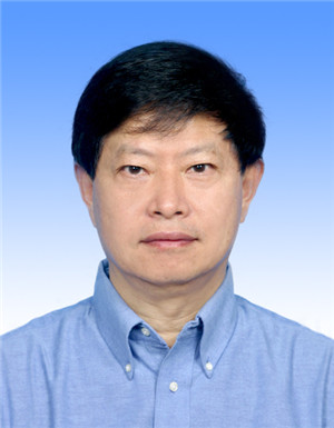 Rongguang Zhang (张荣光)合作论文数National Laboratory of Biomacromolecules, Institute of Biophysics, Chinese Academy of Sciences12Bromberg Raquel合作论文数12Alexei Savchenko合作论文数11Catherine L. Lawson合作论文数11RW SCHEVITZ合作论文数10Yirui Guo合作论文数10
 Qian Cong合作论文数9Marta Orlikowska合作论文数Faculty of Chemistry, University of Gdansk8Aneta Szymańska合作论文数Katedra Chemii Biomedycznej, Wydział Chemii, Uniwersytet Gdański8Jan Konvalinka合作论文数Institute of Organic Chemistry and Biochemistry, Czech Academy of Sciences7
 Frank R. Collart合作论文数Biosciences Division, Argonne National Laboratory7
 Diana R. Tomchick合作论文数Department of Biochemistry, The University of Texas Southwestern Medical Center7Jin-Hui Shen合作论文数6Elina Karimullina合作论文数6Tabitha Emde合作论文数6D L Scott合作论文数Department of Molecular Biophysics and Biochemistry, Yale University, New Haven, Connecticut 06510.6
 George Minasov合作论文数Department of Microbiology-Immunology, Feinberg School of Medicine, Northwestern University6
 Gaya K. Amarasinghe合作论文数Department of Pathology and Immunology, Washington University6Leung Daisy W合作论文数6Jankowska Elżbieta合作论文数Faculty of Chemistry, University of Gdansk6
 Changsoo Chang合作论文数X-Ray Science Division, Argonne National Laboratory;Advanced Photon Source Facility, Argonne National Laboratory6David R. Cooper合作论文数The Cancer Center, University of Virginia6
 Piotr Skowron合作论文数Department of Chemistry, University of Gdansk6
 Christopher F Basler合作论文数Department of Microbiology, Icahn School of Medicine at Mount Sinai6P. Rezacova合作论文数5C. Nakka合作论文数5S Grimshaw合作论文数5R. Rostankowski合作论文数5Janusz Steczko合作论文数5
 Boguslaw Nocek合作论文数Computational Institute Chicago, University of Chicago5M.T. Cymborowski合作论文数4J.T. Bolin合作论文数4A.A. Knapik合作论文数4
 Ronen Marmorstein合作论文数Department of Biochemistry and Biophysics, Perelman School of Medicine, University of Pennsylvania4Ramanan Parameshwaran合作论文数Dept Biochem Biophys & Mol Biol, Iowa State Univ4
 Janusz Petkowski合作论文数Center for Cell Signaling, University of Virginia School of Medicine4
 Jiri Brynda合作论文数4
 Bradley Quade合作论文数Versant NewCo4
 Chad Brautigam合作论文数Department of Biophysics, UT Southwestern Medical Center/Department of Microbiology, UT Southwestern Medical Center4
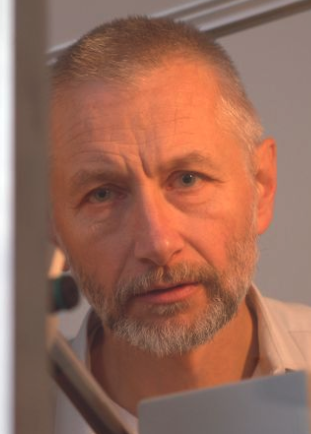 Mariusz Jaskólski合作论文数Department of Crystallography, Faculty of Chemistry, Adam Mickiewicz University, Poznań;Department of Structural Biology of Eukaryotes, Institute of Bioorganic Chemistry, Polish Academy of Sciences4




