Microglia have undergone a fundamental redefinition, transitioning from traditional CNS-restricted scavengers to a highly dynamic and systemically distributed immune lineage. This review synthesizes recent paradigm shifts that challenge long-standing concepts in neuroimmunology. We first discuss the revision of the classical vascular extravasation model by a newly identified integrin-dependent pial surface migration route for embryonic microglial progenitors. Second, we introduce the expanded concept of the “microglial lineage,” which includes transcriptionally and ontogenetically homologous cells residing not only in the CNS but also in peripheral tissues such as the skin, heart, and peripheral nervous system, suggesting broader physiological functions. Third, we highlight the transition from the obsolete M1/M2 polarization model to a microenvironment-driven, dynamic multimodal framework that captures the complex and context-dependent nature of microglial states in health and disease. Finally, we review breakthroughs in regenerative therapy, from pharmacologically induced repopulation to exogenous replacement using iPSC-derived or gene-corrected microglia, offering new hope for genetic microgliopathies such as adult-onset leukoencephalopathy with axonal spheroids and pigmented glia (ALSP). Collectively, these advances position microglia as central mediators of neuro-immune crosstalk and open novel avenues for treating neurodegenerative, neuroinflammatory, and developmental brain disorders.
Once viewed as a homogenous population, microglia are now understood to exist as a dynamic continuum of functionally distinct states whose existence is supported by accumulating evidence from multiple experimental approaches. This heterogeneity, spanning molecular, morphological, and metabolic dimensions, is actively sculpted by brain region, life stage, and a complex microenvironment of neuronal, astrocytic, and systemic signals. In neurological disorders such as Alzheimer’s disease, this plasticity leads to context-dependent diversification into discrete subsets—ranging from protective, phagocytic phenotypes to detrimental, inflammatory ones—that critically influence disease progression. Decoding this heterogeneity through single-cell and spatial omics provides a new pharmacological blueprint: it reveals key druggable nodes (e.g., TREM2, CD14) that govern pathogenic state transitions and informs the rational design of subset-selective delivery systems (e.g., ligand-directed nanocarriers). However, translating these insights requires overcoming persistent challenges, including species differences between mouse and human microglia, a lack of tools for causal subset manipulation, and the integration of spatial with temporal dynamics. By framing microglial heterogeneity as a central targetable axis, this review outlines a pathway for developing precise, state-modulating therapeutics to intervene in neurodegeneration and neuroinflammation.
Central nervous system (CNS) injuries trigger a complex glial response, in which oligodendrocyte precursor cells (OPCs) play a far more dynamic role than previously recognized. Moving beyond their canonical function as a remyelination reservoir, reactive OPCs emerge as plastic signaling hubs whose fate and function are dictated by injury-specific cues. This review synthesizes recent evidence to propose a novel conceptual framework: the "reactive OPC state code." We argue that deciphering this code-the molecular signatures that define pro-regenerative, immunomodulatory, or maladaptive OPC states-is the key to understanding functional heterogeneity in CNS injury. We critically analyze how distinct pathological contexts (trauma, ischemia, neuroinflammation) rewrite this code, leading to diverse outcomes. Finally, we pivot from a generic discussion of OPC-directed therapies to advocate for "state-specific targeting" as the next frontier in translational medicine, offering a roadmap for developing precision interventions that steer reactive OPCs towards repair. This perspective aims to redefine OPC reactivity from a passive response to a central, druggable axis in CNS pathology and repair.
Microglia, the tissue-resident macrophages of the central nervous system (CNS), execute essential functions in neural development, homeostasis, and repair. However, their persistent or dysregulated activation is a hallmark of diverse neurological disorders, where they transition from protective sentinels to drivers of chronic neuroinflammation and tissue damage. This duality has spurred the development of therapeutic strategies aimed not merely at suppressing microglial activity, but at fundamentally resetting the CNS immune landscape. Here, we review the paradigm of transient microglial depletion followed by endogenous repopulation, a strategy that effectively replaces a dysfunctional or pathologically primed microglial compartment with a rejuvenated cohort. Emerging evidence demonstrates that repopulated microglia exhibit a distinct phenotype-characterized by attenuated inflammatory profiles, upregulated homeostatic and neurotrophic gene expression, and enhanced phagocytic capacity-thereby conferring neuroprotection and promoting functional recovery across preclinical models of acute injury, neurodegeneration, and demyelinating disease. We critically evaluate the pharmacological and genetic tools used to achieve depletion, synthesize the context-dependent outcomes across disease spectra, and discuss the mechanistic basis for the superior therapeutic profile of the repopulation phase. Finally, we address the key translational challenges-including therapeutic windows, age- and sex-dependent effects, and the gap between rodent models and human biology-that must be overcome to transform this innovative strategy from a powerful experimental tool into a viable clinical modality for reprogramming CNS immunity.
ABSTRACT As a severe and disabling central nervous system disorder, spinal cord injury (SCI) remains challenging, partly because of the difficulty in addressing secondary injury caused by the blood‐spinal cord barrier (BSCB) disruption. As the neurovascular unit's crucial component, the BSCB regulates the homeostasis of the spinal cord. Inspired by the established protective effect of H2S in pan‐vascular pathologies, we engineered an intravenously administered nanoparticle SPRC@MPDA‐RGD. By utilizing the overexpression of αvβ3 integrin on endothelial cells after SCI, the functionalized peptide c(RGDyK) can guide SPRC@MPDA‐RGD for precise delivery to the BSCB. The MPDA scaffold has the ability to both deliver S‐propargyl‐cysteine (SPRC) and scavenge reactive oxygen species (ROS). Subsequently, the release of SPRC upregulates cystathionine γ‐lyase (CSE) and stimulates endogenous H2S production in injured endothelial cells, thereby protecting the BSCB. We also investigated the biological mechanisms underlying the therapeutic effects of SPRC@MPDA‐RGD. The production of H2S in endothelial cells activates the PI3K/Akt/mTOR pathway, which subsequently suppresses ferritinophagy, reduces ferritin degradation, and ultimately suppresses ferroptosis. In summary, our work proposes a nanotherapeutic strategy that coordinates H2S production and ROS scavenging to inhibit ferritinophagy, thereby promoting BSCB repair, showing significant potential in promoting SCI treatment.
The primary culture of oligodendrocyte progenitor cells (OPCs) provides an indispensable tool for characterizing their biological properties and myelin repair potential. However, the current OPC preparation methods are mainly limited to rat tissues, and it remains a substantial challenge for replicating the primary culture from mouse tissues to generate large quantities of high-quality OPCs. Here, we describe a protocol to successfully establish highly enriched OPC cultures from the cerebral cortex of mice at the age of neonatal 3 days. OPCs were isolated and purified from the bed layer of astrocytes by shaking for 6 h at 250 rpm. Using this protocol, mouse OPCs can be easily produced in bulk and economically without the need for specific cell-surface antibodies and equipment. These mouse OPC cultures were identified by immunocytochemical, immunobloting and RNA-seq analysis. Furthermore, they could be expanded in vitro and differentiate into mature oligodendrocytes. We propose this method as a viable and affordable protocol to obtain mouse OPC culture, which should significantly facilitate studies on OPC lineage progression and their application in myelin-related disease modeling and regenerative medicine.
Traditionally, oligodendrocyte precursor cells (OPCs) were primarily regarded for their differentiation potential to mature oligodendrocytes that ensheath central nervous system (CNS) axons through myelin formation. Recent breakthroughs in single-cell sequencing and in vivo imaging technologies have revolutionized our understanding, revealing that OPCs engage in extensive dynamic interactions with diverse CNS cell populations during neurodevelopment, tissue homeostasis maintenance, and pathological microenvironment remodeling. Notably, while OPCs exhibit relatively conserved phenotypic signatures, their functional plasticity within heterogeneous microenvironments demonstrates significant spatial specificity and disease-context dependence. In this review, we will systematically sort out the molecular interaction mechanism between OPCs and neurons, astrocytes, microglia, and vascular endothelial cells, deeply analyze their dynamic functional profiles, and focus on discussing: (1) the fine-tuning regulatory model of neuronal circuits mediated by OPCs at the developmental stage (2) the bidirectional regulatory mechanism of OPCs involved in maintaining the metabolic-immune balance under homeostasis; (3) OPC functional reprogramming in the pathological process of multiple sclerosis, cerebral ischemia, etc. This review aims to consolidate current evidence into a cohesive perspective on OPC multimodal functions, evaluate non-myelinating contributions, and discuss promising therapeutic targets for neural regenerative medicine.
The axon-wrapping myelin sheath is essential for CNS function. Myelination defects occur in various neurodevelopmental disorders, but the underlying mechanism remains poorly understood. Human solute carrier 44A1 (SLC44A1) deficiency causes a new type of childhood-onset neurodegeneration with cerebellar atrophy and leukoencephalopathy that lacks effective treatment. Here, we show that SLC44A1 is enriched in oligodendrocytes and is required for myelin development in the CNS of zebrafish and rodents. In vivo time-lapse imaging of Slc44a1b-deficient zebrafish reveals impaired oligodendroglial maturation and myelinogenesis. Mechanistically, SLC44A1 deficiency disrupts the expression of genes involved in the phosphatidylcholine production pathway and subsequently inhibits phospholipid biosynthesis and disturbs the lipid composition of myelin sheaths. More importantly, supplementation with citicoline, a natural choline metabolite, restores developmental myelination in SLC44A1-deficient animals. Our findings demonstrate that SLC44A1 is essential for CNS myelination, and citicoline supplementation represents a potential therapy for developmental hypomyelination.
Neuronal reprogramming is an innovative technique for converting non-neuronal somatic cells into neurons that can be used to replace lost or damaged neurons, providing a potential effective therapeutic strategy for central nervous system (CNS) injuries or diseases. Transcription factors have been used to induce neuronal reprogramming, while their reprogramming efficiency is relatively low, and the introduction of exogenous genes may result in host gene instability or induce gene mutation. Therefore, their future clinical application may be hindered by these safety concerns. Compared with transcription factors, small-molecule compounds have unique advantages in the field of neuronal reprogramming, which can overcome many limitations of traditional transcription factor-induced neuronal reprogramming. Here, we review the recent progress in the research of small-molecule compound-mediated neuronal reprogramming and its application in CNS regeneration and repair.
Rationale: As a highly plastic population, parenchymal astrocytes have demonstrated the capacity to become activated and recapitulate neurogenic potential in response to traumatic central nervous system (CNS) injuries, representing a latent reservoir for neuronal regeneration in non-neurogenic brain regions. However, the extrinsic and intrinsic factors regulating this process remain poorly characterized. Elucidating these molecular mechanisms is crucial to harnessing the regenerative potential of reactive astrocytes in CNS repair. Methods: A multidisciplinary approach combining immunostaining, western blotting, RNA interference (RNAi), gene knock out and fate-mapping was used to investigate the role of topoisomerase IIα (TOP2a) in regulation of the stemness response in reactive astrocytes to traumatic brain injury (TBI). Results: Both in vitro and in vivo analyses demonstrated that TBI induces a stem cell-like response in reactive astrocytes concomitant with TOP2a upregulation. Pharmacological inhibition or genetic deletion of TOP2a significantly attenuated this stemness response. Neurosphere culture assay indicates that TOP2a might act as a downstream factor of Sonic Hedgehog (SHH) signaling to mediate the acquisition of stem cell potential. Conclusions: This study identifies TOP2a as a pivotal intrinsic regulator of astrocytic stem cell potential in the injured brain, which will advance our understanding of the molecular underpinnings of the stem cell response and its therapeutic application in neural regeneration.
In the mammalian central nervous system (CNS), astrocytes are the ubiquitous glial cells that have complex morphological and molecular characteristics. These fascinating cells play essential neurosupportive and homeostatic roles in the healthy CNS and undergo morphological, molecular, and functional changes to adopt so-called ‘reactive’ states in response to CNS injury or disease. In recent years, interest in astrocyte research has increased dramatically and some new biological features and roles of astrocytes in physiological and pathological conditions have been discovered thanks to technological advances. Here, we will review and discuss the well-established and emerging astroglial biology and functions, with emphasis on their potential as therapeutic targets for CNS injury, including traumatic and ischemic injury. This review article will highlight the importance of astrocytes in the neuropathological process and repair of CNS injury.
INTRODUCTION:Ectopic expression of transcription factor-mediated in vivo neuronal reprogramming provides promising strategy to compensate for neuronal loss, while its further clinical application may be hindered by delivery and safety concerns. As a novel and attractive alternative, small molecules may offer a non-viral and non-integrative chemical approach for reprogramming cell fates. Recent definitive evidences have shown that small molecules can convert non-neuronal cells into neurons in vitro. However, whether small molecules alone can induce neuronal reprogramming in vivo remains largely unknown. OBJECTIVES:To identify chemical compounds that can induce in vivo neuronal reprogramming in the adult spinal cord. METHODS:Immunocytochemistry, immunohistochemistry, qRT-PCR and fate-mapping are performed to analyze the role of small molecules in reprogramming astrocytes into neuronal cells in vitro and in vivo. RESULTS:By screening, we identify a chemical cocktail with only two chemical compounds that can directly and rapidly reprogram cultured astrocytes into neuronal cells. Importantly, this chemical cocktail can also successfully trigger neuronal reprogramming in the injured adult spinal cord without introducing exogenous genetic factors. These chemically induced cells showed typical neuronal morphologies and neuron-specific marker expression and could become mature and survive for more than 12 months. Lineage tracing indicated that the chemical compound-converted neuronal cells mainly originated from post-injury spinal reactive astrocytes. CONCLUSION:Our proof-of-principle study demonstrates that in vivo glia-to-neuron conversion can be manipulated in a chemical compound-based manner. Albeit our current chemical cocktail has a lowreprogramming efficiency, it will bring in vivo cell fate reprogramming closer to clinical application in brain and spinal cord repair. Future studies should focus on further refining our chemical cocktail and reprogramming approach to boost the reprogramming efficiency.
Background: Direct reprogramming of astrocytes into neurons opens up a new avenue for neuroregenerative medicine. However, the poor understanding of the molecular mechanisms underpinning the latent neurogenic program in astrocytes has largely restricted this strategy towards safe and effective clinical therapies. Methods: Immunocytochemistry, immunohistochemistry, western blotting, qRT-PCR, gene knockdown and fate-mapping are performed to analyze the role of NOTCH1 signaling in regulation of the latent neurogenic program in reactive astrocytes after spinal cord injury. Results: Western blotting analysis highlights that NOTCH1 is a key signaling mediating Ascl1- and Neurog2-driven astrocyte-to-neuron conversion. Inhibition of NOTCH1 signaling in cultured astrocytes by shRNA or DAPT (a NOTCH1 inhibitor) is sufficient to reprogram them into neurons by upregulating the expression of pro-neural transcription factors, including NeuroD1, NeuroD2, Pax6, Lmx1a and Lhx6. In the spinal cord of adult mouse, the expression of Notch1 is detected in resident astrocytes, which was significantly increased after spinal cord injury (SCI). Genetical knockdown of NOTCH1 signaling alone successfully triggers endogenous reactive astrocytes reprogramming into neurons in the injured adult spinal cord. Importantly, pharmacologically blocking NOTCH1 signaling with small molecule DAPT alone can also induce in situ astrocyte-to-neuron conversion after SCI. Conclusions: We identify NOTCH1 as a key common signaling pathway in reactive astrocyte that provides a barrier for cell fate conversion. This proof-of-principle study will significantly expand our molecular understanding of astroglial-lineage reprogramming and overcoming the NOTCH1 gatekeeper with small molecules may provide a transgene-free approach for in vivo chemical neuronal reprogramming with potential clinical application in neuroregeneration.
发育神经生物学作为神经科学的一个重要分支,随着分子生物学的深入研究越来越受到人们的重视.多年来,海军军医大学神经生物学教研室一直致力于神经发育相关内容的教学,在实践中不断总结经验和改进方法,为培养高素质医学、生物学人才发挥着重要力量.文章将分析发育神经生物学教学中的现状和问题,并总结和阐述一系列针对措施,以期获得更高的教学质量,推进基础医学教育的改革与发展.
Topoisomerase IIA (TOP2a) has traditionally been known as an important nuclear enzyme that resolves entanglements and relieves torsional stress of DNA double strands. However, its function in genomic transcriptional regulation remains largely unknown, especially during adult neurogenesis. Here, we show that TOP2a is preferentially expressed in neurogenic niches in the brain of adult mice, such as the subventricular zone (SVZ). Conditional knockout of Top2a in adult neural stem cells (NSCs) of the SVZ significantly inhibits their self-renewal and proliferation, and ultimately reduces neurogenesis. To gain insight into the molecular mechanisms by which TOP2a regulates adult NSCs, we perform RNA-sequencing (RNA-Seq) plus chromatin immunoprecipitation sequencing (ChIP-Seq) and identify ubiquitin-specific protease 37 (Usp37) as a direct TOP2a target gene. Importantly, overexpression of Usp37 is sufficient to rescue the impaired self-renewal ability of adult NSCs caused by Top2a knockdown. Taken together, this proof-of-principle study illustrates a TOP2a/Usp37-mediated novel molecular mechanism in adult neurogenesis, which will significantly expand our understanding of the function of topoisomerase in the adult brain.
Glioblastoma (GBM) is the most lethal primary tumor in the human brain and lacks favorable treatment options. Sex differences in the outcome of GBM are broadly acknowledged, but the underlying molecular mechanisms remain largely unknown. To identify the sex-dependent critical genes in the progression of GBM, raw data from several microarray datasets with the same array platform were downloaded from the Gene Expression Omnibus (GEO) database. These datasets included tumorous and normal tissue from patients with GBM and crucial sex features. Then, the differentially expressed genes (DEGs) in female and male tumors were identified via bioinformatics analysis, respectively. Functional signatures of the identified DEGs were further annotated by Gene Ontology (GO) and pathway enrichment analyses. Venn diagram and functional protein-protein interaction (PPI) network analyses were performed to screen out the sex-specific DEGs. Survival analysis of patients with differences in the expression level of selected genes was then carried out using the data from The Cancer Genome Atlas (TCGA). Here, we showed that ECT2, AURKA, TYMS, CDK1, NCAPH, CENPU, OIP5, KIF14, ASPM, FBXO5, SGOL2, CASC5, SHCBP1, FN1, LOX, IGFBP3, CSPG4, and CD44 were enriched in female tumor samples, whereas TNFSF13B, CXCL10, CXCL8, CXCR4, TLR2, CCL2, and FCGR2A were enriched in male tumor samples. Among these key genes, interestingly, ECT2 was associated with increased an survival rate for female patients, whileTNFSF13B could be regarded as a potential marker of poor prognosis in male patients. These results suggested that sex differences in patients may be attributed to the heterogeneous gene activity, which might influence the oncogenesis and the outcomes of GBM.
NG2-glia are a major type of glial cells that are widely distributed in the central nervous system (CNS). Under physiological conditions, they mainly differentiate into oligodendrocytes and contribute to the myelination of axons, so they are generally called oligodendrocyte progenitor cells. Emerging evidence suggests that NG2-glia not only act as the precursors of oligodendrocytes but also possess many other biological properties and functions. For example, NG2-glia can form synapse with neurons and participate in energy metabolism and immune regulation. Under pathological conditions, NG2-glia can also differentiate into astrocytes, Schwann cells and even neurons, which are involved in CNS injury and repair. Therefore, a deeper understanding of the biological characteristics and functions of NG2-glia under physiological and pathological conditions will be helpful for the treatment of CNS injury and disease. This article reviews the recent advances in the biological characteristics and functions of NG2-glia.
The adult CNS has poor ability to replace degenerated neurons following injury or disease. Recently, direct reprogramming of astrocytes into induced neurons has been proposed as an innovative strategy toward CNS repair. As a cell population that shows high diversity on physiological properties and functions depending on their spatiotemporal distribution, however, whether the astrocyte heterogeneity affect neuronal reprogramming is not clear. Here, we show that astrocytes derived from cortex, cerebellum, and spinal cord exhibit biological heterogeneity and possess distinct susceptibility to transcription factor-induced neuronal reprogramming. The heterogeneous expression level of NOTCH1 signaling in the different CNS regions-derived astrocytes is shown to be responsible for the neuronal reprogramming diversity. Taken together, our findings demonstrate that region-restricted astrocytes reveal different intrinsic limitation of the response to neuronal reprogramming.
Direct conversion of readily available non-neural cells from patients into induced neurons holds great promise for neurological disease modeling and cell-based therapy. Olfactory ensheathing cells (OECs) is a unique population of glia in olfactory nervous system. Based on the regeneration-promoting properties and the relative clinical accessibility, OECs are attracting increasing attention from neuroscientists as potential therapeutic agents for use in neural repair. Here, we report that OECs can be directly, rapidly and efficiently reprogrammed into neuronal cells by the single transcription factor Neurogenin 2 (NGN2). These induced cells exhibit typical neuronal morphologies, express multiple neuron-specific markers, produce action potentials, and form functional synapses. Genome-wide RNA-sequencing analysis shows that the transcriptome profile of OECs is effectively reprogrammed towards that of neuronal lineage. Importantly, these OEC-derived induced neurons survive and mature after transplantation into adult mouse spinal cords. Taken together, our study provides a direct and efficient strategy to quickly obtain neuronal cells from adult OECs, suggestive of promising potential for personalized disease modeling and cell replacement-mediated therapeutic approaches to neurological disorders.
 Cheng He (何成)合作论文数Naval Medical University37Yimin Yuan合作论文数31
 Shangyao Qin合作论文数23Yanling Zhu合作论文数11Zijian Tan合作论文数10Xiao Huang合作论文数10Pu Yingyan合作论文数10
 Hu Xin合作论文数9Yang Qiu合作论文数8Li CAO合作论文数8Yakun Gu合作论文数7Wang Dan合作论文数7Xueyan Cheng合作论文数5Gao Liang合作论文数5Xiu-Jie Liu (刘秀杰)合作论文数5Sun Xiu合作论文数4
 Zhihui Huang (黄智慧)合作论文数School of Pharmacy, Hangzhou Normal University4Aijun Huang合作论文数4
 Jingjing Chen合作论文数School of Medicine, Shanghai JiaoTong University4Feng Zhu合作论文数3
 Hong Liu (柳红)合作论文数University of Chinese Academy of Sciences;Shanghai Institute of Materia Medica, Chinese Academy of Sciences;Nanjing University of Chinese Medicine3Xiang Chen合作论文数2Ying Wang合作论文数2Zheng-hao LI (李政灏)合作论文数2Yan Lu合作论文数2Yuhua Zou合作论文数2Chao Huang合作论文数2
 Wenhao Zhou (周文浩)合作论文数Affiliated Women and Children's Medical Center, Guangzhou Medical University2Shao Qi合作论文数2
 Xiaofeng Lian (连小峰)合作论文数Shanghai Sixth People's Hospital Affiliated to Shanghai Jiao Tong University School of Medicine2
 Chun-Li Zhang合作论文数Graduate School of Biomedical Sciences, UT Southwestern Medical Center;Department of Molecular Biology, UT Southwestern Medical Center;Center for Regenerative Science and Medicine, UT Southwestern Medical Center2
 Xiaofei Gao (高霄飞)合作论文数Shanghai Fourth People's Hospital Affiliated to Tongji University;School of Medicine, Tongji University2
 Xiaobing Yuan (袁小兵)合作论文数Shanghai Key Laboratory of Brain Functional Genomics;East China Normal University2
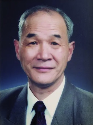 Yizhang Chen (陈宜张)合作论文数Tongji University2
 Mengsheng Qiu (邱猛生)合作论文数Hangzhou Normal University2Lifang Mu合作论文数2Yinyan Pu合作论文数1Mengjia Chen合作论文数1Yuanyuan Li合作论文数1Kefu Chen合作论文数1Jian Wang合作论文数1Yi An合作论文数1Qirong Zhou合作论文数1Wenze Niu合作论文数1Wen-Jing Qian合作论文数1Dazhi Guo合作论文数1Jianfeng Zhou合作论文数1Tao Liu合作论文数1




