Vascular landmark detection plays an important role in medical analysis and clinical treatment. However, due to the complex topology and similar local appearance around landmarks, the popular heatmap regression based methods always suffer from the landmark confusion problem. Vascular landmarks are connected by vascular segments and have special spatial correlations, which can be utilized for performance improvement. In this paper, we propose a multi-task global optimization-based framework for accurate and automatic vascular landmark detection. A multi-task deep learning network is exploited to accomplish landmark heatmap regression, vascular semantic segmentation, and orientation field regression simultaneously. The two auxiliary objectives are highly correlated with the heatmap regression task and help the network incorporate the structural prior knowledge. During inference, instead of performing a max-voting strategy, we propose a global optimization-based post-processing method for final landmark decision. The spatial relationships between neighboring landmarks are utilized explicitly to tackle the landmark confusion problem. We evaluated our method on a cerebral MRA dataset with 564 volumes, a cerebral CTA dataset with 510 volumes, and an aorta CTA dataset with 50 volumes. The experiments demonstrate that the proposed method is effective for vascular landmark localization and achieves state-of-the-art performance.
Vascular structure segmentation plays a crucial role in medical analysis and clinical applications. The practical adoption of fully supervised segmentation models is impeded by the intricacy and time-consuming nature of annotating vessels in the 3D space. This has spurred the exploration of weakly-supervised approaches that reduce reliance on expensive segmentation annotations. Despite this, existing weakly supervised methods employed in organ segmentation, which encompass points, bounding boxes, or graffiti, have exhibited suboptimal performance when handling sparse vascular structure. To alleviate this issue, we employ maximum intensity projection (MIP) to decrease the dimensionality of 3D volume to 2D image for efficient annotation, and the 2D labels are utilized to provide guidance and oversight for training 3D vessel segmentation model. Initially, we generate pseudo-labels for 3D blood vessels using the annotations of 2D projections. Subsequently, taking into account the acquisition method of the 2D labels, we introduce a weakly-supervised network that fuses 2D-3D deep features via MIP to further improve segmentation performance. Furthermore, we integrate confidence learning and uncertainty estimation to refine the generated pseudo-labels, followed by fine-tuning the segmentation network. Our method is validated on five datasets (including cerebral vessel, aorta and coronary artery), demonstrating highly competitive performance in segmenting vessels and the potential to significantly reduce the time and effort required for vessel annotation. Our code is available at: https://github.com/gzq17/Weakly-Supervised-by-MIP.
Anatomical landmark detection plays an important role in cerebrovascular analysis and clinical treatment. However, due to the complex structure and similar local appearance around landmarks, the popular heatmap regression based methods suffer from the landmark confusion problem. In this work, we propose an adversarial learning framework for cerebrovascular landmark detection in MRA images by leveraging cross-modality information. Specifically, we exploit an unpaired large-scale CTA dataset to complement the limited MRA training data. The generator is modified as a U-Net based heatmap regression network, and the discriminator is trained using both MRA and CTA datasets to distinguish between multi-channel heatmap groundtruth and prediction. A relative coordinate matrix and a distance map are introduced to enhance landmark location distribution. Extensive experiments demonstrate the superior and robust performance of our method, even with very limited MRA training data.
Aortic dissection (AD) is a dangerous disease usually diagnosed by computed tomography angiography. Segmentation of true and false lumens of aortic trunk and major branches is very important for the diagnosis and treatment of this disease. In this paper, we proposed a fully automatic vessel analysis algorithm for dissected aorta, which can output centerlines, true lumen, and false lumen of trunk and major branches, and perfusion source of branches. In our experiment, the mean dice similarity coefficient (DSC) of true lumen segmentation was 0.939 for trunk and 0.912 for branch while the mean DSC of whole lumen segmentation was 0.974 for trunk and 0.937 for branch, and the classification accuracy of branch perfusion source was 0.863.
Anatomical landmark detection has important applications in cerebrovascular analysis and clinical treatments, which is challenging due to the complex structure, various natural variations and pathological changes. In this paper, we propose a multi-task deep learning network for accurate detection of 19 landmarks in cerebral Magnetic Resonance Angiography (MRA) images, which is robust to anatomical variations. Besides landmark detection, the network is trained to perform landmark attribute classification, semantic artery segmentation and arterial segment attribute classification simultaneously. The attributes of landmark and arterial segment are defined as local bifurcation appearance and absence variation, respectively, which enhances the contextual information and incorporates the structural prior knowledge explicitly. Experiments on both public and private datasets demonstrate the superior performance of the proposed method.
Airway semantic segmentation, which refers to segmenting airway from background and dividing it into anatomical segments, provides clinically valuable information for lung lobe analysis, pulmonary lesion localization, and comparison between different patients. It is technically challenging due to the complicated tree-like structure, individual variations, and severe class imbalance. We propose a structure-aware graph-based network (SGNet) for airway semantic segmentation directly from chest CT scans. The proposed framework consists of a feature extractor combining a multi-task U-Net with a structure-aware GCN, and an inference module comprised of two convolutional layers. The multi-task U-Net is trained to regress bifurcation landmark heatmaps, binary and semantic segmentation maps simultaneously, providing initial predictions for graph construction. By introducing irregular edges connecting voxels with the sampled points around corresponding bifurcation landmarks, the two-layer GCN incorporates the structural prior explicitly. Experiments on both public and private datasets demonstrate that the SGNet achieves superior and robust performance, even on subjects affected by severe pulmonary diseases.
Bifurcation landmark detection is an important step in automatic analysis of anatomical tree structures, such as airway and vessel. In this paper, we propose a multi-task learning based deep neural network specifically designed for automatically and accurately localizing bifurcation landmarks. Given an input volume, the network is trained to predict landmark confidence maps, branch segmentation maps, and branch orientation fields simultaneously. In this way, we exploit the spatial relationships among landmarks explicitly. The structural prior is also introduced to the architecture, which guides the network to learn more discriminative features. Experiments on airway tree and aorta tree demonstrate that the proposed method is effective for bifurcation landmark detection, and exploiting segmentation and orientation field regression as auxiliary objectives helps to increase accuracy of landmark detection substantially.
Early detection of COVID-19 based on chest CT enables timely treatment of patients and helps control the spread of the disease. We proposed an artificial intelligence (AI) system for rapid COVID-19 detection and performed extensive statistical analysis of CTs of COVID-19 based on the AI system. We developed and evaluated our system on a large dataset with more than 10 thousand CT volumes from COVID-19, influenza-A/B, non-viral community acquired pneumonia (CAP) and non-pneumonia subjects. In such a difficult multi-class diagnosis task, our deep convolutional neural network-based system is able to achieve an area under the receiver operating characteristic curve (AUC) of 97.81% for multi-way classification on test cohort of 3,199 scans, AUC of 92.99% and 93.25% on two publicly available datasets, CC-CCII and MosMedData respectively. In a reader study involving five radiologists, the AI system outperforms all of radiologists in more challenging tasks at a speed of two orders of magnitude above them. Diagnosis performance of chest x-ray (CXR) is compared to that of CT. Detailed interpretation of deep network is also performed to relate system outputs with CT presentations. The code is available at https://github.com/ChenWWWeixiang/diagnosis_covid19 .
Automatic anatomical landmark detection is beneficial to many other medical image analysis tasks. In this paper, we propose a two-stage cascade regression model to make coarse-to-fine landmark detection. Specifically, in the first stage, a Gaussian heatmap regression model customized from U-Net is exploited to make primary prediction, which takes the downsampled entire image as input. In the second stage, we develop a CNN to regress displacements from the primary prediction to the landmarks, using patches in original resolution centered at the previous localization as input. Owing to the different sizes and resolutions of inputs in two stages, the global context information and local appearance can be integrated by our algorithm. The spacial relationships among landmarks can also be exploited by predicting all the landmarks simultaneously. In evaluation on the coronary and aorta CTA images, we show that our proposed method is widely applicable and delivers state-of-the-art performance even with limited training data.
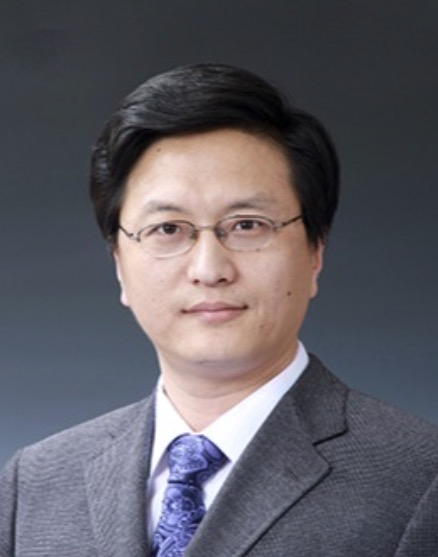 Jie Zhou (周杰)合作论文数Department of Automation, Tsinghua University9
 Jianjiang Feng (冯建江)合作论文数Department of Automation, Tsinghua University9
 Wangsheng Lu合作论文数3Guangming Yang (g yang)合作论文数3Yukun Cao合作论文数Department of Radiology, Union Hospital, Tongji Medical College, Huazhong University of Science and Technology2
 Heshui Shi (史河水)合作论文数Union Hospital, Tongji Medical College, Huazhong University of Science and Technology2Zhanqiang Guo合作论文数1Jinyang Yu合作论文数1Jia Liu合作论文数1Guozhu Shao合作论文数1
 Xin Zhang合作论文数1
 Zhanwei Xu合作论文数Department of Automation, School of Information Science and Technology, Tsinghua University1Yongjie Duan合作论文数1
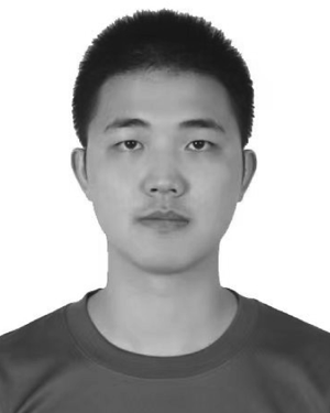 Weixiang Chen合作论文数Tsinghua University;1
 Lei Deng (邓磊)合作论文数Institute of Instrument Science and Technology, Department of Precision Instrument, Tsinghua University;Center for Brain Inspired Computing Research, Tsinghua University1
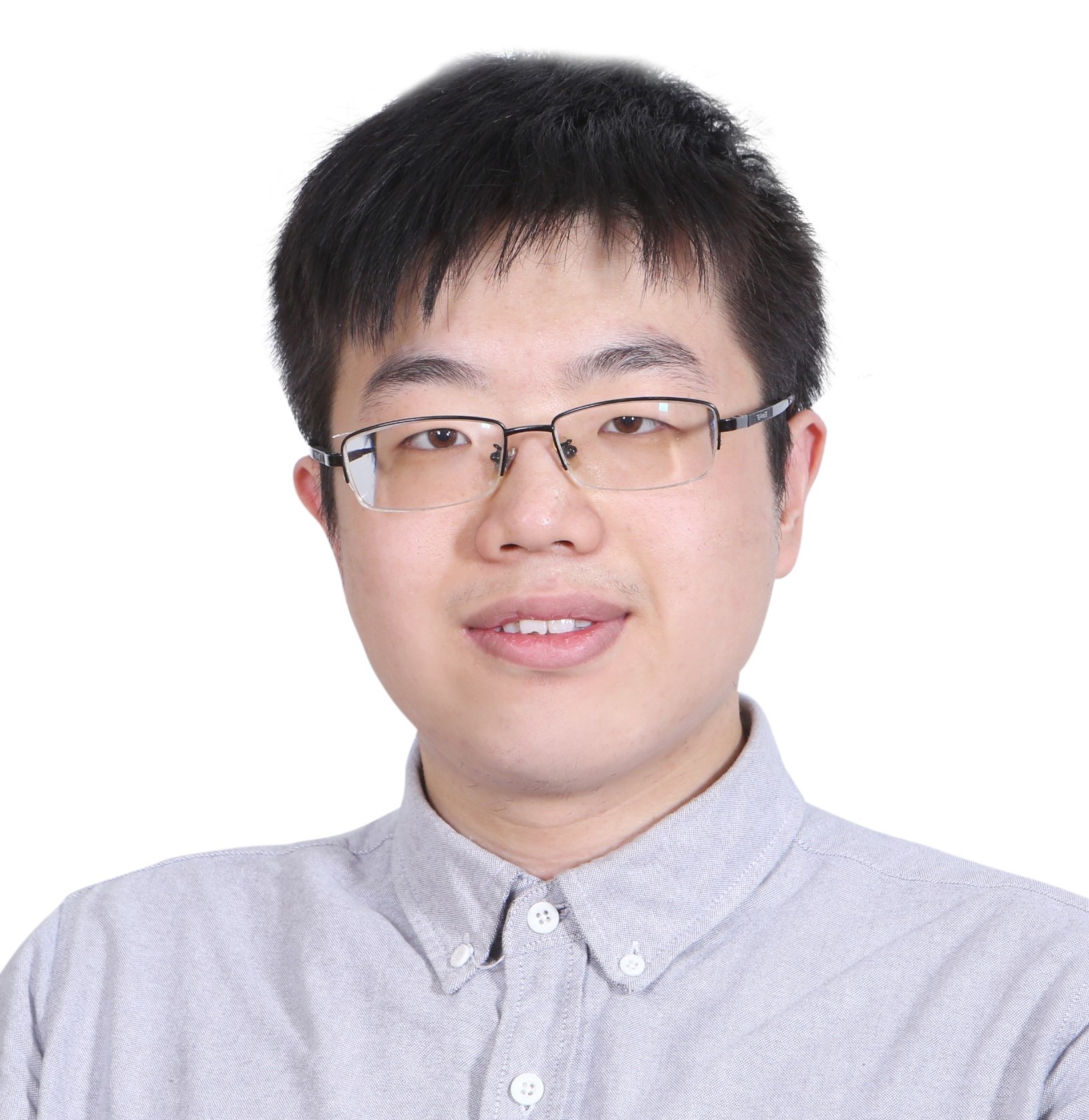 Ziyi Wu (吴紫屹)合作论文数Toronto Intelligent Systems Lab, University of Toronto;Google DeepMind1
 Chuansheng Zheng (郑传胜)合作论文数Union Hospital, Tongji Medical College, Huazhong University of Science and Technology1Guoqiang Zhan合作论文数1




