Neurons are the primary cells responsible for information processing in the central nervous system (CNS). However, they are vulnerable to damage and insult in a variety of neurological disorders. As the most abundant glial cells in the brain, astrocytes provide crucial support to neurons and participate in synapse formation, synaptic transmission, neurotransmitter recycling, regulation of metabolic processes, and the maintenance of the blood-brain barrier integrity. Though astrocytes play a significant role in the manifestation of injury and disease, they do not work in isolation. Cellular interactions between astrocytes and neurons are essential for maintaining the homeostasis of the CNS under both physiological and pathological conditions. In this review, we explore the diverse interactions between astrocytes and neurons under physiological conditions, including the exchange of neurotrophic factors, gliotransmitters, and energy substrates, and different CNS diseases such as Alzheimer’s disease, Parkinson’s disease, stroke, traumatic brain injury, and multiple sclerosis. This review sheds light on the contribution of astrocyte-neuron crosstalk to the progression of neurological diseases to provide potential therapeutic targets for the treatment of neurological diseases.
Inflammation and neuronal apoptosis play a key role in traumatic brain injury (TBI). Quercetin (Que) has been shown to exhibit a neuroprotective effect after TBI, but the underlying molecular mechanism remains unclear. In this study, We established a weight-drop mouse model to illustrate the effects of Que on microglial-induced inflammation in TBI. Mice were divided into four groups: the Sham group, TBI group, TBI+vehicle +vehicle group, and TBI+Que +Que group. The TBI+Que +Que group was treated with Que 30 min after TBI. Brain water content, neurological score, and neuronal apoptosis were measured. Western blotting, TUNEL staining, Nissl staining, quantitative polymerase chain reaction, and immunofluorescence staining were performed to assess the activation of the PGC1 alpha/Nrf2 pathway and nuclear translocation of HDAC3 with Que treatment. The results showed that Que administration alleviated TBI-induced neurobehavioral deficits, encephaledema, and neuron apoptosis. Que also restrained TBI-induced microglial activity and the subsequent expression of the inflammatory factor in the contusion cortex. Moreover, Que treatment activated the PGC-1 alpha/Nrf2 pathway, attributable to the inhibition of HDAC3 translocation to the nucleus. Overall, these results reveal the role of Que in protecting against TBIinduced neuroinflammation and promoting neurological functional recovery, which is achieved through the negative regulation of HDAC3.
Inflammation and neuronal apoptosis play a key role in traumatic brain injury (TBI). Quercetin (Que) has been shown to exhibit a neuroprotective effect after TBI, but the underlying molecular mechanism remains unclear. In this study, We established a weight-drop mouse model to illustrate the effects of Que on microglial-induced inflammation in TBI. Mice were divided into four groups: the Sham group, TBI group, TBI+vehicle group, and TBI+Que group. The TBI+Que group was treated with Que 30min after TBI. Brain water content, neurological score, and neuronal apoptosis were measured. Western blotting, TUNEL staining, Nissl staining, quantitative polymerase chain reaction, and immunofluorescence staining were performed to assess the activation of the PGC-1α/Nrf2 pathway and nuclear translocation of HDAC3 with Que treatment. The results showed that Que administration alleviated TBI-induced neurobehavioral deficits, encephaledema, and neuron apoptosis. Que also restrained TBI-induced microglial activity and the subsequent expression of the inflammatory factor in the contusion cortex. Moreover, Que treatment activated the PGC-1α/Nrf2 pathway, attributable to the inhibition of HDAC3 translocation to the nucleus. Overall, these results reveal the role of Que in protecting against TBI-induced neuroinflammation and promoting neurological functional recovery, which is achieved through the negative regulation of HDAC3.
Ischemic stroke is one of the most significant causes of morbidity and mortality worldwide. However, there is a dearth of effective drugs and treatment methods for ischemic stroke. Significant numbers of circular RNAs (circRNAs) exhibit abnormal expression following ischemic stroke and are considered potential therapeutic targets. CircRNAs have emerged as promising biomarkers due to their stable expression in peripheral blood and their potential significance in ischemic stroke diagnosis and prognosis. This review provides a summary of 31 circRNAs involved in the pathophysiological processes of apoptosis, autophagy, inflammation, oxidative stress, and angiogenesis following ischemic stroke. Furthermore, we discuss the mechanisms of action of said circRNAs and their potential clinical applications. Ultimately, circRNAs exhibit promise as both therapeutic targets and biomarkers for ischemic stroke.
Astrocytes activate and crosstalk with neurons influencing inflammatory responses following ischemic stroke. The distribution, abundance, and activity of microRNAs in astrocytes-derived exosomes after ischemic stroke remains largely unknown. In this study, exosomes were extracted from primary cultured mouse astrocytes via ultracentrifugation, and exposed to oxygen glucose deprivation/re‑oxygenation injury to mimic experimental ischemic stroke. SmallRNAs from astrocyte-derived exosomes were sequenced, and differentially expressed microRNAs were randomly selected and verified by stem-loop real time quantitative polymerase chain reaction. We found that 176 microRNAs, including 148 known and 28 novel microRNAs, were differentially expressed in astrocyte-derived exosomes following oxygen glucose deprivation/re‑oxygenation injury. In gene ontology enrichment, Kyoto encyclopedia of genes and genomes pathway analyses, and microRNA target gene prediction analyses, these alteration in microRNAs were associated to a broad spectrum of physiological functions including signaling transduction, neuroprotection and stress responses. Our findings warrant further investigating of these differentially expressed microRNAs in human diseases particularly ischemic stroke.
Microglia, resident brain immune cells, is critical in inflammation, apoptosis, neurogenesis and neurological recovery during cerebral ischemia/reperfusion (I/R) injury. Mesencephalic astrocyte-derived neurotrophic factor (MANF), a novel identified endoplasmic reticulum stress-inducible neurotrophic factor, can alleviate I/R injury by reducing the inflammatory reaction, but its specific regulatory mechanism on microglia after ischemic stroke has not been fully clarified. To mimic the process of ischemia/reperfusion in vivo and in vitro, middle cerebral artery occlusion/reperfusion (MCAO/R) was induced in C57BL/6J mice and oxygen glucose deprivation/reoxygenation (OGD/R) model was established in BV-2 cells. Moreover, MANF small interfering RNA (siRNA) was used to silence the expression of endogenous MANF, while recombination human MANF protein (rhMANF) acted as an exogenous supplement. Seventy-two hours after MCAO/R, 2,3,5-triphenyltetrazolium staining, neurological scores, brain water content, immunohistochemical staining, immunofluorescent staining, flow cytometry, hematoxylin and eosin staining, quantitative real-time PCR and western blot are applied to evaluate the protective effect and possible mechanism of MANF on cerebral I/R injury. In vitro, cell viability, inflammatory cytokines and the expression of MANF, A20, NF-κB and the markers of microglia were analyzed. The results showed that MANF decreased brain infarct volume, neurological scores, and brain water content. In addition, MANF promoted the polarization of microglia to an anti-inflammatory phenotype both in vivo and in vitro, which are related to A20/NF-κB pathway. In summary, MANF may offer novel therapeutic approaches for ischemic stroke in the process of microglia polarization.
Stroke has become the most disabling and the second most fatal disease in the world. It has been a top priority to reveal the pathophysiology of stroke at cellular and molecular levels. A large number of long non-coding RNAs (lncRNAs) are identified to be abnormally expressed after stroke. Here, we summarize 35 lncRNAs associated with stroke, and clarify their functions on the prognosis through signal transduction and predictive values as biomarkers. Changes in the expression of these lncRNAs mediate a wide range of pathological processes in stroke, including apoptosis, inflammation, angiogenesis, and autophagy. Based on the exploration of the functions and mechanisms of lncRNAs in stroke, more timely, accurate predictions and more effective, safer treatments for stroke could be developed.
T helper 17 (Th17) cells have been involved in the pathogenesis of many autoimmune and inflammatory diseases, like psoriasis, multiple sclerosis (MS), rheumatoid arthritis (RA), and inflammatory bowel disease (IBD). However, the role of Th17 cells in psoriasis has not been clarified completely. Th17-derived proinflammatory cytokines including IL-17A, IL-17F, IL-21, IL-22, and IL-26 have a critical role in the pathogenesis of these disorders. In this review, we introduced the signaling and transcriptional regulation of Th17 cells. And then, we demonstrate the immunopathology role of Th17 cells and functions of the related cytokines in the psoriasis to get a better understanding of the inflammatory mechanisms mediated by Th17 cells in this disease.
ETHNOPHARMACOLOGICAL RELEVANCE:Sjögren's syndrome (SS) is an autoimmune disease and can cause gastrointestinal disorders such as constipation and intestinal inflammation. As a kind of medicinal material, Paeonia lactiflora Pall has a variety of pharmacological effects, and it is also an indispensable component in many pharmaceutical preparations, which has been widely concerned by the medical and pharmaceutical circles. Total glucosides of paeony (TGP) is a mixture of biologically active compounds extracted from the root of Paeonia lactiflora Pall and has therapeutic effects on a variety of autoimmune diseases. AIM OF THE STUDY:To investigate the therapeutic effect of TGP on constipation and intestinal inflammation in mice modeled by SS, and to provide a basis for clinical research. MATERIALS AND METHODS:The SS model was set up by submandibular gland (SMG) immune induction method and then treated with TGP for 24 weeks. The fecal characteristics were observed and the fecal number and moisture content were measured. Colonic pathology was observed by H&E staining. The levels of serum P substance (SP), vasoactive intestinal peptide (VIP), interleukin (IL)-1β, tumor necrosis factor (TNF)-α, nuclear factor (NF)-κB, nitric oxide (NO), and nitric oxide synthase (NOS) were determined by enzyme linked immunosorbent assay (ELISA) and microplate method, respectively. Reverse transcription polymerase chain reaction (RT-PCR) was employed to analyze the mRNA expression of c-kit and stem cell factor (SCF) in colon. RESULTS:Compared with the model group, the dry and rough condition of the feces was improved, and the fecal gloss, number and moisture content significantly increased after the administration of TGP capsules. Meanwhile, TGP treatment improved colonic pathological damage, inhibited the serum concentrations of NO, NOS, IL-1β, TNF-α, NF-κB and SP, increased serum VIP concentration, and up-regulated mRNA expression of SCF and c-kit in colon. CONCLUSIONS:TGP could obviously attenuate SS-mediated constipation and intestinal inflammation in mice by acting on some intestinal motility related factors and inflammatory factors.
BACKGROUND:Sjogren's syndrome (SS) is an inflammatory autoimmune disease whose etiology is complicated. Total glucosides of paeony (TGP) has a variety of pharmacological effects.PURPOSE:To evaluate the therapeutic effects of TGP on SS in mice and anti-inflammatory mechanism.STUDY DESIGN:SS animal model was developed from C57BL/6J mice through immunological induction (SS mice) and NOD/ShiltJNju (NOD) mice. Inflammatory cytokines and other related indicators were measured.METHODS:TGP (720, 360, 180 mg/kg) was intragastrically administered for 6 or 16 weeks for SS mice and NOD mice, respectively. Average food and water intake, average body weight, saliva flow, submandibular gland (SMG) and spleen index, and SMG pathology were measured. ELISA was used to evaluate serum inflammatory cytokines in SS mice and autoantigens in NOD mice. Real-time PCR, Western blot and Luminex liquid suspension chip assay were applied to analyze SMG inflammatory cytokines mRNA and protein expression of NOD mice.RESULTS:Compared with SS mice, TGP treatment improved SMG pathological damage. TGP (720 mg/kg) treatment increased saliva flow, and reduced organ indexes and serum IL-6 and IFN-γ concentration. TGP (360 mg/kg) treatment decreased serum IFN-γ concentration. TGP (180 mg/kg) treatment for 6 weeks decreased average body weight. Compared with NOD mice, TGP treatment increased saliva flow from 9 to 15 weeks, decreased body weight, and alleviated pathological damage of SMG after 2 and 16 weeks. After 2 weeks of administration, TGP treatment inhibited serum concentration of SSB/La, SSA/Ro and α-fodrin, decreased TNF-α, IL-1β and IFN-γ in SMG, and down-regulated protein expressions of BAFF and IL-17A and mRNA expressions of BAFF, TNF-α, IL-17A, CXCL9 and CXCL13 in SMG. After 8 weeks of administration, TGP treatment decreased the concentration of α-fodrin in serum, TNF-α and IL-6 in SMG, and down-regulated mRNA expressions of IL-17A, TNF-α, CXCL9, CXCL13 and BAFF and protein expressions of IL-17A and BAFF in SMG. After 16 weeks of administration, TGP treatment reduced serum SSA/Ro, SSB/La and α-fodrin concentration, and decreased BAFF protein expression and TNF-α, CXCL9, CXCL13, IL-17A, and BAFF mRNA expressions.CONCLUSION:TGP has a certain therapeutic effect on SS mice and NOD mice through inhibiting inflammatory responses.
Ethnopharmacological relevance: Ginkgo biloba L. (Ginkgoaceae) is a traditional Chinese medicine known to treating stroke and other cardio-cerebrovascular diseases for thousands of years in China. Ginkgo diterpene lactones (GDL) attracted much attention because of their neuroprotective properties. Aim of the study: To uncover the effects of GDL, which consist of ginkgolide A (GA), ginkgolide B (GB), and ginkgolide K (GK), on ischemic stroke, as well as the underlying molecular mechanisms. Materials and methods: We used middle cerebral artery occlusion/reperfusion (MCAO/R) and oxygen-glucose deprivation/reoxygenation (OGD/R) models mimicking the process of ischemia/reperfusion in vivo and in vitro, respectively. Anticoagulant effects of GDL were investigated on platelet activating factor (PAF), arachidonic acid (AA) and adenosine diphosphate (ADP)-induced platelet aggregation both in vivo and in vitro. We also evaluated the effects of GDL on lipopolysaccharide (LPS)-induced inflammatory response in primary cultured rats' astrocytes. Infarct size, neurological deficit score, and brain edema were measured at 72h after MCAO. Immunohistochemistry was utilized to analyze neurons necrosis and astrocytes activation. Expression of proinflammatory cytokines, including tumor necrotic factor-alpha (TNF-alpha) and interleukin-1 beta (IL-1 beta) were detected using enzyme-linked immunosorbent assay (ELISA) and real time PCR. The levels of toll-like receptor 4 (TLR4) and nuclear factor kappa B (NF-kappa B) were assessed by real time PCR or Western blot. Results: Compared with MCAO/R rats, GDL significantly reduced infarct size and brain edema, improved neurological deficit score. Meanwhile, GDL suppressed platelet aggregation, astrocytes activation, pro-inflammatory cytokines releasing, TLR4 mRNA expression and transfer of NF-kappa B from cytoplasm to nucleus. Furthermore, GDL alleviated OGD/R injury and LPS-induced inflammatory response in primary astrocytes, characterized by promoting cell viability, decreasing lactate dehydrogenase (LDH) activity, and inhibiting IL-1 beta and TNF-alpha releasing. Conclusions: In summary, GDL attenuate cerebral ischemic injury, inhibit platelet aggregation and astrocytes activation. The anti-inflammatory activity might be associated with the downregulation of TLR4/NF-kappa B signal pathway. Our present findings provide an innovative insight into the novel treatment of GDL in ischemic stroke therapy.
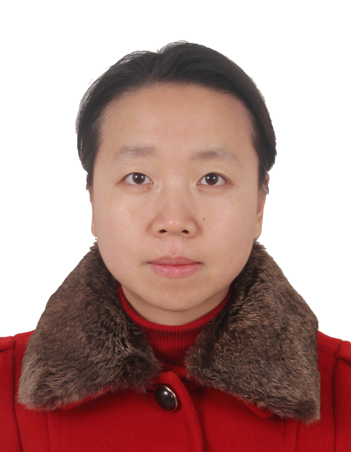 Weirong Fang (方伟蓉)合作论文数中国药科大学生理学教研室10
 Ge Liu合作论文数7Liangliang Huang合作论文数5Li Binbin合作论文数4Xu Fenglian合作论文数4Rui Liu合作论文数3Xueyan Zhao合作论文数3Yan Chen合作论文数3Yi Zhang合作论文数China Medical University Shenyang(China Medical University),Shenyang,China3Xiang Li合作论文数3
 Rong Xie合作论文数3Lele Zixin Yang合作论文数2Xiao-Fu Zhai合作论文数2Hong Yin合作论文数2Yongjian Chen合作论文数2JueMin Gao (高觉民)合作论文数2
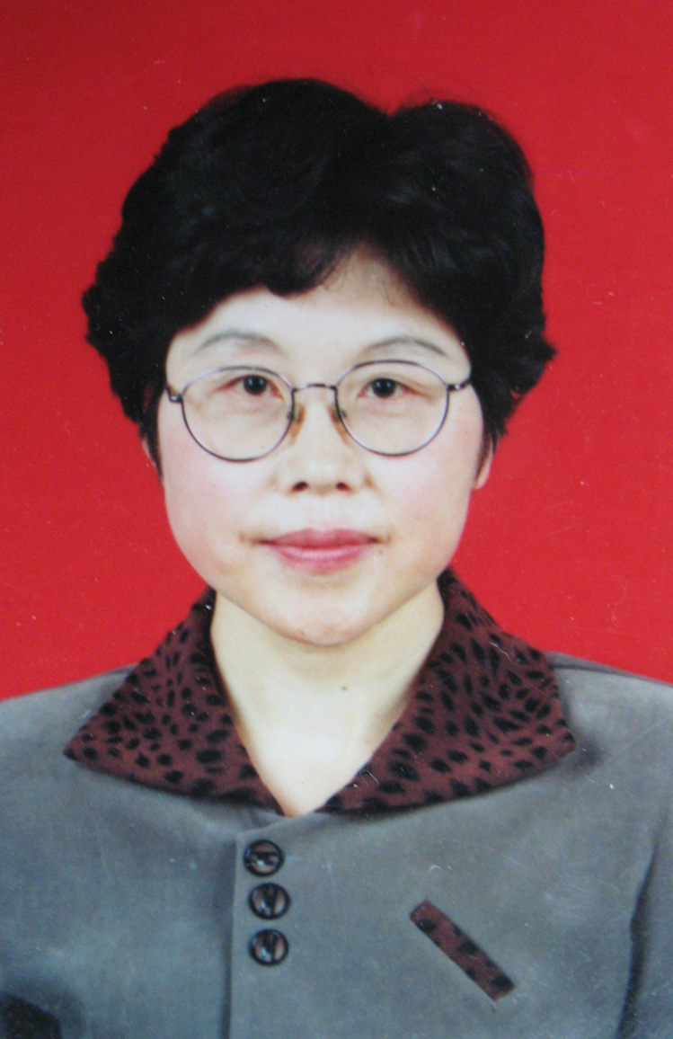 Yunman Li (李运曼)合作论文数State Key Laboratory of Natural Medicines, China Pharmaceutical University2Jiang Wang合作论文数1Shucheng He (贺树成)合作论文数1Zhirui Zhang合作论文数1
 Qiru Fan合作论文数1Dan Han合作论文数1李想合作论文数School of Engineering, China Pharmaceutical University1Fan Wenxiang合作论文数1
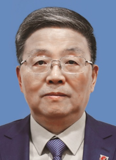 Wei Xiao (肖伟)合作论文数State Key Laboratory on Technologies for Chinese Medicine Pharmaceutical Process Control and Intelligent Manufacture,Nanjing University of Chinese Medicine1
 Baohui Xu (胥宝会)合作论文数Department of Surgery, School of Medicine, Stanford University1




