Abnormal electroencephalography (EEG) asymmetry has been implicated in several psychiatric disorders. However, the underlying mechanisms contributing to its dysregulation remain unclear. In this study, we investigated whether the proportions of leucocyte subpopulations are associated with relative EEG asymmetry across the frontal, central, parietal, and occipital brain regions. We recorded resting-state EEG from 24 healthy participants and calculated relative EEG asymmetry indices following standard preprocessing procedures. Peripheral blood samples were collected from each participant, and DNA was extracted for genome-wide DNA methylation analysis. DNA methylation data were used to estimate the relative proportions of leucocyte subpopulations. Pearson’s correlation was used to identify associations between EEG asymmetry and leucocyte compositions. To account for multiple comparisons, p-values were adjusted using the false discovery rate (FDR) method. We identified several associations between lymphocyte subpopulations and EEG asymmetry. CD8 + T cells were associated with occipital beta asymmetry (p = 0.009), while CD4 + T cells were linked to frontal alpha, frontal delta, occipital alpha, and occipital delta asymmetry (p-values ranging from 0.017 to 0.023). Natural killer cells showed an association with frontal beta asymmetry (p = 0.052), and B cells with occipital beta asymmetry (p <0.001). Granulocytes were associated with occipital beta and occipital delta asymmetry (p-values ranging from 0.01 to 0.032). However, none of these associations remained significant after FDR correction. The identified associations between the leucocyte subpopulation proportions and EEG asymmetry may be partially mediated by cytokine signaling within the central nervous system. Gaining insight into how EEG asymmetry is regulated in healthy individuals may aid future investigations focusing on its dysregulation in psychiatric populations.
BackgroundMild cognitive impairment (MCI), a prodromal stage of Alzheimer's disease and related dementias (ADRD), represents a critical window for intervention. Although mitochondrial dysfunction is increasingly implicated in neurodegeneration, most therapies target downstream protein aggregation. Transcranial photobiomodulation (tPBM) delivers near-infrared light to enhance mitochondrial respiration.ObjectiveWe hypothesized that tPBM in MCI would be safe, feasible, and associated with improvements in cognition, mitochondrial function, and default mode network (DMN) functional connectivity (FC).MethodsWe conducted a single-blind, randomized, sham-controlled pilot trial (NCT05563298) in adults ≥50 years with MCI. Twenty participants were randomized 1:1 to active or sham devices. Active devices delivered pulsed 810-nm light for 20 min per session; shams emitted light for 2 seconds. Stimulation targeted DMN hubs and the olfactory bulb. Participants self-administered treatment at home six days per week for six weeks.ResultsAdherence was high (active 96.9%; sham 94.2%). Adverse events (AEs) were reported by 10 of 20 participants (4 active, 6 sham). No serious AEs occurred. Compared with sham, active tPBM produced greater improvement in global cognition (Mini-Mental State Examination; p = 0.03, d = 1.05) and episodic memory (California Verbal Learning Test-II long-delay recognition; p = 0.02, d = 1.09). Serum pyruvate and lactate increased with a reduced lactate-to-pyruvate (L/P) ratio (p = 0.007, d = -1.37). DMN FC increased (p = 0.014, d = 1.25), and plasma IL-6 declined (p = 0.02, r = -0.52).ConclusionsHome-based tPBM was safe, well tolerated, and feasible, with high adherence and mild AEs. Cognitive, metabolic, and network-level findings are consistent with enhanced mitochondrial efficiency and anti-inflammatory effects. These results support larger, double-blind, multicenter trials to evaluate tPBM as a mitochondria-targeted therapy in early ADRD.
Brain disorders-encompassing neurological, mental, and substance use disorders-account for 10 of the top 25 causes of disability worldwide according to the Global Burden of Disease (GBD) 2021 study. Despite such an impact, they have not been centrally analyzed in prior GBD studies. This paper synthesizes the latest disability-focused GBD study to quantify the prevalence and disability burden of 35 conditions from 2010 to 2021, a period marking the first decline in global health outcomes in three decades. It further incorporates disability metrics from 2021 to 2023 to contextualize post-pandemic trends. The paper covers the prevalence and disability burden of neurological, mental, and substance use disorders along with COVID-19 using disability-adjusted life-years (DALYs) and years lived with disability (YLDs) metrics. From 2010-2021, Parkinson's, Alzheimer's, and migraine (in neurological disorders), major depressive, anxiety, and eating disorders (in mental disorders), and opioid and drug use disorders (in substance use disorders) showed the greatest increases in age-adjusted prevalence rates across both sexes. In 2021, neurological disorders were the largest contributor to DALYs among brain-disorder categories, while depressive and anxiety disorders ranked as the 2nd and 6th leading causes of global YLDs. Alzheimer's disease/dementias, Parkinson's disease, autism spectrum disorder (ASD), depressive and anxiety disorders, and opioid and drug use disorders showed the largest increases in burden within their respective categories between 2010 and 2021. In both 2021 and 2023, females had higher prevalence rates of overall neurological disorders, headache/migraine, multiple sclerosis, depressive/anxiety disorders, and anorexia nervosa, while males had higher rates of stroke, Parkinson's disease, ASD/ADHD, and substance use disorders. In DALY/YLD metrics, females showed higher rates for anorexia nervosa and multiple sclerosis, and males for ASD, certain neurological disorders, COVID-19, and substance use disorders. In the 2021-2023 extension analysis, disability data showed increases in prevalence and disability of several brain disorders, mostly anxiety disorders, while the COVID-19 disability burden declined markedly by 2023. Further sex-specific disability burden metrics, key insights from each disorder, and limitations/confounds are discussed.
OBJECTIVE:Electroencephalography (EEG) microstate analysis has emerged as a tool for investigating the spatial organization and temporal dynamics of large-scale cortical networks. Its potential role in identifying risk and progression of Alzheimer's dementia (AD) remains unclear. We conducted a systematic review and meta-analysis of EEG microstate parameters in AD and mild cognitive impairment (MCI). METHODS:PubMed, PsychINFO, EMBASE, and MEDLINE were searched, identifying 30 eligible studies (16 included in meta-analysis). Random-effects models were used to pool effect sizes and 95% confidence intervals comparing microstate parameters between AD, MCI, and healthy controls. RESULTS:Sixteen studies were included in the meta-analysis. In AD vs controls, microstate A duration (g = 0.41, 95% CI [0.10, 0.72]) and microstate B duration (g = 0.48, 95% CI [0.23, 0.73]) were significantly increased. In MCI vs controls, microstate D duration was significantly decreased (g = -0.26, 95% CI [-0.48, -0.04]) and microstate A occurrence rate was increased (g = 0.40, 95% CI [0.07, 0.74]), while microstate A and B duration were not significantly different. Heterogeneity was substantial for several outcomes. CONCLUSION:Pooled evidence suggests prolonged microstate A/B duration as the most reproducible alteration in AD, with reduced microstate D duration emerging as a modest finding in MCI. However, substantial heterogeneity and possible small-study effects indicate that current evidence is best interpreted as hypothesis-generating pending standardized, longitudinal, and multimodal studies. SIGNIFICANCE:EEG microstate analysis may provide complementary information about large-scale network dysfunction in MCI and AD, but methodological limitations currently constrain clinical biomarker interpretation.
ABSTRACT Background Photobiomodulation (PBM) is an emerging non-invasive light-based brain stimulation technique that can alter cortical oscillations and is currently being pursued for improving cognition and treating neurological and psychiatric conditions. Nearly all human EEG evidence comes from transcranial PBM (tPBM) applied to the forehead, where light must traverse the scalp and thick skull, requiring protocols to compensate with high surface irradiance. Intranasal PBM (iPBM) can reach the anterior skull base at a fraction of that irradiance and has also been shown to modulate cerebrospinal fluid dynamics, yet it has been studied almost exclusively as an adjunct to tPBM, leaving its cortical effects in isolation, and its energy-efficiency relative to the transcranial route, unknown. Objective To define the spatiotemporal EEG response to pulsed iPBM delivered alone, determine whether stimulation parameters or individual biology moderate it, and compare the energy-efficiency of iPBM and tPBM in the same participants. Methods High-density EEG was collected from forty-six healthy young adults during pulsed iPBM and tPBM spanning a parameter space of varying wavelengths, pulsation frequencies, and irradiances. Percent change in band power from a within-session pre-stimulation baseline was tested with spatiotemporal cluster-based permutation tests. Linear mixed-effects models with backward elimination assessed stimulation and biological moderators (sex, nostril-to-cortex distance). Energy-efficiency, defined as the percent change in band power per J/cm 2 of delivered surface energy, was compared between routes within each subject in delivery route-specific cluster regions of interest (ROI) (Wilcoxon signed-rank tests, Benjamini-Hochberg false discovery rate). Results iPBM alone produced significant spatiotemporal clusters in theta, beta, and gamma power, with anterior increases and posterior decreases; no delta or alpha clusters survived correction. Beta and gamma effects appeared at stimulation onset and persisted even after stimulation ended, whereas theta effects strengthened after stimulation ended. No predictor survived elimination in any band, time window, or cluster ROI: response magnitude was independent of wavelength, pulsation frequency, irradiance, sex, and nostril-to-cortex distance. Notably, although iPBM delivered roughly twenty times less surface energy than tPBM (∼0.6-1.1 vs ∼12-24 J/cm 2 ), it produced EEG changes of similar magnitude, and its energy-efficiency exceeded that of tPBM in seven of eight eligible comparisons, with median iPBM-to-tPBM efficiency ratios of 14-32 (all FDR q<0.05) Conclusions Delivered in isolation, pulsed iPBM elicits a robust cortical EEG signature closely resembling that of tPBM, is insensitive to the stimulation parameters and individual factors tested, and achieves this at a small fraction of the delivered surface energy. As a result, delivery route, not surface irradiance alone, should be treated as a primary variable in PBM dose reporting and protocol design.
BACKGROUND Convulsive therapies, including electroconvulsive therapy (ECT) and magnetic seizure therapy (MST), are highly effective for treatment-resistant depression, however, their neural mechanisms remain incompletely understood. We tested whether a data-driven framework applying a massive time-series feature library to electroencephalography (EEG) could reveal novel insights into changes in functional brain dynamics following convulsive therapy and provide preliminary markers of response. METHODS Resting-state EEG was analysed before and after a course of ECT or MST in 42 patients. Data were pooled and reduced to three principal components (PCs) capturing 78.1% of total variance, then >7,000 time-series features per PC were extracted from each patient using the highly comparative time-series analysis ( hctsa ) framework. Linear support vector machines (SVMs) classified pre-versus post-treatment EEG for each PC. A separate linear SVM, was also trained on 18 representative baseline features to predict clinical response. RESULTS hctsa- based classifiers distinguished pre-from post-treatment EEG for each PC (accuracy 73.8-76.2%, pFDR<0.01). Between 986-1,414 features significantly differentiated post-stimulation from baseline time-series across the three PCs (pFDR<0.05). The most discriminative features indexed linear and non-linear autocorrelation, correlation, multiscale entropy, and spectral properties. The baseline SVM combining 18 features showed modest but statistically reliable prediction of treatment response (balanced accuracy=0.69, area under the curve [AUC]=0.61, p=0.014). Single-feature ROC analyses further identified several top features with AUCs=∼0.7. CONCLUSIONS Data-driven analysis using a diverse time-series feature library can uncover novel EEG signatures of brain changes following convulsive therapy. This holds potential for delineating treatment-related mechanisms and developing predictive biomarkers to support precision psychiatry. ### Competing Interest Statement In the last 3 years PBF has received equipment for research from Neurosoft and Nexstim. He has served on a scientific advisory board for Magstim and received speaker fees from Otsuka. He has also acted as a founder and board member for TMS Clinics Australia and Resonance Therapeutics. PBF is supported by a National Health and Medical Research Council of Australia Investigator grant (1193596). ZJD has received research and equipment in-kind support for an investigator-initiated study through Brainsway Inc and Magventure Inc and industry-initiated trials through Magnus Inc. He also currently serves on the scientific advisory board for Brainsway Inc. His work has been supported by the National Institutes of Mental Health (NIMH), the Canadian Institutes of Health Research (CIHR), Brain Canada and the Temerty Family, Grant and Kreutzcamp Family Foundations. DMB receives research support from CIHR, NIMH, Wellcome Trust, Brain Canada and the Temerty Family through the CAMH Foundation and the Campbell Family Research Institute, has received research grant support and in-kind equipment support for an investigator- initiated study from Brainsway Ltd, was the site principal investigator for three sponsor-initiated studies for Brainsway Ltd, has received in-kind equipment support from Magventure for investigator- initiated studies, has received medication supplies for an investigator-initiated trial from Indivior, is a scientific advisor for Sooma Medical and is a member of the Clinical Standards Committee of the Clinical TMS Society (unpaid). ATH, KG, JAGL, RZ, and NWB have nothing to declare. ### Clinical Trial NCT01596608 ### Funding Statement This study did not receive any funding. ### Author Declarations I confirm all relevant ethical guidelines have been followed, and any necessary IRB and/or ethics committee approvals have been obtained. Yes The details of the IRB/oversight body that provided approval or exemption for the research described are given below: Ethics Committee of the Center for Addiction and Mental Health gave ethical approval for the study in accordance with the declaration of Helsinki. I confirm that all necessary patient/participant consent has been obtained and the appropriate institutional forms have been archived, and that any patient/participant/sample identifiers included were not known to anyone (e.g., hospital staff, patients or participants themselves) outside the research group so cannot be used to identify individuals. Yes I understand that all clinical trials and any other prospective interventional studies must be registered with an ICMJE-approved registry, such as ClinicalTrials.gov. I confirm that any such study reported in the manuscript has been registered and the trial registration ID is provided (note: if posting a prospective study registered retrospectively, please provide a statement in the trial ID field explaining why the study was not registered in advance). Yes I have followed all appropriate research reporting guidelines, such as any relevant EQUATOR Network research reporting checklist(s) and other pertinent material, if applicable. Yes Data presented in the study is not available at this time.
The PACt-MD study demonstrated that combined cognitive remediation (CR) and transcranial direct current stimulation (tDCS) slows global cognitive decline in individuals with mild cognitive impairment (MCI) or remitted major depressive disorder (rMDD) over a median follow-up of four years. Prefrontal theta-gamma coupling (TGC), measured via electroencephalography (EEG), is a marker of prefrontal cortical function and may index cognitive compensation, the mechanism thought to underlie CR+tDCS effects. This secondary analysis investigated whether baseline TGC influenced the efficacy of CR+tDCS, hypothesizing that participants with high baseline TGC-indicating better prefrontal function-would benefit more from CR+tDCS than those with low baseline TGC. TGC was assessed during an N-back task at baseline in 260 participants (mean age=71.9, SD = 6.0) and dichotomized by median split into high vs low groups. Cognition was evaluated two months post-baseline and annually up to six years. Baseline TGC significantly moderated the effects of CR+tDCS on global cognition (χ²=12.46, p = 0.006), verbal memory (χ²=16.93, p = 0.0007), and executive function (χ²=18.57, p = 0.0003). In the high-TGC group, global cognition declined more slowly with active CR+tDCS compared to sham. No such difference was observed in the low-TGC group. Lower baseline TGC may reflect reduced capacity for cognitive compensation, limiting CR+tDCS effectiveness in at-risk older adults. Higher TGC may identify those most likely to benefit from this intervention. ClinicalTrials.gov Identifier: NCT02386670.
Background:Post-COVID-19 condition (PCC) affects millions globally, with cognitive dysfunction ("brain fog") impairing daily functioning in up to 88% of patients. No effective treatments exist for PCC-related cognitive impairment. Photobiomodulation (PBM), a non-invasive therapy delivering near-infrared light, enhances mitochondrial function and reduces neuroinflammation, showing promise in neurological disorders. This study aimed to evaluate the efficacy and safety of home-based intranasal and transcranial PBM (itPBM) for PCC cognitive dysfunction. Methods:This randomized, double-blind, sham-controlled pilot trial in the USA (ClinicalTrials.govNCT05857124) enrolled 43 adults (18-65 years) with PCC cognitive symptoms ≥12 weeks post-infection. Participants were randomized 1:1, stratified by age (<45 vs ≥ 45 years), using computer-generated assignment to 8 weeks of daily 20-min itPBM, 6 days per week, with the Vielight Neuro RX Gamma device or sham, targeting the brain's default mode network, followed by 4 weeks of observation. Participants, investigators, and assessors were masked to group assignment. The primary outcome was mean change in Creyos cognitive battery composite score at Day 56. Secondary outcomes included fatigue, quality of life, and safety. Analyses used mixed-model repeated measures in the per-protocol population. Findings:The trial was completed, with 43 participants randomized (23 active, 20 sham) and 41 analyzed (21 active, 20 sham). They were recruited between July 5, 2023, and September 1, 2024. Active itPBM improved composite cognitive scores more than sham (mean difference 0.043, 95% CI -0.007 to 0.092, p = 0.088), with significant gains in participants <45 years (prespecified but exploratory, p = 0.028). Attention tasks improved consistently (p < 0.050 at multiple timepoints). Secondary outcomes mobility favored sham (p = 0.007), and fatigue also favored sham. No serious adverse events occurred; compliance was high (median 55 days, interquartile range 2 days). Interpretation:Home-based itPBM is safe and feasible, showing potential cognitive benefits for PCC brain fog, particularly in younger adults. Larger trials are needed to confirm efficacy and optimize parameters. Funding:Vielight Inc.
Background Difficult-to-treat depression (DTD) in older adults is associated with high rates of disability, hospitalization, and mortality. As the effectiveness of once daily theta burst stimulation (TBS) has been demonstrated in older adults, we aimed to explore the feasibility and clinical effects of an accelerated bilateral TBS protocol. Methods This single arm, open-label trial enrolled outpatients aged 60 years and over with a current major depressive episode and nonresponse to at least one antidepressant. Participants received continuous TBS (600 pulses) to the right dorsolateral prefrontal cortex (DLFPC) followed by intermittent TBS (600 pulses) to the left DLPFC, 8 times daily for 5 consecutive days. The primary outcome measures were proportion of participants retained in the study and change in Montgomery-Åsberg Depression Rating Scale (MADRS). Secondary outcomes included rates of remission defined by MADRS ≤ 10, changes in measures of anxiety and suicidality, and measures of tolerability including number of serious adverse events. Results 79 participants enrolled in the study over a period of 2.5 years, 78 (67.7 ± 5.9 yrs, 75.6 % female) of whom received majority of TBS sessions. MADRS scores decreased a mean ( ± SD) 8.5 ± 6.7 points following treatment completion (F(2,154) = 61.6, p < 0.0001) and this improvement persisted 4 weeks later (p < 0.0001). The remission rate was 18% at treatment end and increased to 24 % 4 weeks later. Symptoms of anxiety (p < 0.01) and suicidal ideation (p = 0.03) also improved at treatment end. Treatment was well-tolerated and no serious adverse events were reported. Conclusions Accelerated TBS is feasible in older patients with DTD; its efficacy is comparable to once daily TBS; and it is well-tolerated. If these results are confirmed in a larger randomized trial, accelerated TBS may become a preferred treatment option for older depressed patients. Clinicaltrials.gov Identifier NCT05119699
Mild Cognitive Impairment (MCI) is a frequent precursor to Alzheimer’s dementia (AD). Mitochondrial dysfunction, marked by reduced cytochrome c oxidase (CCO) activity and lower ATP production, is linked to these neurodegenerative diseases. This study evaluates transcranial photobiomodulation (tPBM), a non-invasive technique using near-infrared light to stimulate mitochondrial CCO, potentially enhancing neuronal energy and cognitive function in individuals with MCI. Twenty patients with mild cognitive impairment (MCI) were randomly assigned to an active treatment group (n = 10) or a sham control group (n = 10) using visually identical devices to maintain blinding. Participants completed daily home-based tPBM sessions for 6 weeks. Pre- and post-treatment assessments included cognitive tests (MMSE, TMT-A & B, CVLT-II) and biomarker evaluations (blood samples via ELISA). Neuroimaging included proton magnetic resonance spectroscopy (¹H-MRS) of the posterior cingulate cortex (PCC) and whole-brain structural and resting-state functional MRI. Change scores were calculated by subtracting baseline from post-treatment values. Compliance exceeded 98% in both groups, and tPBM was well-tolerated. The active tPBM group showed significantly greater post-treatment improvements from baseline (p < 0.05) compared to the sham group, including: (1) better recognition memory (higher long-delay hits, fewer false positives on the CVLT-II); (2) improved cognition (higher MMSE); (3) faster processing speed (shorter TMT-B times); (4) decreased plasma IL-6 levels; (5) higher choline/creatine ratio and a trend toward increased myoinositol/creatine in the PCC; (6) increased left nucleus-accumbens volume; and (7) enhanced functional connectivity within the DMN and between the caudate and DMN, along with decreased FC within the limbic network. Daily, home-based tPBM is a well-tolerated and feasible intervention that led to significant improvements in both cognitive function and biological markers in individuals with MCI. The active tPBM group demonstrated enhanced cognition, recognition memory, and processing speed, alongside reductions in inflammation and structural and functional brain changes, including increased neuroplasticity and alterations in brain connectivity. These findings suggest that tPBM may promote neuronal function and brain network modifications, particularly within the default mode network and limbic regions, providing evidence for its potential as a therapeutic approach in the early stages of Alzheimer's disease.
Objective: This study aimed to optimally evaluate the effect of the long-interval intracortical inhibition (LICI) in the dorsolateral prefrontal cortex (DLPFC) through transcranial magnetic stimulation combined with electroencephalography (TMS-EEG) by eliminating the volume conductance with signal source estimation and using a realistic sham coil as a control. Methods: We compared the LICI effects from the DLPFC between the active and sham stimulation conditions in 27 healthy participants. Evoked responses between the two conditions were evaluated at the sensor and source levels. Results: At the sensor level, a significant LICI effect was confirmed in the active condition in the global mean field power analysis; however, in the local mean field power analysis focused on the DLPFC, no LICI effect was observed in the active condition. However, in the signal source estimation analysis for the DLPFC, we could reconfirm a significant LICI effect (p = 0.023) in the interval 30-250 ms post-stimulus, compared to the sham condition. Conclusions: Our results demonstrate that application of realistic sham stimulation condition and source estimation method allows for a robust and optimal identification of the LICI effect in the DLPFC. Significance: The optimal DLPFC-LICI effect was identified by the use of the sophisticated sham coil.
BACKGROUND:GABA(B) receptor-mediated inhibitory dysfunction is implicated in the pathophysiology of treatment-resistant depression (TRD); however, the underlying mechanisms remain unclear. Long-interval intracortical inhibition (LICI) paradigm using transcranial magnetic stimulation combined with electroencephalography (TMS-EEG) enables the evaluation of GABA(B) receptor-mediated cortical inhibition from the cortex. OBJECTIVE:We aimed to evaluate the LICI effect in the left dorsolateral prefrontal cortex (DLPFC) of TRD using combined TMS-EEG. METHODS:Thirty individuals with TRD and age- and sex-matched healthy controls (HC) underwent the LICI TMS-EEG, which consisted of single- and paired-pulse TMS in the left DLPFC, with an interstimulus interval of 100 ms. The LICI effects were evaluated by comparing time-series and time-frequency signals at the sensor and source levels within- and between-groups (TRD vs. HC). RESULTS:Within-group comparisons showed inhibitory changes at the signal source level for the HC group in the time-series analysis, and inhibitory changes at the sensor and signal source levels for both groups in the time-frequency analysis. Between-group comparisons showed a significant reduction of the LICI effect in the time-series signal (35-87 ms) and θ-β band power at the source level in TRD compared to HC. CONCLUSION:Our findings suggest that reduced LICI in the DLPFC may contribute to the pathophysiological basis of TRD.
BACKGROUND:Cortical excitability has been proposed as a novel neurophysiological marker of neurodegeneration in Alzheimer's dementia (AD). However, the link between cortical excitability and structural changes in AD is not well understood. OBJECTIVE:To assess the relationship between cortical excitability and motor cortex thickness in AD. METHODS:In 62 participants with AD (38 females, mean ± SD age = 74.6 ± 8.0) and 47 healthy control (HC) individuals (26 females, mean ± SD age = 71.0 ± 7.9), transcranial magnetic stimulation resting motor threshold (rMT) was determined, and T1-weighted MRI scans were obtained. Skull-to-cortex distance was obtained manually for each participant using MNI coordinates of the motor cortex (x = -40, y = -20, z = 52). RESULTS:The mean skull-to-cortex distances did not differ significantly between participants with AD (22.9 ± 4.3 mm) and HC (21.7 ± 4.3 mm). Participants with AD had lower motor cortex thickness than healthy individuals (t(92) = -4.4, p = <0.001) and lower rMT (i.e., higher excitability) than HC (t(107) = -2.0, p = 0.045). In the combined sample, rMT was correlated positively with motor cortex thickness (r = 0.2, df = 92, p = 0.036); however, this association did not remain significant after controlling for age, sex and diagnosis. CONCLUSIONS:Patients with AD have decreased cortical thickness in the motor cortex and higher motor cortex excitability. This suggests that cortical excitability may be a marker of neurodegeneration in AD.
Background Multiscale dispersion entropy (MDEnt) is a nonlinear EEG measure that quantifies brain complexity across time scales, reflecting both local and global brain dynamics. Previous research indicates lower complexity at short time scales in Alzheimer's disease (AD) compared to mild cognitive impairment (MCI) and healthy controls (HCs), with MCI also showing lower values than HCs. Major depressive disorder (MDD) has also been preliminarily linked to reduced complexity during acute episodes.Objective To assess whether MDEnt at short time scales can distinguish AD from MCI and HCs, and to examine complexity differences across additional groups, remitted MDD (rMDD) and rMDD + MCI, while exploring associations with cognitive performance.Methods The study included 316 older adults: 44 HCs, 46 with rMDD, 114 with MCI, 71 with rMDD + MCI, and 41 with AD. Resting-state, eyes-closed EEGs were analyzed using MDEnt at 24 ms (short) and 60 ms (long) time scales. Cognitive function was measured with the Montreal Cognitive Assessment and a composite cognitive score.Results Short time scale complexity was lowest in AD, followed by MCI, and highest in HCs; rMDD presence had no impact. Only AD showed reduced complexity at long time scales. Complexity at both time scales was significantly correlated with cognitive performance.Conclusions This study highlights the value of MDEnt to assess complexity at short time scale and differentiate individuals with AD, MCI, or HCs. Reduced complexity in these individuals may underlie their cognitive impairment. In contrast, our study suggests that any MDD impact on complexity is likely related to active depressive symptoms.
Previous literature has identified slowing of resting state electroencephalography (EEG) rhythm and abnormal cortical excitation in Alzheimer’s Dementia (AD). However, the relationship between these two divergent functional abnormalities and cognitive symptoms of AD are not well understood. Resting state EEG signal was recorded in participants with AD and HCs for 5 minutes with eyes closed. Relative resting state EEG power was measured for the five frequency bands. Participants underwent a single pulse transcranial magnetic stimulation (TMS) combined with EEG. Cortical evoked activity (CEA) was assessed using TMS-evoked potential (TEP) rectified area under the curve (AUC) from 25 to 80 ms post-TMS stimulus, and the TEPs peak amplitudes were calculated by taking the maximal peak in the following time windows: 25 – 35 ms (P30), 40 – 50 ms (N45), and 55 – 65 ms (P60). Compared to 32 HC (18 females; mean ± SD age: 69.3 ± 7.9 years), 52 participants with AD (32 females; 74.2 ± 8.5 years) had higher relative theta power than HCs (t(66.6) = 5.34, p<0.001). AD participants also had a significantly lower alpha power (t(76) = -3.03, p=0.003) and beta power (t(76) = -2.51, p=0.014) than HCs. Controlling for sex, age and years of education, AD participants showed a positive association between theta power and CEA (r partial =0.573, p=0.008); and an inverse association between alpha power and CEA (r partial =-0.471, p=0.036). Theta power in AD participants showed a positive association with P60 (r partial =-0.637, p=0.003) and an inverse association with N45 (r partial =-0.512, p=0.025), while alpha power was inversely associated with P60 (r partial =-0.531, p=0.016). This study showed a positive association between resting state EEG slowing and increased cortical excitability in AD, indicating a possible shared mechanistic pathway between these abnormalities.
Mild Cognitive Impairment (MCI) is a clinical prodromal stage of Alzheimer’s disease. Enhancing executive functions in patients with MCI could optimize cognitive compensatory mechanisms and slow cognitive decline. The prefrontal cortex (PFC) and its connections to the hippocampus support executive functions, including working memory. Transcranial alternating current stimulation (tACS) can modulate these connections by engaging theta-gamma coupling (TGC) and may thereby strengthen working memory. This study, “tACS to engage theta-gamma coupling and enhance working memory in MCI” (tACS-MCI), will assess the feasibility and cognitive effects of EEG and MRI-guided individualized tACS. The stimulation will target the prefrontal and temporal cortices in 20 MCI participants. Participants will be randomized to receive either individualized tACS or sham tACS for 10 days. tACS individualization will involve adjusting the theta frequency, tACS electrode locations, and current intensity for each participant. Cognitive and functional assessments will occur at baseline and post-intervention. We aim to determine: 1) the feasibility of individualized tACS in MCI, including recruitment and retention; 2) whether tACS engages TGC by assessing its increase in response to tACS; and 3) changes in N-back working memory performance following tACS, as well as whether changes in TGC mediate the changes in performance. The tACS-MCI study will employ an EEG and MRI-guided individualized approach to promote synchronization between frontal and temporal cortices, using participant’s unique brain structure and neurophysiology. We aim to assess the feasibility of this novel intervention as a potential approach to more effectively prevent cognitive decline.
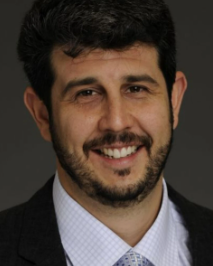 Daniel Blumberger合作论文数Temerity Centre for Therapeutic Brain Intervention, Temerty Faculty of Medicine, University of Toronto;Department of Psychiatry, Temerty Faculty of Medicine, University of Toronto;Late-Life Mood Disorders Clinic, Centre for Addiction and Mental Health, University of Toronto140
 Jeff Daskalakis合作论文数Institute of Medical Science, Temerty Faculty of Medicine, University of Toronto113
 Tarek Rajji合作论文数Centre for Addiction & Mental Health, Department of Psychiatry, Temerty Faculty of Medicine, University of Toronto110
 Benoit Mulsant合作论文数Department of Psychiatry, Temerty Faculty of Medicine, University of Toronto;Centre for Addiction and Mental Health49
 Sanjeev Kumar合作论文数Institute of Medical Science, Temerty Faculty of Medicine, University of Toronto43
 Bruce G. Pollock合作论文数Department of Pharmacology and Toxicology, Temerty Faculty of Medicine, University of Toronto;Centre for Addiction and Mental Health, University of Toronto33
 Daphne Voineskos合作论文数Division of Neuroscience & Clinical Translation, Department of Psychiatry, Temerty Faculty of Medicine, University of Toronto;Krembil Research Institute;Temerty Centre for Therapeutic Brain Intervention, Campbell Family Mental Health Research Institute, Centre for Addiction and Mental Health;rTMS Clinic;Poul Hansen Family Centre for Depression, University Health Network29
 Mera S. Barr合作论文数Centre for Addiction and Mental Health, University of Toronto/Department of Psychiatry, Temerty Faculty of Medicine, University of Toronto/Otsuka Canada Pharmaceutical Inc.25
 Yoshihiro Noda (野田賀大)合作论文数Multidisciplinary Translational Research Lab, Department of Neuropsychiatry, School of Medicine, Keio University22
 Robert Chen合作论文数Institute of Medical Sciences, Temerty Faculty of Medicine, University of Toronto21
 I. Hadas合作论文数Department of Clinical Pharmacology, Ben-Gurion University of the Negev20
 Hill Aron T合作论文数*Neuroscience Department, The Alfred Hospital;Monash Medical Centre, Monash University;The Alfred Hospital, Monash University18
 Faranak Farzan合作论文数School of Mechatronic Systems Engineering, Simon Fraser University17
 Pushpal Desarkar合作论文数Department of Psychiatry, Temerty Faculty of Medicine, University of Toronto;Division of Adult Psychiatry, Centre for Addiction and Mental Health;Azrieli Adult Neurodevelopment Centre, Centre for Addiction and Mental Health16
 Nathan Herrmann合作论文数Sunnybrook Health Sciences Centre;Department of Psychiatry, Temerty Faculty of Medicine, University of Toronto16
 Corinne Fischer合作论文数Division of Geriatric Psychiatry, University of Toronto15
 Linda Mah合作论文数Department of Psychiatry, Temerty Faculty of Medicine, University of Toronto15
 Lew Lim合作论文数Vielight Inc.14Mina Mirjalili合作论文数14
 Alastair J. Flint合作论文数Department of Psychiatry, Temerty Faculty of Medicine, University of Toronto14
 Christoph Zrenner合作论文数Network Biology Research Laboratories, Technion - Israel Institute of Technology Haifa14Michelle Goodman合作论文数13
 Corinne E. Fischer合作论文数Department of Psychiatry, Temerty Faculty of Medicine, University of Toronto12Genane Loheswaran合作论文数12
 Sushmit Das合作论文数11
 Aristotle Voineskos合作论文数Department of Psychiatry, University of Toronto;Slaight Family Centre for Youth in Transitionm, Centre for Addiction and Mental Health;Schizophrenia Division, Centre for Addiction and Mental Health11
 Robin Cash合作论文数Department of Biomedical Engineering, School of Chemical and Biomedical Engineering, Faculty of Engineering and Information Technology, The University of Melbourne10
 Paul B. Fitzgerald合作论文数School of Medicine and Psychology, The Australian National University;College of Health and Medicine, The Australian National University10
 Zaid Ghazala合作论文数9
 Jonathan Downar合作论文数Institute of Medical Science, Temerty Faculty of Medicine, University of Toronto;Department of Psychiatry, Temerty Faculty of Medicine, University of Toronto;MRI-Guided rTMS Clinic, University Health Network;Toronto Western Research Institute9
 Jennifer Lissemore合作论文数Brain Stimulation Lab, Stanford University9
 Tony P. George合作论文数Institute of Medical Sciences, Temerty Faculty of Medicine, University of Toronto;CAMH’s Institute for Mental Health Policy Research9
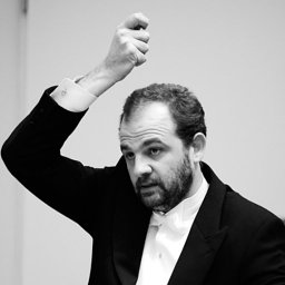 Pantelis Lioumis合作论文数Helsinki University Central Hospital9Mohsen Poorganji合作论文数8Heather Brooks合作论文数8
 Rashidi-Ranjbar Neda合作论文数8
 Christopher R. Bowie合作论文数Departments of Psychology and Psychiatry, Queen’s University8
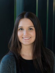 Melissa Kirkovski合作论文数The Alfred and Central Clinical School, Monash University7Helmut Kröger合作论文数7
 Krista Lanctôt合作论文数Department of Pharmacology and Toxicology, Temerty Faculty of Medicine, University of Toronto;Department of Psychiatry, Temerty Faculty of Medicine, University of Toronto;Sunnybrook Health Sciences Centre7Yuliya Knyahnytska合作论文数Department of Psychiatry, Temerty Faculty of Medicine, University of Toronto6
 Colin Hawco合作论文数Department of Psychiatry, Temerty Faculty of Medicine, University of Toronto;Kimel Family Translational Imaging-Genetics Laboratory, Centre for Addiction & Mental Health6
 Peter Enticott合作论文数The Alfred and Central Clinical School, Monash University6
 Reza Rostami合作论文数Faculty of Psychology and Education, University of Tehran6
 Hamed Azami合作论文数Department of Electrical Engineering, Iran University of Science and Technology6
 Nigel Rogasch合作论文数Turner Institute for Brain and Mental Health, School of Psychological Sciences, Monash University6
 Anastasios Daskalakis Daskalakis合作论文数5
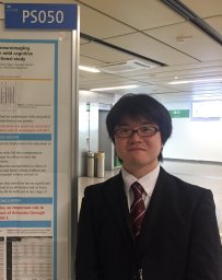 Masataka Wada合作论文数Dept Neuropsychiat, Keio Univ5Mayuko Takano合作论文数Teijin5




