Supplementary Figure 1. EGFR specifically co-immunoprecipitates with TROY, but not Fn14.
Apolipoprotein E (APOE) and clusterin (CLU) are critical in the pathogenesis of Alzheimer’s disease (AD). Common coding variants in APOE (e.g. APOE4) impart at least 3-fold higher risk of developing AD, while common noncoding variants in CLU impart smaller but still highly significant risk for AD. Using genetically engineered mouse models lacking either APOE or CLU, previous studies from our lab and others have shown that both are critical in the formation of amyloid. However, only a single published study has examined the combined effects of APOE and CLU in a mouse model of amyloidosis. We generated mice that lacked both murine APOE and CLU (double knockout mice) on the APP/PS1 background of AD amyloidosis. We assessed these mice using histological analysis as well as single-cell RNAseq profiling from brain tissue. Compared to APP/PS1 controls, we found that double knockout mice for both APOE and CLU had a striking reduction in the amount of amyloid plaques in the cortex. Moreover, we found that these double-knockout mice were completely void of amyloid plaques in the hippocampus. Single-cell RNAseq experiments revealed a strong reduction in the amount of activated microglia in these double knockout mice. Contrary to a previous study that reported increased amyloid load in APOE-/-;CLU-/- double knockout mice, we found a dramatic reduction in amyloid plaques and a concomitant loss of activated microglia when using genetically inbred (C57BL/6J) mouse lines. Further studies are needed to determine whether these effects are cell-type specific (e.g. astrocyte-derived APOE and CLU conditional knockouts) or whether these lipoproteins have synergistic roles in tau pathology.
The choroid plexus, a tissue responsible for producing cerebrospinal fluid, is found predominantly in the lateral and fourth ventricles of the brain. This highly vascularized and ciliated tissue is made up of specialized epithelial cells and capillary networks surrounded by connective tissue. Given the complex structure of the choroid plexus, this can potentially result in contamination during routine tissue dissection. Bulk and single-cell RNA sequencing studies, as well as genome-wide in situ hybridization experiments (Allen Brain Atlas), have identified several canonical markers of choroid plexus such as Ttr, Folr1, and Prlr. We used the Ttr gene as a marker to query the Gene Expression Omnibus database for transcriptome studies of brain tissue and identified at least some level of likely choroid contamination in numerous studies that could have potentially confounded data analysis and interpretation. We also analyzed transcriptomic datasets from human samples from Allen Brain Atlas and the Genotype-Tissue Expression (GTEx) database and found abundant choroid contamination, with regions in closer proximity to choroid more likely to be impacted such as hippocampus, cervical spinal cord, substantia nigra, hypothalamus, and amygdala. In addition, analysis of both the Allen Brain Atlas and GTEx datasets for differentially expressed genes between likely "high contamination" and "low contamination" groups revealed a clear enrichment of choroid plexus marker genes and gene ontology pathways characteristic of these ciliated choroid cells. Inclusion of these contaminated samples could result in biological misinterpretation or simply add to the statistical noise and mask true effects. We cannot assert that Ttr or other genes/proteins queried in targeted assays are artifacts from choroid contamination as some of these differentials may be due to true biological effects. However, for studies that have an unequal distribution of choroid contamination among groups, investigators may wish to remove contaminated samples from analyses or incorporate choroid marker gene expression into their statistical modeling. In addition, we suggest that a simple RT-qPCR or western blot for choroid markers would mitigate unintended choroid contamination for any experiment, but particularly for samples intended for more costly omic profiling. This study highlights an unexpected problem for neuroscientists, but it is also quite possible that unintended contamination of adjacent structures occurs during dissections for other tissues but has not been widely recognized.
BACKGROUND:Many studies have shown an elevated level of cholesterol in colon tumors as compared to normal tissue. Obesity and high low-density lipoprotein cholesterol (LDL-C) are known risk factors for colon cancer. However, the role of LDL-C in colon cancer patients with normal body mass index (BMI) remains elusive.METHODS:Levels of serum cholesterol and oxysterols were quantified by ultra-performance liquid chromatography-tandem mass spectrometer (UPLC-MS/MS) from 129 individuals with normal BMI, including 32 with solitary polyp, 36 with multiple polyps, and 31 with adenocarcinoma as well as 32 healthy controls. In vitro, colon cancer cells were treated with LDL-C and assayed for chemokines via RNA-Seq and mitochondrial morphology via transmission electron microscopy and immunofluorescence. Additionally, correlation analysis was performed between LDL-C-induced chemokines and the overall survival of colon cancer patients from the Cancer Genome Atlas (TCGA), the Genotype-Tissue Expression (GTEx), and the Human Protein Atlas (HPA) database.RESULTS:The serum cholesterol level was significantly higher in colon adenocarcinoma patients with normal BMI than that in healthy controls (P<0.001). LDL-C potentiated colon cancer cell invasion and resistance to glucose-deprivation in vitro via chemokine-mediated signaling, mainly upregulation of CC chemokine ligand (CCL) 5 and downregulation of CCL 11. By analyzing the RNA expression data of colorectal cancer from TCGA, GTEx, and HPA, we demonstrated that the CCL5 level in colorectal adenocarcinoma tissues was significantly increased relative to adjacent normal tissues (P=0.01) while the CCL11 level was decreased (P=0.01). Both increased CCL5 and decreased CCL11 showed a negative correlation with the 5-year overall survival in tumor node metastasis (TNM) stage II colon cancer patients (P=0.0032, 0.026 for CCL5 and CCL11, respectively).CONCLUSIONS:Our study supports the idea that LDL-C regulates the expression of CCL5 and CCL11 chemokines, which may have predictive values for survival in colon cancer patients with normal BMI, especially for patients in TNM stage II.
Glioblastoma (GBM) is the most common primary malignant brain cancer in adults. A hallmark of GBM is aggressive invasion of tumor cells into the surrounding normal brain. The current standard of care therapy, as well as targeted therapies, has largely failed to specifically address this issue. Therefore, identifying key regulators of GBM cell migration and invasion is of particular interest. The leukemia-associated Rho guanine nucleotide exchange factor (LARG) has previously been implicated in cell invasion in other tumor types; however, the role of LARG in GBM pathobiology remains undefined. Herein, we report that the expression level of LARG, RhoC, and RhoA increases with glial tumor grade and is highest in GBM. LARG and RhoC protein expression is more prominent in the invading cells, whereas RhoA expression is largely restricted to cells in the tumor core. Knockdown of LARG by siRNA inhibits GBM cell migration in vitro and invasion ex vivo in organotypic brain slices. Moreover, siRNA-mediated silencing of RhoC suppresses GBM cell migration in vitro and invasion ex vivo, whereas depletion of RhoA enhances GBM cell migration and invasion, supporting a role for LARG and RhoC in GBM cell migration and invasion. Depletion of LARG increases the sensitivity of GBM cells to temozolomide treatment. Collectively, these results suggest that LARG and RhoC may represent unappreciated targets to inhibit glioma invasion.
Cancer stem cells (CSCs) are cancer cells (found within tumors or hematological cancers) that possess characteristics associated with normal stem cells, specifically the ability to give rise to all cell types found in a particular cancer sample. CSCs are therefore tumorigenic (tumor-forming), perhaps in contrast to other non-tumorigenic cancer cells. CSCs may generate tumors through the stem cell processes of self-renewal and differentiation into multiple cell types. Such cells are hypothesized to persist in tumors as a distinct population and cause relapse and metastasis by giving rise to new tumors. Therefore, development of specific therapies targeted at CSCs holds hope for improvement of survival and quality of life of cancer patients, especially for patients with metastatic disease.
Glioblastoma (GBM) is the most common primary malignant brain tumor in adults and carries a discouraging prognosis. Its aggressive and highly infiltrative nature renders the current standard treatment of maximal surgical resection, radiation, and chemotherapy relatively ineffective. Identifying the signaling pathways that regulate GBM migration/invasion and resistance is required to develop more effective therapeutic regimens to treat GBM. Expression of TROY, an orphan receptor of the TNF receptor superfamily, increases with glial tumor grade, inversely correlates with patient overall survival, stimulates GBM cell invasion in vitro and in vivo, and increases resistance to temozolomide and radiation therapy. Conversely, silencing TROY expression inhibits GBM cell invasion, increases sensitivity to temozolomide, and prolongs survival in a preclinical intracranial xenograft model. Here, we have identified for the first time that TROY interacts with JAK1. Increased TROY expression increases JAK1 phosphorylation. In addition, increased TROY expression promotes STAT3 phosphorylation and STAT3 transcriptional activity that is dependent upon JAK1. TROY-mediated activation of STAT3 is independent of its ability to stimulate activity of NF-κB. Inhibition of JAK1 activity by ruxolitinib or knockdown of JAK1 expression by siRNA significantly inhibits TROY-induced STAT3 activation, GBM cell migration, and decreases resistance to temozolomide. Taken together, our data indicate that the TROY signaling complex may represent a potential therapeutic target with the distinctive capacity to exert effects on multiple pathways mediating GBM cell invasion and resistance.
Glioblastoma (GBM) is the most common primary malignant brain cancer in adults. A hallmark of GBM is aggressive invasion of tumor cells into the surrounding normal brain. Both the current standard of care and targeted therapies have largely failed to specifically address this issue. Therefore, identifying key regulators of GBM cell migration and invasion is important. The leukemia-associated Rho guanine nucleotide exchange factor (LARG) has previously been implicated in cell invasion in other tumor types; however, its role in GBM pathobiology remains undefined. Herein, we report that the expression levels of LARG and ras homolog family members C (RhoC), and A (RhoA) increase with glial tumor grade and are highest in GBM. LARG and RhoC protein expression is more prominent in invading cells, whereas RhoA expression is largely restricted to cells in the tumor core. Knockdown of LARG by siRNA inhibits GBM cell migration in vitro and invasion ex vivo in organotypic brain slices. Moreover, siRNA-mediated silencing of RhoC suppresses GBM cell migration in vitro and invasion ex vivo, whereas depletion of RhoA enhances GBM cell migration and invasion, supporting a role for LARG and RhoC in GBM cell migration and invasion. Depletion of LARG increases the sensitivity of GBM cells to temozolomide treatment. Collectively, these results suggest that LARG and RhoC may represent unappreciated targets to inhibit glioma invasion.
PURPOSE:Dyslipidemia was associated with gastric adenocarcinoma or neuroendocrine tumors, but its role in a more malignant entity, gastric cancer with neuroendocrine immunophenotypes (GCNEI), was unclarified. This study sought to explore the relationship between serum lipid levels and the biological behaviors of gastric cancer with neuroendocrine immunophenotypes (GCNEI).METHODS:Patients with neuroendocrine carcinoma (NEC), GC with NEC components (GC-NEC), or GC expressing NE marker(s) but no NE morphology (GC-NENM) were enrolled from three centers. Their preoperative serum lipid levels, demographic, and clinicopathological information were analyzed and compared with those of patients with pure adenocarcinoma (PAC) or a background population selected from 10,061 health-check people by propensity-score matching.RESULTS:A total of 342 GCNEI patients were enrolled. Compared with the background population, total cholesterol (TCHO) and high-density lipoprotein cholesterol (HDL-C) levels were lower in GCNEI. Compared with PAC, GC-NENM and GC-NEC showed lower triglyceride (TG) levels, while, carcinoma with NE morphology showed higher low-density lipoprotein cholesterol (LDL-C) levels. Among GCNEI subtypes, GC-NEC differed from the others by higher LDL-C and non-HDL-C levels. A higher LDL-C level and(or) lower TG, HDL-C levels correlated to higher stages or large tumor sizes in GC-NENM, and a lower HDL-C level correlated to large tumor sizes in GC-NEC. A higher LDL-C level, lower TG, HDL-C, and non-HDL levels increased the risk of GC-NEC, and lower TG, and HDL-C levels increased the risk of GC-NENM and NEC.CONCLUSION:GCNEI had distinct and heterogeneous serum lipid patterns, which correlated to tumor development and progression.
Cancer stem cells (CSCs) play a critical role in tumor development and progression and are involved in cancer metastasis. The role of reactive oxygen species (ROS) in CSCs and cancer metastasis remains controversial. The aim of the present study was to investigate the correlation between ROS level of CSCs and cancer metastasis and to explore the possible underlying molecular mechanisms. Four different cell lines were used to isolate tumor spheres and to analyze intrinsic properties of tumor sphere cells including proliferation, self-renewal potential, differentiation, drug-resistance and cancer metastasis in vitro and in vivo. ROS assays were used to detect the intracellular ROS level of tumor spheres cells. Gene expression analysis and western blot were used to investigate the underlying mechanisms of ROS in regulating cancer metastasis. Tumor spheres possessed the characteristic features of CSCs, and ROS-high tumor spheres (RH-TS) displayed elevated mitochondrial ROS level exclusively drove metastasis formation. The gene expression analysis showed elevated fatty acid β-oxidation, downregulation of epithelial marker upregulation of mesenchymal markers, and the activation of MAP kinase cascades. Furthermore, 14 up-regulated genes in RH-TS cells were associated with reduced overall survival of different cancer patients. Our findings demonstrate that CSCs characterized by elevated mitochondrial ROS level potentiate cancer metastasis. Mechanistically, elevated mitochondrial ROS via fatty acid β-oxidation, activates the MAPK cascades, resulting in the epithelial-mesenchymal transition (EMT) process of RH-TS cells, thereby potentiating caner invasion and metastasis. Therefore, targeting mitochondrial ROS might provide a promising approach to prevent and alleviate cancer metastasis induced by RH-TS cells.
Abstract Glioblastoma (GBM) is the most common and deadly malignancy of the central nervous system in adults with a median survival of about 15 months after diagnosis. Its aggressive and highly infiltrative nature renders the current standard treatment of maximal surgical resection, radiation, and chemotherapy relatively ineffective. Identifying the molecular events that regulate GBM migration/invasion is required to develop more effective therapeutic regimens to treat GBM. Expression of TROY, an orphan receptor of the TNFR superfamily, increases with increasing glial tumor grade, inversely correlates with patient survival, stimulates GBM cell invasion in vitro and in vivo, and increases resistance to temozolomide and radiation therapy. Conversely, silencing TROY expression inhibits GBM cell invasion, increases sensitivity to temozolomide, and prolongs survival in a preclinical intracranial xenograft model. Thus, the TROY signaling pathway represents an attractive therapeutic target. We previously reported that increased TROY expression strongly activates NF-kB. We have identified for the first time that TROY forms a unique signaling complex with JAK1. Interaction with TROY stimulates phosphorylation of JAK1 and significantly increases the activation of STAT3. TROY specifically associates with JAK1 as it does not associate with other JAK family members. Co-immunoprecipitation analysis demonstrated that TROY interacts with JAK1 through its cytoplasmic domain as a TROY variant without its extracellular domain associates with JAK1 while a TROY variant without a cytoplasmic domain fails to bind JAK1. Luciferase reporter assay confirmed that full length TROY or a variant lacking the extracellular domain are able to induce STAT3 activation, but not a TROY variant lacking the cytoplasmic domain. Pharmacological inhibition of JAK1 by the FDA approved inhibitor ruxolitinib or knockdown of JAK1 by siRNAs significantly inhibited TROY-induced STAT3 activation and GBM cell migration. Together, our data indicate that the TROY-JAK1 complex represents an unappreciated therapeutic target to inhibit glioma invasion and decrease therapeutic resistance.
The presence of FMS-like receptor tyrosine kinase-3 internal tandem duplication (FLT3-ITD) mutations in patients with acute myeloid leukemia (AML) is associated with poor clinical outcome. FLT3 tyrosine kinase inhibitors (TKIs), although effective in kinase ablation, do not eliminate primitive FLT3-ITD+ leukemia cells, which are potential sources of relapse. Thus, understanding the mechanisms underlying FLT3-ITD+ AML cell persistence is essential to devise future AML therapies. Here, we show that expression of protein arginine methyltransferase 1 (PRMT1), the primary type I arginine methyltransferase, is increased significantly in AML cells relative to normal hematopoietic cells. Genome-wide analysis, coimmunoprecipitation assay, and PRMT1-knockout mouse studies indicate that PRMT1 preferentially cooperates with FLT3-ITD, contributing to AML maintenance. Genetic or pharmacological inhibition of PRMT1 markedly blocked FLT3-ITD+ AML cell maintenance. Mechanistically, PRMT1 catalyzed FLT3-ITD protein methylation at arginine 972/973, and PRMT1 promoted leukemia cell growth in an FLT3 methylation-dependent manner. Moreover, the effects of FLT3-ITD methylation in AML cells were partially due to cross talk with FLT3-ITD phosphorylation at tyrosine 969. Importantly, FLT3 methylation persisted in FLT3-ITD+ AML cells following kinase inhibition, indicating that methylation occurs independently of kinase activity. Finally, in patient-derived xenograft and murine AML models, combined administration of AC220 with a type I PRMT inhibitor (MS023) enhanced elimination of FLT3-ITD+ AML cells relative to AC220 treatment alone. Our study demonstrates that PRMT1-mediated FLT3 methylation promotes AML maintenance and suggests that combining PRMT1 inhibition with FLT3 TKI treatment could be a promising approach to eliminate FLT3-ITD+ AML cells.
Myelodysplastic syndrome (MDS), a largely incurable hematological malignancy, is derived from aberrant clonal hematopoietic stem/progenitor cells (HSPCs) that persist after conventional therapies. Defining the mechanisms underlying MDS HSPC maintenance is critical for developing MDS therapy. The deacetylase SIRT1 regulates stem cell proliferation, survival, and self-renewal by deacetylating downstream proteins. Here we show that SIRT1 protein levels were downregulated in MDS HSPCs. Genetic or pharmacological activation of SIRT1 inhibited MDS HSPC functions, whereas SIRT1 deficiency enhanced MDS HSPC self-renewal. Mechanistically, the inhibitory effects of SIRT1 were dependent on TET2, a safeguard against HSPC transformation. SIRT1 deacetylated TET2 at conserved lysine residues in its catalytic domain, enhancing TET2 activity. Our genome-wide analysis identified cancer-related genes regulated by the SIRT1/TET2 axis. SIRT1 activation also inhibited functions of MDS HSPCs from patients with TET2 heterozygous mutations. Altogether, our results indicate that restoring TET2 function through SIRT1 activation represents a promising means to target MDS HSPCs.
Glioblastoma multiforme (GBM) is the most common type of malignant brain tumors in adults and has a dismal prognosis. The highly aggressive invasion of malignant cells into the normal brain parenchyma renders complete surgical resection of GBM tumors impossible, increases resistance to therapeutic treatment, and leads to near-universal tumor recurrence. We have previously demonstrated that TROY (TNFRSF19) plays an important role in glioblastoma cell invasion and therapeutic resistance. However, the potential downstream effectors of TROY signaling have not been fully characterized. Here, we identified PDZ-RhoGEF as a binding partner for TROY that potentiated TROY-induced nuclear factor kappa B activation which is necessary for both cell invasion and survival. In addition, PDZ-RhoGEF also interacts with Pyk2, indicating that PDZ-RhoGEF is a component of a signalsome that includes TROY and Pyk2. PDZ-RhoGEF is overexpressed in glioblastoma tumors and stimulates glioma cell invasion via Rho activation. Increased PDZ-RhoGEF expression enhanced TROY-induced glioma cell migration. Conversely, silencing PDZ-RhoGEF expression inhibited TROY-induced glioma cell migration, increased sensitivity to temozolomide treatment, and extended survival of orthotopic xenograft mice. Furthermore, depletion of RhoC or RhoA inhibited TROY- and PDZ-RhoGEF–induced cell migration. Mechanistically, increased TROY expression stimulated Rho activation, and depletion of PDZ-RhoGEF expression reduced this activation. Taken together, these data suggest that PDZ-RhoGEF plays an important role in TROY signaling and provides insights into a potential node of vulnerability to limit GBM cell invasion and decrease therapeutic resistance.
Systemic inflammatory parameters are associated with poor outcomes in malignant patients. Several inflammation-based cumulative prognostic score systems were established for various solid tumors. However, there is few inflammation based cumulative prognostic score system for patients with diffuse large B cell lymphoma (DLBCL).
The current view is that treatment failures of AML patients are due to persistence of leukemia stem cells (LSCs). The presence of FMS-like tyrosine kinase-3 (FLT3) Internal tandem duplication (ITD) is associated with poor prognosis. But, FLT3 tyrosine kinase inhibitors (TKI) demonstrate transient clinical activity in FLT3-ITD+ AML patients. Persistent FLT3-ITD+ AML LSC represent a source of relapse. There is a pressing need to target LSC and improve outcomes for FLT3-ITD+ AML patients.
1 Department of Internal Medicine, St. Joseph’s Hospital and Medical Center, Phoenix, AZ, USA 2 Department of Biochemistry and Molecular Biology, Mayo Clinic Arizona, Scottsdale, AZ, USA Baylor University, Waco, TX, USA University of Arizona Cancer Center at Dignity Health St. Joseph's, Phoenix, AZ, USA 3 Baylor University, Waco, TX, US 4 University of Arizona Cancer Center at Dignity Health St. Joseph’s, Phoenix, AZ, US
Glioblastoma multiforme (GBM) is the most frequent primary brain tumor in adults with a 5-year survival rate of 5% despite intensive research efforts. The poor prognosis is due, in part, to aggressive invasion into the surrounding brain parenchyma. Invasion is a complex process mediated by cell-intrinsic pathways, extrinsic microenvironmental cues, and biophysical cues from the peritumoral stromal matrix. Recent data have attributed GBM invasion to the glioma stem-like cell (GSC) subpopulation. GSCs are slowly dividing, highly invasive, therapy resistant, and are considered to give rise to tumor recurrence. GSCs are localized in a heterogeneous cellular niche, and cross talk between stromal cells and GSCs cultivates a fertile environment that promotes GSC invasion. Pro-migratory soluble factors from endothelial cells, astrocytes, macrophages, microglia, and non-stem-like tumor cells can stimulate peritumoral invasion of GSCs. Therefore, therapeutic efforts designed to target the invasive GSCs may enhance patient survival. In this review, we summarize the current understanding of extrinsic pathways and major stromal and immune players facilitating GSC maintenance and survival.
 Nhan Tran合作论文数Translational Genomics Research Institute11
 Joseph C. Loftus合作论文数11Jean Kloss合作论文数9Bin Zhang合作论文数5Ying-Hui Zhu (朱颖慧)合作论文数Collaborat Innovat Ctr Canc Med, Sun Yat Sen Univ5Dandan Zhao合作论文数City Hope Med Ctr, Hematol Malignancies & Stem Cell Transplantat Ins, Gehr Family Ctr Leukemia Res, Duarte, CA 91010 USA5
 Marc Symons合作论文数Feinstein Institute for Medical Research5Haojie Dong合作论文数5Rosamaria Ruggieri合作论文数4Herman Wu合作论文数4
 Alison Roos合作论文数Mayo Clin Arizona, Dept Canc Biol, Scottsdale, AZ 85259 USA4Xin He合作论文数City Hope Med Ctr, Dept Hematol Malignancies Translat Sci, Gehr Family Ctr Leukemia Res,Beckman Res Inst, Hematol Malignancies & Stem Cell Transplantat Ins, 1500 E Duarte Rd, Duarte, CA 91010 USA4
 Ling Li合作论文数Department of Hematologic Malignancies Translational Science, Beckman Research Institute, City of Hope4
 Xun Hu (胡讯)合作论文数School of Medicine, Zhejiang University;Zhejiang University Cancer Center4
 Guido Marcucci合作论文数Department of Hematologic Malignancies Translational Science, City of Hope;Division of Leukemia, City of Hope;Gehr Family Center for Leukemia Research, City of Hope4
 Harshil Dhruv合作论文数4zhiwan dong合作论文数3xun hu合作论文数3Patrick Pirrotte合作论文数3Hanying Wang合作论文数School of Medicine, Zhejiang University3Yu-Ping Yang合作论文数Dept Biochem & Mol Biol, Univ Miami3Shannon Fortin Ensign合作论文数3
 Lifeng Feng (冯利锋)合作论文数Sir Run Run Shaw Hospital, School of Medicine, Zhejiang University3
 Lei Zhang合作论文数Wenzhou Medical College, Wenzhou Medical College3
 Ya-Huei Kuo合作论文数3
 biao Zhang合作论文数3
 Juan Du合作论文数3Eschbacher Jennifer合作论文数3Chunpeng Zhu合作论文数3
 Tuncali Serdar合作论文数3
 Ravi Bhatia合作论文数O'Neal Comprehensive Cancer Center, School of Medicine, The University of Alabama at Birmingham;Center for Clinical and Translational Science, General Clinical Research Center, School of Medicine, The University of Alabama at Birmingham;General Surgery Gastrointestinal Section, Department of Medicine, School of Medicine, The University of Alabama at Birmingham3
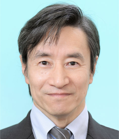 Kaoru Tohyama (通山薫)合作论文数Department of Medical Technology, Faculty of Health Science and Technology, Kawasaki University of Medical Welfare3
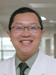 Yimei Feng (冯一梅)合作论文数The Second Affiliated Hospital of Army Medical University3
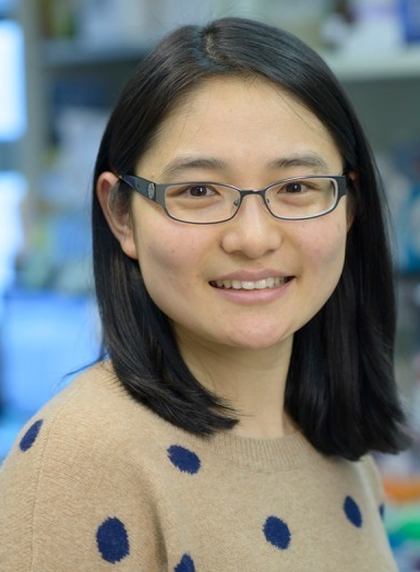 Jie Sun (孙洁)合作论文数School of Basic Medical Sciences, School of Medicine, Zhejiang University3
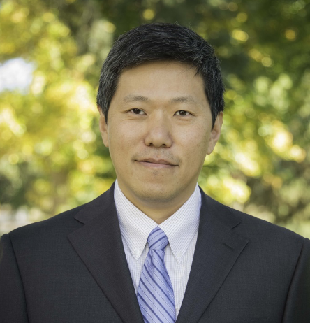 Xiaochun Yu (俞晓春)合作论文数Genomic Instability and Tumorigenesis Laboratory, School of Life Sciences, Westlake University3
 Samer Khaled合作论文数Orlando Health Cancer Institute3
 Philipp Oberdoerffer合作论文数Receptor Biology and Gene Expression, The Center for Cancer Research3Zhiyu Yan合作论文数3Hemragul Sabit合作论文数3
 Mitsutoshi Nakada合作论文数Kanazawa University3
 Hongchuan Jin (金洪传)合作论文数The Affiliated Sir Run Run Shaw Hospital, Zhejiang University School of Medicine3
 Lianjun Zhang合作论文数2Valentina Rubio合作论文数2Yi Zou合作论文数2zhen liu合作论文数2
 Yudao Shen (沈玉道)合作论文数School of Pharmaceutical Sciences, Shanghai Jiao Tong University2Feifei Sun合作论文数2




