DHODH-dependent respiration delays acquisition of mtDNA in ρ0 cells. A, 4T1 ρ0 and 4T1 ρ0 AOX cells were subcutaneously injected into Balb/c mice at 106 per animal. On days 3, 5, 10, 15, 20, and 25, tumor tissue was retrieved from the mice, and sublines were established. Parental cells and sublines derived from 4T1 ρ0 and 4T1 ρ0 AOX cells were assessed for mtDNA using qPCR. B, Parental, ρ0 D15, and 4T1 ρ0 AOX D25 cells were assessed for the distribution of mtDNA polymorphism in tRNAArg, D-loop, and 16S rRNA in single cells of lines. C, Heatmap illustrates mRNA expression of mitochondrial and nuclear-encoded OXPHOS genes in the cell lines indicated. The patterns were generated by the median of clustering analysis of bulk RNA sequencing data. The vertical axis represents individual samples, whereas the horizontal axis shows individual transcripts. D, Protein expression of AOX was assessed in parental cells, and sublines were derived from ρ0 AOX. E and F, Routine and DHODH-dependent respiration of parental, ρ0, ρ0 AOX, and the derived sublines was evaluated using the Oxygraph. G and H, DHODH activity was assessed in parental and ρ0, ρ0 D3, ρ0 D15, and ρ0 AOX sublines and NAD+/NADH ratio for parental ρ0, ρ0 AOX. I, Parental and ρ0, ρ0 D3, ρ0 D5, ρ0 D15, ρ0 AOX, ρ0 AOX D15, ρ0 AOX D20, and ρ0 AOX D25 cells were evaluated by native blue gel electrophoresis for the assembly of respiratory complexes and supercomplexes using antibodies to relevant subunits as shown. J, Balb/c mice were grafted subcutaneously with parental, ρ0, and ρ0 AOX cells (106 per animal), and tumor volume was assessed on days as indicated using calipers (n = 3 for each group). Data are derived from at least three independent experiments. Statistical analysis was performed using ordinary one-way ANOVA and the GraphPad Prism 8 software, considering differences with the P value of ≤0.05 as statistically significant. *, P value of 0.01 to 0.05; **, P value of 0.001 to 0.01; ***, P value of 0.0001 to 0.001; ****, P < 0.0001.
The field of horizontal mitochondrial transfer (HMT), also referred to as intercellular mitochondrial transfer, has recently gained momentum due to an increasing number of publications that go well beyond diseases such as cancer. From co-culture experiments to in vivo evidence in mouse cancer models, noncancerous diseases, and normal tissue and organ homeostasis and development, it is becoming increasingly clear that HMT is a fundamental physiological phenomenon broadly relevant to complex organisms. Recent methodological advances, epitomized by ultra-high-resolution microscopy and spatial and single-cell multiomics technologies, allow for research that strongly supports HMT as an emerging area of cell biology.
MSCs are recruited early after grafting ρ0 cells. A, Tumor tissue derived from parental and ρ0 cells, as shown, was evaluated by FCM for MSCs as a percentage of nonimmune, CD45− cells, as cells positive for Sca1 and CD90. B, Tumor tissue derived from parental and ρ0 cells on days shown was evaluated by confocal microscopy for Sca1+ and CD90+ cells following staining with relevant antibodies. DAPI was used for the detection of nuclei. C–E, Heatmap illustrating diverse mRNA expression of positive regulation of microtubule polymerization or depolymerization, negative regulation of G1–S transition of the mitotic cell cycle, and positive regulation of cell growth in parental, ρ0, ρ0 D3, ρ0 D5, and ρ0 D15 cells after 72-hour cultivation without uridine. Patterns were generated through clustering analysis of bulk RNA sequencing data. The vertical axis represents individual samples, whereas the horizontal axis delineates the various mRNA transcripts. F, MSCs isolated from mito::mKate2 mice were combined with GFP-positive ρ0 cells at 1:3 and grafted in Geltrex in Balb/c mice. After 3 days, mice were sacrificed. The tissue derived from the grafted cell mixture was placed in culture flasks, and 24 and 48 hours later, the cells were inspected by confocal microscopy for red and green fluorescence. G, Parental and ρ0 cells were grafted in Balb/c mice (106 per animal), and the tumor tissue was evaluated by FCM for the presence of cancer cells in the stroma, as the percentage of CD24+/CD49f+ (breast cancer) cells of CD45− (nonimmune) cells. H, Tumor volume was evaluated using calipers. Six mice were used for each group. The presented data reflect mean values with SEM for groups. *, P value of 0.01 to 0.05; **, P value of 0.001 to 0.01; ***, P value of 0.0001 to 0.001; ****, P < 0.0001.
Cancer cells with severe defects in mitochondrial DNA (mtDNA) can import mitochondria via horizontal mitochondrial transfer to restore respiration. Mitochondrial respiration is necessary for the activity of dihydroorotate dehydrogenase (DHODH), an enzyme of the inner mitochondrial membrane that catalyzes the fourth step of de novo pyrimidine synthesis. In this study, we investigated the role of de novo synthesis of pyrimidines in driving tumor growth in mtDNA-deficient (ρ0) cells. Although ρ0 cells grafted in mice readily acquired mtDNA, this process was delayed in cells transfected with alternative oxidase (AOX), which combines the functions of mitochondrial respiratory complexes III and IV. The ρ0 AOX cells were glycolytic but maintained normal DHODH activity and pyrimidine production. Deletion of DHODH in a panel of tumor cells completely blocked or delayed tumor growth. The grafted ρ0 cells rapidly recruited tumor-promoting/stabilizing cells of the innate immune system, including protumor M2 macrophages, neutrophils, eosinophils, and mesenchymal stromal cells (MSC). The ρ0 cells recruited MSCs early after grafting, which were potential mitochondrial donors. Grafting MSCs together with ρ0 cancer cells into mice resulted in mitochondrial transfer from MSCs to cancer cells. Overall, these findings indicate that cancer cells with compromised mitochondrial function readily acquire mtDNA from other cells in the tumor microenvironment to restore DHODH-dependent respiration and de novo pyrimidine synthesis. The inhibition of tumor growth induced by blocking DHODH supports targeting pyrimidine synthesis as a potential widely applicable therapeutic approach. SIGNIFICANCE:Mitochondrial complexes III and IV promote tumor progression by supporting de novo pyrimidine synthesis, requiring cancer cells devoid of mitochondrial DNA to recruit mitochondria from source cells to restore respiration in order to form tumors.
Recent research has shown that mtDNA-deficient cancer cells (ρ0 cells) acquire mitochondria from tumor stromal cells to restore respiration, facilitating tumor formation. We investigated the role of Miro1, an adaptor protein involved in movement of mitochondria along microtubules, in this phenomenon. Inducible Miro1 knockout (Miro1KO) mice markedly delayed tumor formation after grafting ρ0 cancer cells. Miro1KO mice with fluorescently labeled mitochondria revealed that this delay was due to hindered mitochondrial transfer from the tumor stromal cells to grafted B16 ρ0 cells, which impeded recovery of mitochondrial respiration and tumor growth. Miro1KO led to the perinuclear accumulation of mitochondria and impaired mobility of the mitochondrial network. In vitro experiments revealed decreased association of mitochondria with microtubules, compromising mitochondrial transfer via tunneling nanotubes (TNTs) in mesenchymal stromal cells. Here we show the role of Miro1 in horizontal mitochondrial transfer in mouse melanoma models in vivo and its involvement with TNTs.
BACKGROUND:Mitochondrial transfer is becoming recognized as an important immunomodulatory mechanism used by mesenchymal stem cells (MSCs) to influence immune cells. While effects on T cells and macrophages have been documented, the influence on B cells remains unexplored. This study investigates the modulation of B lymphocyte fate by MSC-mediated mitochondrial transfer. METHODS:MSCs labelled with MitoTracker dyes or derived from mito::mKate2 transgenic mice were co-cultured with splenocytes. Flow cytometry assessed mitochondrial transfer, reactive oxygen species (ROS) levels, apoptosis and mitophagy. Glucose uptake was measured using the 2-NBDG assay. RNA sequencing analysed gene expression changes in CD19+ mitochondria recipients and nonrecipients. Pathway analysis identified affected processes. In an LPS-induced inflammation model, mito::mKate2 MSCs were administered, and B cells from different organs were analysed for mitochondrial uptake and phenotypic changes. MSC-derived mitochondria were also isolated to confirm uptake by FACS-sorted CD19+ cells. RESULTS:MSCs transferred mitochondria to CD19+ cells, though less than to other immune cells. Transfer correlated with ROS levels and mitophagy induction. Mitochondria were preferentially acquired by activated B cells, as indicated by increased CD69 expression and glycolytic activity. Bidirectional transfer occurred, with immune cells exchanging dysfunctional mitochondria for functional ones. CD19+ recipients exhibited increased viability, proliferation and altered gene expression, with upregulated cell division genes and downregulated antigen presentation genes. In vivo, mitochondrial acquisition reduced B cell activation and inflammatory cytokine production. Pre-sorted B cells also acquired isolated mitochondria, exhibiting a similar anti-inflammatory phenotype. CONCLUSIONS:These findings highlight mitochondrial trafficking as a key MSC-immune cell interaction mechanism with immunomodulatory therapeutic potential.
Horizontal transfer of mitochondria from the tumour microenvironment to cancer cells to support proliferation and enhance tumour progression has been shown for various types of cancer in recent years. Glioblastoma, the most aggressive adult brain tumour, has proven to be no exception when it comes to dynamic intercellular mitochondrial movement, as shown in this study using an orthotopic tumour model of respiration-deficient glioblastoma cells. Although confirmed mitochondrial transfer was shown to facilitate tumour progression in glioblastoma, we decided to investigate whether the related electron transport chain recovery is necessary for tumour formation in the brain. Based on experiments using time-resolved analysis of tumour formation by glioblastoma cells depleted of their mitochondrial DNA, we conclude that functional mitochondrial respiration is essential for glioblastoma growth in vivo, because it is needed to support coenzyme Q redox cycling for de novo pyrimidine biosynthesis controlled by respiration-linked dihydroorotate dehydrogenase enzyme activity. We also demonstrate here that astrocytes are key mitochondrial donors in this model.
Although controversial, cancer stem cells (CSCs) are thought to be one tumor component, being characterized by their strong self-renewal and survival properties. Cancer cells, CSCs included, are thought to rely mostly on glycolysis, even in the presence of oxygen, which confers them adaptive advantages. Adenine nucleotide translocator 2 (ANT2), responsible for the exchange of ADP and ATP in the mitochondrial inner membrane, has been correlated with a higher glycolytic metabolism and is known to be overexpressed in cancer cells. Using P19 embryonal carcinoma stem cells, we inhibited ANT2 translation by using siRNA. ANT2 protein levels were shown to be overexpressed in P19 undifferentiated cells (P19SCs) when compared to their differentiated counterparts (P19dCs). Furthermore, we showed here that the OXPHOS machinery and mitochondrial membrane potential are compromised after ANT2 depletion, leading to a metabolic adaptation towards a less oxidative phenotype. Interestingly, hexokinase II levels were downregulated, which was also accompanied by decreased cell growth, and reduced ability to form spheroids. Our findings underscore ANT2 as a key regulator of metabolic remodeling and cell survival of cancer stem-like cells, suggesting its potential as a therapeutic target for controlling CSC-driven tumor progression.
Complex II (CII) activity controls phenomena that require crosstalk between metabolism and signaling, including neurodegeneration, cancer metabolism, immune activation, and ischemia-reperfusion injury. CII activity can be regulated at the level of assembly, a process that leverages metastable assembly intermediates. The nature of these intermediates and how CII subunits transfer between metastable complexes remains unclear. In this work, we identify metastable species containing the SDHA subunit and its assembly factors, and we assign a preferred temporal sequence of appearance of these species during CII assembly. Structures of two species show that the assembly factors undergo disordered-to-ordered transitions without the appearance of significant secondary structure. The findings identify that intrinsically disordered regions are critical in regulating CII assembly, an observation that has implications for the control of assembly in other biomolecular complexes.
Mammalian genes were long thought to be constrained within somatic cells in most cell types. This concept was challenged recently when cellular organelles including mitochondria were shown to move between mammalian cells in culture via cytoplasmic bridges. Recent research in animals indicates transfer of mitochondria in cancer and during lung injury in vivo, with considerable functional consequences. Since these pioneering discoveries, many studies have confirmed horizontal mitochondrial transfer (HMT) in vivo, and its functional characteristics and consequences have been described. Additional support for this phenomenon has come from phylogenetic studies. Apparently, mitochondrial trafficking between cells occurs more frequently than previously thought and contributes to diverse processes including bioenergetic crosstalk and homeostasis, disease treatment and recovery, and development of resistance to cancer therapy. Here we highlight current knowledge of HMT between cells, focusing primarily on in vivo systems, and contend that this process is not only (patho)physiologically relevant, but also can be exploited for the design of novel therapeutic approaches.
Supplemental Figure 1. Next-generation sequencing of PDXs and primary MCL cells Supplemental Figure 2. IHC analysis of primary MCL samples and murine xenografts Supplemental Figure 3. Array comparative genomic hybridization of 24 primary MCL samples Supplemental Figure 4. Sensitivity of HBL2 and MAVER-1 cells resistant to venetoclax to BCL-XL inhibitors WEHI-539 and A1155463 Supplemental Table 1. Complete list of protein coding variants Supplemental Table 2. FISH analyses of the established murine PDXs of MCL Supplemental Table 3. Baseline characteristics of patients Supplemental Table 4. IHC analysis of MCL samples and xenografts Supplemental Table 5. Tumor and spleen weights at the end of in vivo experiments
Gamete fusion is a critical event of mammalian fertilization. A random one-bead one-compound combinatorial peptide library represented synthetic human egg mimics and identified a previously unidentified ligand as Fc receptor–like 3, named MAIA after the mythological goddess intertwined with JUNO. This immunoglobulin super family receptor was expressed on human oolemma and played a major role during sperm-egg adhesion and fusion. MAIA forms a highly stable interaction with the known IZUMO1/JUNO sperm-egg complex, permitting specific gamete fusion. The complexity of the MAIA isotype may offer a cryptic sexual selection mechanism to avoid genetic incompatibility and achieve favorable fitness outcomes.
BACKGROUND:Pheochromocytoma and paraganglioma (PPGL) are neuroendocrine tumors with frequent mutations in genes linked to the tricarboxylic acid cycle. However, no pathogenic variant has been found to date in succinyl-CoA ligase (SUCL), an enzyme that provides substrate for succinate dehydrogenase (SDH; mitochondrial complex II [CII]), a known tumor suppressor in PPGL. METHODS:A cohort of 352 patients with apparently sporadic PPGL underwent genetic testing using a panel of 54 genes developed at the National Institutes of Health, including the SUCLG2 subunit of SUCL. Gene deletion, succinate levels, and protein levels were assessed in tumors where possible. To confirm the possible mechanism, we used a progenitor cell line, hPheo1, derived from a human pheochromocytoma, and ablated and re-expressed SUCLG2. RESULTS:We describe 8 germline variants in the guanosine triphosphate-binding domain of SUCLG2 in 15 patients (15 of 352, 4.3%) with apparently sporadic PPGL. Analysis of SUCLG2-mutated tumors and SUCLG2-deficient hPheo1 cells revealed absence of SUCLG2 protein, decrease in the level of the SDHB subunit of SDH, and faulty assembly of the complex II, resulting in aberrant respiration and elevated succinate accumulation. CONCLUSIONS:Our study suggests SUCLG2 as a novel candidate gene in the genetic landscape of PPGL. Large-scale sequencing may uncover additional cases harboring SUCLG2 variants and provide more detailed information about their prevalence and penetrance.
 Jiri Neuzil合作论文数School of Medical Science, Griffith University26
 Renata Zobalova合作论文数17
 Jaromir Novak合作论文数12
 Jakub Rohlena合作论文数Biotechnology and Biomedicine Centre of the Academy of Sciences and Charles University12
 Sarka Dvorakova合作论文数10Boukalova Stepana合作论文数10Ladislav Anděra合作论文数7
 Jaroslav Truksa合作论文数Department of Molecular and Experimental Medicine;Scripps Research Institute;Department of Molecular and Experimental Medicine, Scripps Research Institute7
 Radislav Sedlacek合作论文数6
 Paulo J. Oliveira合作论文数Center for Neuroscience and Cell Biology, University of Coimbra;MitoTAG;MitoXT-Services6Karel Chalupsky合作论文数5Orlando Patrick合作论文数Department of Clinical and Dental Sciences, Polytechnic University of Marche5
 Michael Berridge合作论文数Babraham Institute5
 Luca Tiano合作论文数Department of Biology MCA, University of Camerino5
 Katerina Hadrava Vanova合作论文数4
 G. Oliveira合作论文数CIBB Ctr Innovat Biomed & Biotechnol, Univ Coimbra4Zdenek Lansky合作论文数Czech Acad Sci, Inst Biotechnol, Vestec, Czech Republic4
 Anderova Miroslava合作论文数Dept Cellular Neurophysiol, Acad Sci Czech Republ4
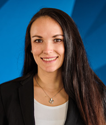 Sona Vodenkova合作论文数Department of Molecular Biology of Cancer, Institute of Experimental Medicine, Czech Academy of Sciences;Biomedical Center, Charles University4
 Jan Procházka合作论文数Faculty of Agriculture, University of South Bohemia4Jan Soukup合作论文数3
 Pavel Klener合作论文数3Diana Tuskova合作论文数3Michal Kraus合作论文数3Jana Karolova合作论文数3Linda Krobova合作论文数Czech Acad Sci, Inst Biotechnol, Prague 25250, Czech Republic3Zuzana Ezrova合作论文数3
 Marek Trneny合作论文数3E Pokorná合作论文数3
 Olena Butenko合作论文数3Hubackova Sona合作论文数3
 Dzamba David合作论文数3Ghayee Hans K合作论文数3Kriska Jan合作论文数3Klanova Magdalena合作论文数3Forsterova Kristina合作论文数3Vockova Petra合作论文数3Prukova Dana合作论文数3
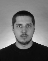 Jiri Cerny合作论文数Institute of Organic Chemistry and Biochemistry, Academy of Sciences of the Czech Republic.3Thanh-Truc Huynh合作论文数National Institute of Neurological Disorders and Stroke Clinical Neurocardiology Section USA3
 Karel Pacak合作论文数Charles University3Vladimir Korinek合作论文数Institute of Molecular Genetics;Academy of Sciences of the Czech Republic3
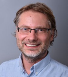 Ondrej Havranek合作论文数Charles University in Prague, General University Hospital in Prague3
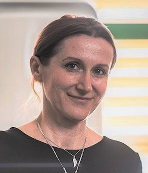 Eva Froňková合作论文数Department of Paediatric Haematology and Oncology, Second Faculty of Medicine, Charles University3L. Andera合作论文数2D. Kolenicova合作论文数2P. Honsa合作论文数2L. Janeckova合作论文数2Verena Puttrich合作论文数2




