The different methods in differentiating biliary atresia (BA) from non-BA-related cholestasis were evaluated in order to provide a prnctical basis for a rapid, early and accurate differential diagnosis of !he diseases. 396 infauts with cholestatic jaundice were studied prospectively during !he period of May 2007 to Iune 2011. The liver function in ali subjects was tested. Ali cases underwent abdominal ultrasonography aud duodenal fluid examination. Most cases were subjected to hepatobiliary scintigraphy, magnetic resonance cholaugiopancreatography (MRCP) and a percutaneous liver biopsy. The diagnosis of BA was finally made by cholangiography or histopathologic examination. The accuracy, sensitivity, specificity and predictive values of these various methods were compared. 178 patients (108 males and 70 females with a mean age of 58±30 days) were diagnosed as having BA. 218 patients (136 males and 82 females with a mean age of 61±24 days) were diagnosed as having non-BA etiologies of cholestasis jaundice during 1he follow-up period in which jaundice faded after treatruent with medica! 1herapy. For diagnosis of BA, clinical evaluation, hepatomegaly, stool color, serum gamma-glutamyltranspeptidase (GGT), duodenal juice color, bile acid in duodenal juice, ultrasonography (gallbladder), ultrasonography (griangular cord or strip-apparent hyperechoic foci), hepatobiliary scintigraphy, MRCP, liver biopsy hadan accuracy of 76.0%, 51.8%, 84.3%, 70.0%, 92.4%, 98.0%, 90.4%, 67.2%, 85.3%, 83.2% and 96.6%, a sensitivity of 83.l %, 87.6%, 96.1%, 73.7%, 90.4%, 100%, 92.7%, 27.5%, 100%, 89.0% and 97.4%, a specificity of70.2%, 77.5%, 74.8%, 67.0%, 94.0%, 96.3%, 88.5%, 99.5%, 73.3%, 75.4% and 94.3%, a positive predictive value of 69.0%, 72.6%, 75.7%, 64.6%, 92.5%, 95.7%, 86.8%, 98.0%, 75.4%, 82.6% and 98.0%, anda negative predictive value of 83.6%, 8.5%, 95.9%, 75.7%, 92.3%, 100%, 84.2%, 93.7%, 100%, 84.0% and 92.6%, respectively. It was concluded that all 1he differential diagnosis methods are useful. The test for duodenal drainage and elemeuts is fast and accurate. It is helpful in the differential diagnosis ofBA and non-BA etiologies of cholestasis. It shows good prnctical value clinically.
The different methods in differentiating biliary atresia (BA) from non-BA-related cholestasis were evaluated in order to provide a practical basis for a rapid, early and accurate differential diagnosis of the diseases. 396 infants with cholestatic jaundice were studied prospectively during the period of May 2007 to June 2011. The liver function in all subjects was tested. All cases underwent abdominal ultrasonography and duodenal fluid examination. Most cases were subjected to hepatobiliary scintigraphy, magnetic resonance cholangiopancreatography (MRCP) and a percutaneous liver biopsy. The diagnosis of BA was finally made by cholangiography or histopathologic examination. The accuracy, sensitivity, specificity and predictive values of these various methods were compared. 178 patients (108 males and 70 females with a mean age of 58±30 days) were diagnosed as having BA. 218 patients (136 males and 82 females with a mean age of 61 ±24 days) were diagnosed as having non-BA etiologies of cholestasis jaundice during the follow-up period in which jaundice faded after treatment with medical therapy. For diagnosis of BA, clinical evaluation, hepatomegaly, stool color, serum gamma-glutamyltranspeptidase (GGT), duodenal juice color, bile acid in duodenal juice, ultrasonography (gallbladder), ultrasonography (griangular cord or strip-apparent hyperechoic foci), hepatobiliary scintigraphy, MRCP, liver biopsy had an accuracy of 76.0%, 51.8%, 84.3%, 70.0%, 92.4%, 98.0%, 90.4%, 67.2%, 85.3%, 83.2% and 96.6%, a sensitivity of 83.1%, 87.6%, 96.1%, 73.7%, 90.4%, 100%, 92.7%, 27.5%, 100%, 89.0% and 97.4%, a specificity of 70.2%, 77.5%, 74.8%, 67.0%, 94.0%, 96.3%, 88.5%, 99.5%, 73.3%, 75.4% and 94.3%, a positive predictive value of 69.0%, 72.6%, 75.7%, 64.6%, 92.5%, 95.7%, 86.8%, 98.0%, 75.4%, 82.6% and 98.0%, and a negative predictive value of 83.6%, 8.5%, 95.9%, 75.7%, 92.3%, 100%, 84.2%, 93.7%, 100%, 84.0% and 92.6%, respectively. It was concluded that all the differential diagnosis methods are useful. The test for duodenal drainage and elements is fast and accurate. It is helpful in the differential diagnosis of BA and non-BA etiologies of cholestasis. It shows good practical value clinically.
Biliary atresia (BA) is the most common cause of persistent jaundice in infant,it can rapidly advance to biliary cirrhosis and death.Surgical treatment in time can decrease mortality,so it is essential to be differentiated from other diseases as early as possible.Main diagnosis methods of BA include clinical feature of patients,liver biopsy,and high-frequen cy ultrasonography,MR cholangiopancreatography (MRCP),hepatobiliary scintigraphy.High-frequency ultrasonography is superior to other methods with high diagnostic specificity and sensitivity.The progresses of ultrasonography in diagnosis of BA were reviewed in this article.
Objective:To study the correlation between the features of ultrasonography,histopathology and ultrastructural pathologic changes in children with non-biliary atresia (BA) cholestatic diseases.Methods:26 children with non-biliary atresia (BA) cholestatic diseases were received liver biopsy guided by ultrasonography.The children were scanned by ultrasound before the biopsy,and the tissues obtained by biopsy were send for pathology or gene examination.Results:Ultrasonography of 26 children showed hyperechoic liver parenchyma in 8 cases,Glisson system wall thickening in 4 cases,coarse parenchyma in 3 cases,hepatomegaly in 5 cases,and no special changes in other 6 cases.Ordinary pathologic reports included 18 cases of inflammatory cell infiltration,14 hepatocyte hydropic degeneration,13 cholestasis,and others included hepatocyte swelling,fatty degeneration and fiber tissues proliferation.Ultrastructural pathologic changes showed 20 cases of hepatocyte cholestasis,17 dilation of cholangiole and 15 increased glycogen.Other findings were as follows:fatty degeneration,fibrous tissues proliferation and inflammatory cell infiltration.Conclusion:US features can reflect the liver pathological state,and these can guide clinical diagnosis and estimate prognosis.The diagnosis of hereditary metabolic disorders could be made by liver biopsy,especially the ultrastructure pathology.
Objective To evaluate the differential diagnostic value and superior of biliary atresia(BA)in the infants with cholestatic hepatopathy by high-frequency ultrasonography (HUS).Methods After 4 hours fasting,124 infants with cholestatic hepatopathy were scanned with high-frequency US.The data of hepatic size and parenchyma,gallbladder,triangular cord (TC) sign,bile duct,right hepatic artery (RHA)and portal vein (PV) were observed and measured.Meanwhile,the other data were collected,which included the clinical diagnosis,blood biochemical tests,the MRCP and dynamic duodenal liquid color check finding,the pathological results after liver puncture biopsy and so on.Results In 124 infants with cholestatic hepatopathy,BA was found in 61 infants and ruled out in 63.TC thickness,RHA diameter,and gallbladder length and width exhibited significant differences between the group with BA and the group non-BA(all P <0.001).The correctness for the diagnosis of BA was 90.3% by the combination of TC sign and abnormal gallbladder morphology,and 83.1 % by stool color,81.5 % by γ-GT,47.5 % by MRCP,83.3 % by dynamic duodenal liquid color check,95.2% by the pathology after liver puncture biopsy,respectively.Conclusions HUS is superior to other diagnostic methods in BA with higher accuracy rate,noninvasion,simplicity and economy.
Objective To investigate whether ultrasound(US) irradiation combined with nano/microbubble(NMB) can enhance the inhibiting effects of adriamycin on H22 hepatocarcinoma in mice.Methods 50 models of transplanted subcutaneously tumors in female KM mice were randomly divided into five groups,ten mice in every group:the control group,ADM group,ADM+NMB group,ADM+US group and ADM+NMB+US group.The mice were treated every other day for five times.The growth of the tumor was monitored by ultrasound,the necrosis area and the microvascular density of the extracted tumor were obtained,the remained mice were fed as usual to observe the survival time.Results Compared with the control group,treatment group tumors grew slowly,the lifespan lengthened,the tumor's necrosis area increased,the microvascular density decreased(P<0.05),and the difference of the ADM+NMB+US group is more significant compared with the other experience groups(P<0.05).Conclusions Ultrasound combined with nano/microbubbles can enhance the inhibiting effect of adriamycin on H22 hepatocarcinoma in mice.
Objective To investigate whether ultrasound mediated nano/microbubbles(NMB) could reduce the dosage and cardiotoxicity of adriamycin(ADM) on the treatment of H22 transplanted mice model.Methods Thrity models of transplanted subcutaneously tumors in female KM mice were randomly divided into 5 groups(n =6):the control group,ADM 1.0 mg/kg,1.5 mg/kg,2.5 mg/kg groups and 1.0 mg/kg ADM + NMB + US group.The mice were treated every other day for five times.After treatment the mice were sacrificed and the growth of the tumor was monitored by ultrasound and drawn tumor growth curve diagram.The tumor was extracted for HE staining,CD34 and ki67 immumohistochemical staining to observe and count the necrosis area,the microvascular density and the proliferation of tumor cells.The myocardial enzymes (AST,CK,LDH) were assayed to evaluate the cardiac toxicity of each treatment in mice.Results Compared with the control group,the all experiment groups were with a tumor growth slowly,necrosis area enlargement,microvascular density and the tumor cell proliferating phase decreased (P<0.05).The tumor's volume,microvascular density and the tumor cell proliferating phase in the 1.0 mg/kg ADM+ NMB+ US group were less than those in other groups(P <0.05) expect for 2.5 mg/kg ADM group(P >0.05).The values of AST,CK and LDH in 1.0 mg/kg ADM + NMB + US group were less than those in 2.5 mg/kg ADM group(P <0.05).The CK in 1.0 mg/kg ADM + NMB + US group was less than that in 1.5 mg/kg ADM group(P =0.002).Conclusions Ultrasound irradiation combined with nano/microbubbles can keep the therapeutic effect of adriamycin but decrease its dose and toxicity on H22 transplanted mice.
Objective To investigate the optimal ultrasound exposure parameter on H22 neoplasms of mice meditated by ultrasound exposure combined with self‐made nanobubbles ,and then observe their therapeutic effect combined with cisplatin and their possible mechanism of anti‐tumor . Methods Thirty mice engrafts models with subcutaneous H22 neoplasms were established and divided into 6 groups randomly ,which received ultrasound exposure at different intensity and exposure time . The contrast enhanced ultrasound imaging ( CEUS ) was performed in every group at the four time points of before treatment and at 0 h ,24 h ,72 h after treatment . To obtain the optimal ultrasound parameters ,the tumor inhibitory effect was assessed by enhanced intensity ( EI) and microvascular density ( MVD) . The H22 tumor were treated by ultrasound exposure nanobubbles combined with cisplatin to observe their tumor growth inhibition rate ,and the microvessels density and nuclear associated antigen Ki‐67 proliferation index were measured by immunohistochemical staining . Results There was a statistically difference in enhanced intensity (EI) between the experimental groups and control group ( P < 0 .05) . With the increasing of ultrasound intensity and exposure time ,the tumor inhibitory effect was more obvious ,with an increasing side reactions . Except the simple ultrasound group ,there was a statistical difference in tumor inhibition ,the mean MVD and the tumor cell proliferation index (KI‐67) between control group and the other ultrasound therapy groups ( P<0 .05) . The tumor inhibitory rate was the highest ( tumor inhibition rate 70 .0% ) and the mean MVD and KI‐67 expression were the lowest ( P <0 .05) in the combination group comparing with the others . Conclusions The ideal ultrasound exposure parameter of tumor inhibition showed that exposure intensity chose 1 W/cm2 and exposure time chose 1 min or 3 min intermittence . The ultrasound exposure self‐made nanobubbles combined with cisplatin could enhance the tumor inhibitory effect .Its mechanism may be related to the decrease of microvascular density ,the inbition of tumor cell proliferation and the increase of tumor cell necrosis .
Objective To summarize the sonographic characteristics of high frequency ultrasound and elastography of thyroid microcarcinoma(TMC),and to analyze the causes of misdiagnosis.Methods The preoperative ultrasonic data of 245 suspicious TMCs in 202 patients,as confirmed by operation pathology,were retrospectively analyzed.Results Preoperative ultrasonography accurately diagnosed 221 TMCs,the diagnosis rate was 90.2%,and the misdiagnosis rate was 9.8%.Among the missed diagnosed lesions,18 lesions were nodular goiter,others were 3 nodular goiter with focal papillary hyperplasia of follicular epithelium,2 nodular goiter with adenomatous hyperplasia,1 focal lymphocytic thyroiditis,respectively.The thyroid lesions≤0.5 cm in diameter were more easily to misdiagnose.According to the importance of the ultrasonographic features of TMC,the order were aspect ratio (A/T) ≥ 1,irregular-shape,microcalcifications,low or very-low echo.Based on the above corresponding characteristics and considering other features together,the diagnostic accuracy rate were 94.1 %,93.9%,92.4% and 90.5%,respectively.Less blood supply and ill-defined boundary were the secondary sonographic signs of TMC.The elastographic scores of TMC were most showed 4 to 5 points.Diagnosis of TMC relied on elastography alone is less effective,but when elastograph diagnosis based on high frequency ultrasound,the diagnostic accuracy is much higher,especially when there is no calcification in the lesions.Conclusions High-frequency ultrasound has a very important value in the diagnosis of TMC,while elastography has certain assistant value on the basis of high-frequency ultrasonic diagnosis.
超声分子成像以靶向超声微泡造影剂为显影剂,能够对体内组织器官病变进行微观的探测与显像,且随着这项技术的进步,已使疾病的诊断及治疗成为一体。国内外学者在造影剂表面或内部载入基因或药物,使超声造影剂成为一种安全、便捷的载体,靶向释放药物或基因,从而达到治疗疾病的目的。现就超声联合微泡在肿瘤治疗中的研究进展作一综述。
目的 通过超声检测重症全身性感染患者肾血流的变化,探讨其在重症全身性感染患者急性肾损伤早期诊断中的意义.方法 选取2011年4月-2012年6月入住ICU诊断为重症全身性感染患者27例,其中急性肾损伤的患者17例(急性肾损伤组),非急性肾损伤的患者有10例(非急性肾损伤组);于患者入院的第1天和第5天血流动力学稳定时,采用超声测量肾脏大小以及肾脏上中下三级叶间动脉的收缩期峰值流速以及舒张期峰值流速,计算肾动脉阻力指数以及肾血管搏动指数,并且常规检测血肌酐的数值;采用SPSS11.0统计软件进行数据分析.结果 患者入院第1天急性肾损伤患者组的肾动脉阻力指数显著高于非急性肾损伤患者组,分别为0.77、0.62(P<0.01);人院第5天急性肾损伤患者组肾叶间动脉的肾动脉阻力指数较非急性肾损伤患者组显著升高,分别为0.75、0.70(P<0.01).结论 超声检测肾动脉阻力指数可以作为重症全身性感染患者急性肾损伤早期诊断的一种方法.
Objective To investigate the value of serum interleukin-6 (IL-6) level in predicting white matter injury in preterm very low birth weight infants (VLBWI) and extremely low birth weight infants (ELBWI).Methods Totally,98 VLBWI and ELBWI were admitted to Tongji Hospital between June 2012 and June 2013,and were divided into two groups according to the neuroimaging findings:46 infants with white matter injury as white matter injury group and 52 infants without white matter injury as control group.Serum IL-6 levels within one hour and at day 7 after birth were detected by enzyme-linked immunosorbent assay.Receiver operating characteristic curve was used to analyze the sensitivity and specificity of serum IL-6 levels in predicting white matter injury.Two-sample t test,Chi-square test and Wilcoxon rank sum test were used for statistical analysis.Results The mean gestational age and mean birth weight of all infants were (29.8± 1.8) weeks (26-34 weeks) and (1 261 ±162) g (780-1 490 g).Compared with the control group,the white matter injury group infants had lower 5 min Apgar score (4.6±2.1 vs 6.2± 1.5,t=1.983) and higher incidence of intrauterine infection [56.5% (26/46) vs 19.2% (10/52),x2=14.605],intubation-surfactantextubation [67.4% (31/46) vs 32.7% (17/52),x2=11.760],and Ⅰ-Ⅱ and Ⅲ-Ⅳ grade of periventricularintraventricular hemorrhage [60.9% (28/46) vs 34.6% (18/52),x2=6.755; 28.3% (13/46) vs 11.5% (6/52),x2=4.367; all P<0.05].The serum IL-6 levels within one hour after birth were significantly increased in the white matter injury group compared with the control group [130.7 pg/ml (38.2-1 020.0 pg/ml) vs 46.3 pg/ml (13.6-336.0 pg/ml),Z=-6.929,P=0.000].Receiver operating characteristic curve showed that,if the cut-off point of serum IL-6 level was 55.0 pg/ml,the sensitivity and specificity of IL-6 within one hour after birth were 93.5% and 72.3%,respectively,in predicting white matter injury in VLBWI and ELBWI,with the area under the curve of 0.907 (95%CI:0.844-0.970,P=0.000).There were no significant differences of IL-6 levels between the two groups at day 7 after birth [18.2 pg/ml (3.0-231.0 pg/ml) vs 16.4 pg/ml (1.0-121.0 pg/ml),Z=-0.246,P=0.806].Conclusions Serum IL 6 level within one hour after birth can be used as a biomarker to predict whitez matter injury in VLBWI and ELBWI.
BACKGROUND: It is a globally challenging problem to differentially diagnose biliary atresia (BA) from other disease processes causing infantile cholestatic jaundice. The high frequency ultrasonography (HUS) yields much improved spatial resolution and therefore, might show better image in BA diagnostic examination. The present study was to evaluate the HUS on the diagnosis of BA in infants with jaundice. METHODS: Fifty-one infants with neonatal jaundice were scanned with ultrasonography. Images included gallbladder, bile duct, right hepatic artery (RHA), portal vein (PV) and triangular cord (TC) sign, magnetic resonance imaging and additionally laboratory tests and histopathology reports were assessed. RESULTS: Twenty-three BA and 28 non-BA cases were con firmed. The sensitivity, specificity, and accuracy of HUS were 91.3%, 92.9%, and 92.2%, respectively. All of these indices were significantly higher than those of conventional ultrasonography (P<0.01) and MR cholangiopancreatography (P<0.05). The HUS features, included a positive TC sign, an increased RHA diameter and RHA-diameter to portal-vein-diameter ratio (RHA/PV) and abnormal gallbladder, were important in the diagnosis of BA. CONCLUSION: HUS provided better imaging of BA and should be considered as a primary modality in the differential diagnosis of infantile jaundice.
Objective To observe self-made cationic nanobubbles as non-viral gene carrier to transfer green fluorescent protein reporter gene into HepG2 cell in vitro.Methods Cationic nanobubbles(PNB) were prepared by sonicating liposomes、polyethylenimine and perfluoropropan.The surface potential and the size of nanobubbles were assessed by laser particle analyzer.HepG2 cells were incubated with DNA,nanobubbles with or without ultrasound exposure.The transfection efficiency was evaluated by flow cytometer and the cell viability by cell counting Kit-8.Results The mean diameter of PNB was (834.57 ± 6.4) nm and the surface charge was (4.15± 0.98)mV.The PNB-DNA complexes,which blocked by the Agarose gel electrophoresis,could effectively transfer HepG2 cells,and the ultrasound exposure could enhance the transfection efficiency further significantly (P < 0.05).Conclusions The new PNB could effectively combine with pDNA to enhance gene delivery and ultrasound exposure could improve its efficiency further in HepG2 cell in vitro.
Objective To investigate whether ultrasound-mediated microbubble destruction(UMD) could enhance cationic liposome (CL) induced plasmid DNA delivery or not,and optimize the transfection conditions.Methods Multiple parameters were explored to obtain the optimal transgene efficiency by means of with or without serum in culture medium,various CL or nano-liposomal bubble(NB) concentrations,different time point of ultrasonic irradiation.The transfection efficiency was assessed by fluorescence microscopy and flow cytometer,and cell viability was evaluated by Cell Counting Kit-8 (CCK-8) assay.Results The serum could protect the cells but show little impact on transfection efficiency induced by CL.CL and plasmid DNA at a weight ratio of 4 ∶ 1 exhibited high transfection efficiency of (17.71-± 0.79)% and high cell viability of (91.28 ± 0.76) %.CL combining with ultrasonic irradiation at the time point of 1 hour could increase the transfection efficiency to (24.85 ± 0.78)% (P <0.01).Higher transfection rate (32.47 ± 4.01) % was obtained by adding NB at the concentration of 10 % (P <0.05).Conclusions UMD accompanied with CL could enhance gene delivery effectively,which would provide a new method for gene therapy.
Objective Different diagnostic methods of biliary atresia (BA) and infantile intrahepatic cholestasis disease were investigated in order to find a simple,fast,practical,economic and non invasive differential diagnostic method. Methods A total of 584 cases of infantile cholestasis were collected from May 2006 to June 2012 for persistent jaundice,pale yellow or white shit who lived in Department of Pediatric Digestion and Infection,Tongji Hospital,Tongji Medical College,Huazhong University of Science & Technology (HUST) . Seven methods including clinical diagnosis,blood biochemical tests,liver and gallbladder ultrasonography,dynamic continuous duodenal liquid bile check,nuclide hepatic imaging,magnetic resonance imaging and histology were applied for differential diagnosis and the results were analyzed. Results The correctness of clinical diagnosis method was 74.5%,sensitivity 81.6%,specificity 69.9%;liver size: 49.0%,89.0%,and76.9%;stool color:83.2%,96.1% and96.7%;blood total bilirubin:63.0%,93.1% and91.2%;serum γ-GT:79.7%,71.1% and78.7%;dynamic duodenal liquid color check:93.3%,91.7% and92.7%;bile acid of duodenal liquid:97.8%,100.0% and 100.0%;B graphy :89.7%,91.7%and94.3%;porta fibre block check: 72.1%,29.4%and 68.7%;nuclide hepatic imaging:60.5%,100.0% and 100.0%;MRCP:88.3%,96.5%and94.4%;liver pathology:97.4%,98.2% and 94.9%. Conclusion Differential diagnosis in 1 week can help differentiate biliary atresiafrom infantile intrahepatic cholestasis. B-ultrasonography and dynamic duodenal fluid test are simple,fast practical,economic and noninvasive as differential diagnostic methods.
Objective:To assess the incidence of focal spared areas around bed of gall baldder in hepatic steatosis.Methods:543 patients were diagnosed hepatic steatosis on ultrasound including 310 male and 233 female.Mean age was 45.6 years(range,13~69 years).All patients underwent routine ultrasound examination by the same sonographer.More attention was paid to areas around bed of gall bladder.Results:The incidence of focal spared areas around bed of gall bladder in patients with hepatic steatosis was 81.9%.The incidence was 81.2% and 87.1% in patients with mild hepatic steatosis and moderate-severe hepatic steatosis respectively.The incidence was 83.5% and 79.8%for men and women in patients with hepatic steatosis respectively.No correlation was found between degree of hepatic steatosis(P=0.226)and gender(P=0.265).Conclusion:There exists high incidence of focal spared areas around bed of gall bladder in hepatic steatosis.These focal sapred areas should be considered in the differential diagnosis of primary or secondary lesions of liver.
Ultrasound-triggered phase transition sensitive nanodroplets with multimodal imaging functionality were prepared via premix Shirasu porous glass (SPG) membrane emulsification method. The nanodroplets with fluorescence dye DiR and SPIO nanoparticles (DiR-SPIO-NDs) had a polymer shell and a liquid perfluoropentane (PFP) core. The as-formed DiR-SPIO-NDs have a uniform size of 385 ± 5.0 nm with PDI of 0.169 ± 0.011. The TEM and microscopy imaging showed that the DiR-SPIO-NDs existed as core-shell spheres, and DiR and SPIO nanoparticles dispersed in the shell or core. The MTT and hemolysis studies demonstrated that the nanodroplets were biocompatible and safe. Moreover, the proposed nanodroplets exhibited significant ultrasound-triggered phase transition property under clinical diagnostic ultrasound irradiation due to the vaporization of PFP inside. Meanwhile, the high stability and R2 relaxivity of the DiR-SPIO-NDs suggested its applicability in MRI. The in vivo T2-weighted images of MRI and fluorescence images both showed that the image contrast in liver and spleen of rats and mice model were enhanced after the intravenous injection of DiR-SPIO-NDs. Furthermore, the ultrasound imaging (US) in mice tumor as well as MRI and fluorescence imaging in liver of rats and mice showed that the DiR-SPIO-NDs had long-lasting contrast ability in vivo. These in vitro and in vivo findings suggested that DiR-SPIO-NDs could potentially be a great MRI/US/fluorescence multimodal imaging contrast agent in the diagnosis of liver tissue diseases.
Objective To made a kind of nano-liposomal bubbles(NB) and observe its base physical properties and ultrasound contrast enhanced effect,and compared with SonoVue in vitro and in vivo.Methods The liposome was made with the method of reverse phase evaporation,and then the liposome was sonieated to prepare NB.After the physical properties of NB such as its morphous,average particle size,surface potential and concentration were observed and calculated,200 μl of saline and two contrast agents (NB and SonoVue) were injected into degassed PBS (10 ml) respectively to observe their enhancing effects.For in vivo study,a healthy rabbit heart and liver were imaged before and after intravenous injection of 2.0 ml/kg of saline and two contrast agents in succession to compare their dynamic enhancing effects in the grey scale imaging subjectively.Results The self-made NB distributed uniformly and its size ranged from 133.1 ~199.5 nm with a mean diameter of (171.60 ± 30.82) nm.Its surface potential and concentration were -(1.92± 0.65)mV and (3.8 ~ 5.6) × 108/ml individually.These basic characteristics were not observed changing dramatically after placement one week to one month under room temperature.The NB and SonoVue all displayed significantly enhancing effect comparing with saline no matter in vitro or in vivo.Conclusions The self-made NB are stable and distribute uniformly,which display the same contrast enhancing effect as SonoVue in vitro and in vivo.The self made NB would have a more potential use in ultrasound molecular imaging and gone or drug delivery.
With the development of imaging technology and ultrasound contrast agents,ultrasound microbubbles are not only used for diagnosis,but also for therapy.Recently,more attention has been paid to use microbubbles as a new carrier of drug or gene.Meanwhile,ultrasound mediated microbubbles destruction can release the therapeutic drug or gene locally and suggest a certain advantage for target therapy.The application and development of ultrasound and microbubbles in liver carcinoma targeted therapy were reviewed in this article.
 Qingping Zhang (张青萍)合作论文数Tongji Hospital, Tongji Medical College, Huazhong University of Science and Technology34
 Kaiyan Li (李开艳)合作论文数Tongji Hospital, Tongji Medical College of Huazhong University of Science and Technology22
 Daozhong Huang (黄道中)合作论文数Tongji Hospital, Tongji Medical College of Huazhong University of Science and Technology19
 Naxiang Liu合作论文数9Kai Hong合作论文数Huazhong University of Science and Technology(Huazhong University of Science and Technology,Huazhong Univ. of Sci. and Technol.),Wuhan,China9
 Zhihui Wang (王志辉)合作论文数Tongji Hospital, Tongji Medical College, Huazhong University of Science and Technology8Lu Ding合作论文数6Xiao-ling Liu合作论文数6Fen Wang (王芬)合作论文数6Ting Xiao (肖婷)合作论文数6
 Zhihua Huang (黄志华)合作论文数Tongji Hospital, Tongji Medical College, Huazhong University of Science and Technology5JiuJun Chen (陈九军)合作论文数4Wei Zhu (朱蔚)合作论文数4
 Liangjun Hu合作论文数4
 Hongchang Luo (罗鸿昌)合作论文数Tongji Hospital, Tongji Medical College, Huazhong University of Science and Technology4Yuan Deng (邓远)合作论文数3Xian Cui (崔贤)合作论文数3
 Youbin Deng (邓又斌)合作论文数Tongji Hospital, Tongji Medical College of Huazhong University of Science and Technology3Guirong Le (乐桂蓉)合作论文数2Fen Xu (徐芬)合作论文数2Yu Liu (刘玉)合作论文数2Yan Liu (刘艳)合作论文数2Na Liu (刘娜)合作论文数2
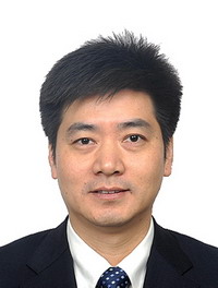 Xiaoping Luo (罗小平)合作论文数Tongji Hospital, Tongji Medical College of Huazhong University of Science and Technology2
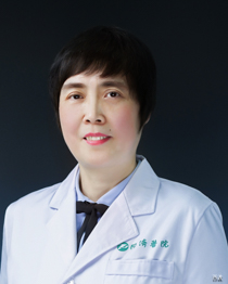 Chunlei Li (黎春雷)合作论文数Tongji Hospital, Tongji Medical College, Huazhong University of Science and Technology2Hui-Yun Zhu合作论文数1Jie Wan合作论文数1CHEN Ling (陈玲)合作论文数1renhui li (李仁会)合作论文数1Jingzhou Yang (杨静洲)合作论文数1Jingjing Shi合作论文数1yingliang li (李英亮)合作论文数1Haiying Song (宋海英)合作论文数1LongHai Yi (衣龙海)合作论文数1Chen Dong (董琛)合作论文数1BinHua Luo (罗斌华)合作论文数1YuanYuan Zhou (周元媛)合作论文数1Yang Guan (官阳)合作论文数1JinBing Li (李进兵)合作论文数1ZhiHua Li (李志花)合作论文数1AiYun Zhou (周爱云)合作论文数1HuanMing Wu (吴焕明)合作论文数1Ping Li (黎萍)合作论文数1HaiXia Li (李海霞)合作论文数1Chen Chen (陈琛)合作论文数1




