INTRODUCTION:Obesity is characterized by hypothalamic dysfunction and metabolic dysregulation. While high-intensity interval training (HIIT) effectively treats obesity, the underlying central mechanisms remain unclear. This study aims to investigate HIIT's effects onmetabolic parameters and hypothalamic gene expression, exploring possible pathways linking central and peripheral adaptations. MATERIAL AND METHODS:Our study evaluated HIIT-induced metabolic changes in male diet-induced obese (DIO) mice. Body weightand caloric intake were recorded throughout the intervention period, and glucose regulation was assessed via glucose tolerance test(GTT) and insulin tolerance test (ITT). Following the HIIT intervention, blood was collected for cytokine assessment and hypothalamictissue was harvested for molecular characterization. RNA sequencing (RNA-seq) was employed to analyze gene expression, with enrichmentanalysis to identify biological pathways, and reverse transcription quantitative polymerase chain reaction (RT-qPCR) to validate keyfindings. Protein expression was examined through Western blotting, and circulating cytokine levels were quantified using enzyme-linkedimmunosorbent assay (ELISA). RESULTS:High-intensity interval training significantly reduced caloric intake while improving glucose metabolism and insulin sensitivity in DIO mice, accompanied by hypothalamic transcriptomic remodeling. RNA-seq identified 178 upregulated and 292 downregulated genes post-HIIT,with pathway enrichment analysis revealing activation of interleukin 17 (IL-17) signaling. Notably, HIIT increased hypothalamic lipocalin 2(Lcn2) mRNA and protein expression, which correlated with decreased hypothalamic apoptosis and elevated hypothalamic pro-opiomelanocortin(POMC) expression in DIO mice. Consistently, circulating IL-17 levels were elevated following HIIT intervention. CONCLUSIONS:Collectively, our findings identify hypothalamic Lcn2 as a prominent molecular responder to short-term HIIT training, whoseupregulation parallels enhanced IL-17 signaling and coincides with the recovery of POMC neurons.
AIMS:To evaluate the efficacy and safety of low-frequency repetitive transcranial magnetic stimulation (LF-rTMS) on the right dorsolateral prefrontal cortex (DLPFC) in patients with post-stroke depression (PSD), and to explore the possible neural mechanism of repetitive transcranial magnetic stimulation (rTMS) using functional near-infrared spectroscopy (fNIRS). METHODS:28 patients were randomly assigned to the rTMS group (n = 14) and control group (n = 14). The treatment effect was assessed by the Hamilton Depression Scale (HAMD), Hamilton Anxiety Scale (HAMA), National Institutes of Health Stroke Scale (NIHSS), and Barthel Index (BI). In addition, fNIRS was used to detect changes in blood oxygen concentration in the frontal cortex. RESULTS:After treatment, HAMD scores in the rTMS group decreased significantly more than those in the control group (p < 0.05). Oxyhemoglobin (HbO) in CH17 increased and deoxyhemoglobin (HbR) in CH6 decreased after rTMS (p < 0.05). HbR in CH6 and CH8 decreased significantly, while HbR in CH12 and CH15 increased significantly (p < 0.05). The rTMS group also showed significantly greater improvements in HAMA, NIHSS, and Barthel Index scores (p < 0.05). CONCLUSION:The findings from this pilot study suggest that LF-rTMS on the right DLPFC combined with conventional treatment may be a safe approach to alleviate depressive symptoms in patients with PSD. Furthermore, fNIRS revealed that the mechanism of action may be related to improving left-right brain tissue blood flow and activation balance.
China has the largest population requiring assistive products and the greatest market potential in the world. The demand for assistive product fit services among functionally impaired groups, such as the elderly, individuals with disabilities, and patients with chronic diseases, is increasing steadily. Currently, healthcare institutions play an important role in the assistive products service system. However, problems still exist in service delivery, including unclear service content, non-standardized procedures, and insufficient professional capacity, which hinder the improvement of service quality and industry development. To promote the standardization and normalization of assistive products fitting services and to enhance the service capacity of healthcare institutions, this document systematically specifies the terms and definitions, service principles, service resources, service procedures, service content, documentation management, service management, and quality management and complaint handling associated with assistive products fitting services. It also defines the operational requirements for the entire service process, from reception, assessment, plan formulation, configuration, training, and delivery to follow-up. The issuance and implementation of this specification will provide healthcare institutions with a scientific, systematic, and operational guide for assistive products fitting services, promote the uniformity and improvement of service quality, and contribute to the enhancement and development of the assistive products service system.
Tophi, inflammatory granulomas formed by intra-articular monosodium urate (MSU) crystal deposition, remain challenging due to limited drug efficacy. While arthroscopic surgery removes tophi rapidly, mechanical debridement risks injury, and intraoperative MSU release can trigger severe oxidative stress induced inflammatory storm. To overcome this, we developed a cascade nanozyme, Ce-MOF@Pt, enabling synergistic MSU clearance and ROS scavenging for MSU-induced gouty arthritis. Pt nanoparticles possess oxidase (OXD)-like activity, dissolving deeply embedded MSU crystals without mechanical injury and incidentally producing anti-inflammatory allantoin. The Ce-MOF matrix supports Pt and duplicates catalase (CAT)/superoxide dismutase (SOD)-like activities, cascading H2O2 decomposition and MSU-induced ROS elimination. As UA degradation elevates the local pH values, its dominant activity intelligently switches from OXD-like to SOD-like mode, while the CAT-like activity remains consistently high, ensuring continuous clearance of the H2O2 byproduct and offering integrated anti-oxidative therapy. In vitro, Ce-MOF@Pt reduced inflammation via ROS scavenging and macrophage reprogramming. In vivo, it alleviated pain symptoms and joint inflammation in MSU-induced gouty arthritis mice. Transcriptomics revealed Ce-MOF@Pt mitigated the inflammatory storm by alleviating endoplasmic reticulum stress. This novel Ce-MOF@Pt strategy offers minimally invasive tophi dissolution and prevents MSU-induced inflammation, providing a promising therapeutic approach for tophi.
The loss of endogenous bioelectrical rhythms in bone is a critical driver of osteoporosis initiation and progression. Reconstructing bioelectrical rhythms synchronized with mechanical stimulation is critical for restoring bone homeostasis. Here, we propose a motion-activated skeletal "bioelectrical pacemaker" strategy that microfluidically encapsulates alendronate-modified BaTiO3 piezoelectric nanoparticles (BTO@ALN) within chitosan/alginate core-shell hydrogel microspheres, enabling gastric protection, pH-responsive intestinal release, and noninvasive oral delivery with systemic skeletal enrichment. Upon skeletal enrichment, the piezoelectric units compensate for impaired electrophysiological responsiveness in osteoporotic bone and are activated by physiological mechanical stimuli during daily motion to generate movement-synchronized dynamic bioelectrical signals. This process reconstructs endogenous bone bioelectrical rhythms systemically, thereby restoring bone homeostasis. Experimentally, BTO@ALN exhibited 6.8-fold higher skeletal accumulation than controls; oral administration led to a 52% increase in bone piezoelectric performance within 24 h, accompanied by concurrent suppression of osteoclast activity and promotion of osteogenesis. When combined with low-intensity exercise, motion-triggered piezoelectric output further amplified therapeutic efficacy, resulting in a 2.4-fold increase in osteogenesis and a 1.5-fold stronger suppression of bone resorption. Collectively, this closed-loop strategy integrating oral delivery, bone-targeted enrichment, and motion-synchronized electrical activation systematically re-establishes endogenous bone bioelectrical rhythms, introducing a strategy for osteoporosis based on electrophysiological regulation of bone.
The key to treating infectious bone defects (IBD) is regulating reactive oxygen species (ROS) balance to achieve “anti-infective first, then osteogenesis” temporal sequential repair. However, sensing microenvironmental changes to regulate ROS remains challenging. This study prepared a nanoparticle (PBD-ICG nanoparticlces) capable of sensing pH changes to enable protonation and deprotonation with “triple-element synergy” acid-base-driven assembly method, and loaded onto GelMA/HAMA hydrogel microsphere established a functional-responsiveness hydrogel microsphere (PGI) based on microenvironment sensing. With ultrasonic cavitation, indocyanine green transition to excited state, enabling electron transfer or energy transfer with H2O or 3O₂ in microenvironment to generate ROS. Under 1 W ultrasound, ROS release 74
Background Mirror therapy (MT)-specifically paradigms using mirror visual feedback (MVF)-is widely used in neurorehabilitation; however, mechanistic implementations vary substantially in movement content, rhythmicity and attentional demands. This protocol describes an acute mechanistic, within-participant fNIRS screening study designed to compare three prespecified upper-limb mirror-therapy task paradigms and to quantify associated subjective experience after each condition in healthy adults during a single visit. Methods and analysis This is a single-centre, within-participant, randomised crossover study conducted at Wuhan Wuchang Hospital (Wuhan, China). Healthy adults aged 18-35 years will complete three task conditions once each in a counterbalanced order using a 3*3 Latin-square scheme: UMT1 (task-oriented rhythmic functional movement), UMT2 (open-ended free movement with auditory control), and UMT3 (non-functional rhythmic movement). fNIRS will be acquired using the NirSmart-6000A system during a standardised block design. The primary outcome is ROI-level HbO activation quantified as GLM-derived β estimates within the prespecified primary ROIs (bilateral SM1/M1 and bilateral PMC). Secondary outcomes include ROI-level windowed ΔHbO (5-20 s post-onset relative to the immediately preceding rest; descriptive only), ROI-level ΔHbR, and post-condition subjective ratings (illusion, immersion, confusion and fatigue; 1-7 Likert). Condition effects will be analysed using linear mixed-effects models with fixed effects for condition and period and prespecified multiplicity-adjusted pairwise contrasts. Ethics and dissemination Ethics approval was obtained from the Ethics Committee of Wuchang Hospital Affiliated to Wuhan University of Science and Technology (Approval No.: 2025-112-01; approved on 2025-08-21). The study is expected to be minimal risk. Findings will be disseminated through publication of this protocol manuscript and subsequent results manuscripts and conference presentations. Trial registration number Chinese Clinical Trial Registry (ChiCTR2600116634). This study is conducted as a prespecified mechanistic sub-study under the overarching registered project. ### Competing Interest Statement The authors have declared no competing interest. ### Clinical Trial ChiCTR2600116634 ### Author Declarations I confirm all relevant ethical guidelines have been followed, and any necessary IRB and/or ethics committee approvals have been obtained. Yes The details of the IRB/oversight body that provided approval or exemption for the research described are given below: Ethics committee of Wuchang Hospital Affiliated to Wuhan University of Science and Technology gave ethical approval for this work I confirm that all necessary patient/participant consent has been obtained and the appropriate institutional forms have been archived, and that any patient/participant/sample identifiers included were not known to anyone (e.g., hospital staff, patients or participants themselves) outside the research group so cannot be used to identify individuals. Yes I understand that all clinical trials and any other prospective interventional studies must be registered with an ICMJE-approved registry, such as ClinicalTrials.gov. I confirm that any such study reported in the manuscript has been registered and the trial registration ID is provided (note: if posting a prospective study registered retrospectively, please provide a statement in the trial ID field explaining why the study was not registered in advance). Yes I have followed all appropriate research reporting guidelines, such as any relevant EQUATOR Network research reporting checklist(s) and other pertinent material, if applicable. Yes De-identified participant data will be made available in accordance with ethics approval and institutional policy via a public repository after publication of the primary results; access will require a methodologically sound proposal and a data use agreement prohibiting re-identification Wuhan Natural Science Foundation Key Clinical Research Program for Municipal Medical Institutions, 2026020301040188
Thrombosis and its associated disorders remain critical global health threats, often causing acute organ injury and chronic complications that impair long-term prognosis. Despite clinical advances, current therapies face significant limitations, necessitating safer and more effective interventions. Ultrasound-Targeted Microbubble Destruction (UTMD) has emerged as a promising innovation, utilizing microbubble cavitation to facilitate localized drug delivery. By leveraging both stable and inertial cavitation, UTMD disrupts thrombus architecture and enhances the penetration of thrombolytic agents, thereby reducing required dosages and mitigating systemic toxicity. This review summarizes the microbubble characteristics and ultrasound parameters essential to UTMD, alongside its preclinical applications in thrombolysis. While clinical translation is currently hindered by challenges in stability, targeting precision, and biocompatibility, future research focusing on thrombus-specific targets, multimodal imaging, and optimized material design will be vital for the clinical adoption of UTMD.
INTRODUCTION:Poststroke motor dysfunction places a heavy burden on individuals and society. Virtual reality (VR) offers enhanced motor skill transfer and active rehabilitation by overcoming the scenario-specific constraints of conventional therapies. Validating the efficacy of VR rehabilitation could lead to scalable and cost-effective solutions, potentially enabling home-based rehabilitation. However, the widespread clinical application remains constrained by the lack of rehabilitation-specific VR and multidimensional quantitative assessments. The aim of this study was to investigate the multidimensional effects and neural mechanisms of VR rehabilitation in poststroke motor recovery. METHODS AND ANALYSIS:This study is a prospective, randomised, controlled clinical trial protocol designed to evaluate the effects of multisensory VR training on motor dysfunction in patients who had a stroke using multidimensional assessments. The trial consists of a baseline assessment, a 4-week intervention period and an endpoint assessment. A total of 40 patients who had a stroke will be randomly allocated in a 1:1 ratio to either a VR combined with treadmill group or a treadmill-only group. The primary outcome measure is the Fugl-Meyer Assessment of Lower Extremity score, while secondary outcomes include three-dimensional gait analysis, the Berg Balance Scale score, the activities of daily living score and functional near-infrared spectroscopy results. Safety will be evaluated by monitoring the incidence of adverse events. This study aims to determine whether VR rehabilitation offers superior efficacy in improving motor function in patients who had a stroke by using a multidimensional assessment approach, including neural coupling function, muscle movement mechanics and clinical performance. The findings will provide robust, high-quality evidence to support the broader application of VR in clinical practice. ETHICS, REGISTRATION AND DISSEMINATION:The trial was approved by the Ethics Committee of the First Affiliated Hospital of Chongqing Medical University (2022-155). This study protocol was registered with the clinicaltrials.gov (NCT06275516). The results will be published in a peer-reviewed journal or presented at a conference. TRIAL REGISTRATION NUMBER:NCT06275516.
Objective: To assess alterations in peripheral nerves subsequent to a stroke by utilizing shear wave elastography (SWE) alongside microvascular imaging (MVI) technology, and to investigate the relationship between ultrasound parameters and clinical assessments. Design setting and participants: This cross-sectional investigation involved 41 individuals who experienced a stroke resulting in unilateral hemiplegia. Interventions and main outcome measures: Clinical and ultrasonographic assessments were conducted, which included the Erasmus modification of the Nottingham Sensory Assessment (Em-NSA), the Fugl-Meyer Motor Assessment (FMA), and various ultrasonographic metrics such as cross-sectional area (CSA), Young's modulus (YM), and shear wave velocity (SWV) of the median and tibial nerves, in addition to microvascular imaging of the median nerve. A comparative analysis was performed between the ultrasonographic parameters of the hemiplegic and non-hemiplegic sides. Furthermore, the correlation between ultrasonographic parameters on the hemiplegic side and clinical functionalities was examined. Results: The CSA of the median and tibial nerves on the hemiplegic side demonstrated a marked decrease in comparison to the non-hemiplegic side (P < 0.01). Conversely, the YM and SWV of the median and tibial nerves on the hemiplegic side were markedly elevated compared to those on the non-hemiplegic side (P < 0.01). Additionally, microvascular perfusion in the median nerve on the hemiplegic side was considerably less than that on the contralateral side (P < 0.05). Pearson correlation analysis demonstrated that both YM and SWV of the tibial nerve exhibited a negative correlation with Em-NSA and FMA scores (P < 0.05). Conclusions: There is an augment in peripheral nerve stiffness on the hemiplegic side, accompanied by a decrease in microvascular perfusion. Moreover, YM and SWV of the tibial nerve are associated with sensory and motor deficits. The integration of SWE and MVI techniques may serve as a novel biomarker for evaluating peripheral nerve lesions following a stroke.
Tendon-bone interface (TBI) injuries cause excessive deposition of fibrous scar tissue, leading to irreversible destruction of the complex gradient hierarchy and markedly deteriorating mechanical-functional properties, presenting a persistent repair dilemma in clinical practice. Tissue engineering offers a promising strategy for the regeneration of functional interface tissue, with its core principle being the achievement of effective tissue healing through biomimicry of the gradient structure in the TBI rather than mere anatomical reconnection. Given the heterogeneity of the interface, biomaterials are often designed with stratified structures to replicate the characteristic variations across different regions. These materials, by mimicking the gradient structures of natural tissue, can guide directional cell migration and differentiation, thereby promoting the regeneration of gradient interface tissue. Additionally, they can serve as delivery vehicles for stem cells and bioactive molecules, both of which have been demonstrated as key players in tissue repair. This review summarizes the applications of biomaterials in interface tissue regeneration. First, it introduces the structural and functional aspects of interface tissue and its developmental maturation process. Next, it discusses current therapeutic approaches for TBI healing and highlights the unique advantages of biomaterials in this field. Emphasis is placed on biomimetic strategies for constructing interfacial gradients through material-based approaches, including mechanical gradients, cellular gradients, extracellular matrix (ECM) gradients, structural gradients, and electrical field gradients. Finally, the current challenges and future research directions in the application of biomaterials for interface tissue regeneration are outlined.
Neurogenic bone regeneration is essential for the effective restoration of bone tissue functionality, with exosomes derived from Schwann cells regionalized in bone injury tissue playing a crucial role in this process. However, precisely regulating the secretion of Schwann cells localized in bone injury tissue to enhance neurogenic bone regeneration remains a considerable challenge. In this study, an injectable, ultrasound-responsive piezoelectric conductive short fiber network (US@SFG) was innovatively developed using uniform short fiber homogenization techniques and multifunctional chemical modifications, enabling precise acoustic–electrical conversion that regionally activated the secretion of miRNAs from Schwann cell-derived exosomes, thereby promoting neurogenic bone regeneration. The incorporation of the piezoelectric polymer glycine imparts superior piezoelectric characteristics to the fiber network, while the conjugated π-electron motion within the conductive graphene network enhances internal electron transfer efficiency, thereby facilitating electrical conductivity. Compared with traditional piezoelectric fiber networks, acousto-electric conversion fiber networks demonstrated a 1.7-fold increase in piezoelectric performance and a 30-fold increase in conductivity, facilitating precise electrochemical regulation under ultrasound stimulation. In vitro studies revealed that acousto-electric conversion fiber networks precisely modulate the secretion of localized Schwann cell exosomal miRNAs (miRNA-494-3p, miRNA-381-3p, and miRNA-369-3p), activating the phosphatidylinositol 3-kinase/protein kinase B and Wnt signaling pathways in bone marrow mesenchymal stem cells, and thereby promoting osteogenic differentiation. Furthermore, in vivo experiments confirmed that under ultrasound imaging guidance, acousto–electric conversion fiber networks could be directed precisely to bone defects, where precise control of ultrasound parameters facilitated acoustic–electrical conversion and electrical signal modulation, markedly promoting the formation of neural networks and bone tissue regeneration. In this study, for the first time, an injectable acousto-electric conversion fiber network was constructed to activate Schwann cell exosomes in bone injury tissue regionally, providing a novel therapeutic strategy and potential molecular targets for neurogenic bone regeneration.
With the intensification of global population aging, osteoarthritis (OA) has emerged as a major socioeconomic burden requiring urgent therapeutic interventions. Low-intensity pulsed ultrasound (LIPUS), a non-invasive physical therapy modality, delivers pulsed acoustic energy to target tissues with negligible thermal effects. Accumulating evidence from preclinical studies and randomized controlled trials has demonstrated its potential to decelerate OA progression. This systematic review synthesizes current knowledge on LIPUS-mediated OA management, elucidates mechanistic pathways through biomechanical and molecular analyses, strategies combining LIPUS with biomaterials to improve its efficacy, evaluates clinical translation challenges, and proposes standardized treatment protocols to optimize therapeutic outcomes.
Impaired functional regeneration following tendon-to-bone interface (TBI) injury is a major challenge in sports medicine. The rigidity and limited efficacy of existing rehabilitation strategies remain significant constraints. Physical modalities, leveraging advantages such as non-invasiveness, spatiotemporal controllability, and low immunogenicity, offer effective intervention options for the long-term management of TBI healing. However, conventional physical modalities struggle to address the complex pathological microenvironment involved in TBI healing. In contrast, therapeutic strategies utilizing physical energy-responsive biomaterials enable programmable, precise, and dynamic regulation, potentially integrating these advantages while overcoming inherent limitations, thereby opening new and effective therapeutic avenues for TBI healing. This review systematically examines the benefits, current applications, and shortcomings of physical modalities for TBI injuries, with particular focus on parameter-response relationships and underlying biological effector mechanisms. Furthermore, it summarizes the design strategies and application progress of energy-responsive biomaterials in TBI healing and discusses future directions and promising prospects, aiming to address the core therapeutic challenges in achieving robust TBI healing.
Pressure ulcers (PUs) pose a significant challenge in the care of bedridden patients, to which automated tilt nursing beds have emerged as a promising solution. However, the lack of effective models to elucidate the mechanical responses of deep tissue during assisted repositioning and identify the optimal tilt angle has hindered the implementation of effective automatic assisted repositioning systems for long-term care patients. Therefore, this study developed a novel computational model that integrates the buttocks with a support mattress to simulate automatic assisted repositioning, thereby analyzing deep tissue responses and optimizing tilt angles for effective load offloading. Inverse modeling was used to reconstruct the 3D shape of the buttocks, nodal equivalence techniques were employed to simplify the mesh and accurately represent internal tissue contacts, and soft tissue parameters were optimized using Response Surface Methodology (RSM). Finally, finite element (FE) analysis was conducted to evaluate the biomechanical responses and optimize the repositioning strategies. Model validation demonstrated a deformation error of $6.93~\pm ~7.41$ mm (mean ± standard deviation) and interface pressure differences within 22.4%, demonstrating the efficacy and bio-fidelity of the system. Repositioning simulations at angles from 0° to 30° showed a 20% reduction in total soft tissue strain, with peak equivalent stress decreasing by 22.27% at the mattress- to-buttock interface and by 20.43% at the muscle-to-adipose tissue interface. These simulations suggest that a 30° turning angle is beneficial for alleviating pressure concentration, which may inspire the design and optimization of automatic assisted repositioning strategies in rehabilitation practices.
BACKGROUND:Post-stroke cognitive impairment (PSCI) is a common complication after stroke, and blood pressure management is vital for stroke survivors. However, the association between diastolic blood pressure (DBP) during post-stroke rehabilitation and the risk of PSCI remains unclear. This study aims to evaluate this association in a real-world stroke cohort. METHODS:A total of 508 consecutive stroke patients admitted between May 2022 and March 2024 were included in this study. Univariate and multivariate logistic regression analyses were conducted to identify risk factors for PSCI. Restricted cubic spline (RCS) analysis was performed to explore the association between DBP and PSCI. Logistic regression models were used to evaluate the risk of PSCI in different DBP groups. Subgroup analysis was employed to assess the effect of DBP across different subgroups. RESULTS:During a median follow-up of 5.5 months, 41% of patients were identified as having PSCI. Univariate and multivariate logistic regression analyses indicated that low DBP is an independent risk factor for PSCI (odds ratio [OR] = 0.960 by 1 mmHg, 95% confidence interval [CI] = 0.933-0.988, p < 0.01). RCS analysis revealed a nonlinear relationship between DBP and PSCI incidence, with an inflection point of around 80 mmHg (P overall < 0.001, P nonlinear = 0.002). Logistic regression models suggested a higher risk of PSCI in the group with DBP < 75 mmHg compared with the group with 75-80 mmHg (OR = 2.606, 95% CI = 1.445-4.699, p < 0.01). CONCLUSION:DBP during post-stroke rehabilitation is associated with the incidence of PSCI in a non-linear manner. Specifically, a low DBP level during this period is an independent risk factor for PSCI. Maintaining a DBP above 80 mmHg during rehabilitation is beneficial for preventing PSCI.
BACKGROUND:Synovial inflammation is a pivotal factor in the pathogenesis of osteoarthritis (OA). Platelet-rich plasma-derived Exosomes (PRP-Exos), known for their low immunogenicity, have demonstrated efficacy in modulating chondrocyte function. However, the specific effects and mechanisms of PRP-Exos in synovial inflammation remain unclear. This study aimed to investigate the therapeutic effects and mechanisms of PRP-Exos in synovial inflammation induced by destabilization of the medial meniscus (DMM) in mice. RESULTS:PRP-Exos were extracted via ultracentrifugation. In vivo experiments revealed that PRP-Exos alleviated pain behaviors and synovial inflammation in DMM mice. Furthermore, it was discovered that PRP-Exos enhanced the synovial lymphatic function in DMM mice and promoted lymphangiogenesis. Meanwhile, the therapeutic effect of PRP-Exos on synovial inflammation was attenuated after inhibition of lymphatic function. In vitro studies demonstrated that PRP-Exos enhanced the proliferation, migration, and tube formation ability of lymphatic endothelial cells (LECs), via regulating the PI3K/Akt signaling pathway. CONCLUSIONS:This research is the first to reveal that PRP-Exos alleviate pain behaviors and synovial inflammation in DMM mice through activation of the PI3K/Akt signaling pathway in LECs, thereby enhancing synovial lymphatic function and promoting the clearance of inflammatory cells and associated cytokines. These findings offer a novel theoretical foundation for the treatment of synovial inflammation and other inflammation-associated disorders.
The main reasons why the current treatment methods for rheumatoid arthritis are not efficient in treating RA at present are insufficient drug control and the inability of surgery to repair the damage. In this study, a multifunctional hydrogel was constructed by cross-linking β-cyclodextrin with sodium alginate to synergistically utilize the photodynamic effect and arthritis therapeutic activity of curcumin. Based on the cuprous oxide/calcium peroxide complex system, the ROS produced by photoexcitation of curcumin can trigger the conversion of cuprous oxide to copper oxide, while the continuous oxygen supply of calcium peroxide enhances the efficacy of PDT. The combined effect jointly inhibits the differentiation of osteoclasts. This system forms a self-regulating collaborative treatment system through the linkage mechanism of “ROS generation – oxygen supply – bone repair”. This catalytic composite hydrogel exhibits excellent photoresponse characteristics. Compared with the second-generation traditional photosensitizers, it significantly reduces the subsequent generation of ROS, confirming its precisely controllable ROS generation ability. Moreover, by synergistic regulating the GPX4-mediated ferroptosis pathway, it promotes ferroptosis in osteoclasts in vitro. In the rat model of collagen-induced arthritis, the precise controlled-release characteristics of this system effectively inhibited osteoclast differentiation, significantly improving the pathological progression of the joints.
Osteoarthritis (OA) is a degenerative disease which places an enormous burden on society, effective treatments are still limited. As a non-invasive and safe physical therapy, low-intensity pulsed ultrasound (LIPUS) can alleviate OA progression, but the underlying mechanism is not fully understood, especially the mechanical transduction between LIPUS and the organism. In this pioneering study, the biomechanical effects of LIPUS on living mice chondrocytes and living body zebrafish are investigate by using fluorescence imaging technology, to dynamically "visualize" its invisible mechanical stimuli in the form of calcium oscillations. It is also confirmed that LIPUS maintains cartilage homeostasis by promoting chondrocyte autophagy in a calcium-dependent manner. In addition, chondrocyte ion channels are screened by scRNA-seq and confirm that the mechanosensitive ion channel transient receptor potential vanilloid 4 (TRPV4) mediated the biological effects of LIPUS on chondrocytes. Finally, it is found that a combination of pharmacologically induced and LIPUS-induced Ca2+ influx in chondrocytes enhances the cartilage-protective effect of LIPUS, which may provide new insights for optimizing LIPUS in the treatment of OA.
Objective To assess the therapeutic efficacy of low-intensity focused ultrasound(LIFU)on knee arthrofibrosis in rats and explore its underlying mechanisms.Methods Fifteen male SD rats(8 weeks old,weighing 250~300 g)were randomly divided into a blank control group,a model group,and an ultrasound treatment group,with 5 animals in each group.Rat model of knee joint stiffness was established in the model and ultrasound treatment group.The rats in the ultrasound group received LIFU intervention(frequency:1 MHz,power:1.5 W,20 min per session,5 times/week)for 4 weeks.The knee ranges of motion(ROM,flexion and extension)were measured in all 3 groups at 0,14 and 28 d after intervention.Histological analysis was conducted for the morphological changes in the posterior capsular area of the knee.After the synovial fibroblasts were primary isolated from rat knee joints,the cells were divided into blank control,LIFU,TGF-β(10 ng/mL),and TGF-β+LIFU groups.Changes in fibrosis-related indicators and the TGF-β/Smad signaling pathway were assessed in each group.Results In the animal experiments,LIFU intervention for 14 d resulted in significantly improved flexion-extension ROM in knee joints when compared with the rats in the model group(P<0.05),and the improvement was more obvious at 28 d after intervention(P<0.05).After 28 d of LIFU intervention,the fibrosis of the posterior capsule of the knee joint was notably improved in the ultrasound group than the model group,and the expression levels of fibrosis-related indicators(α-SMA,TGF-β)were decreased(P<0.05).In the cellular experiments,the TGF-β group exhibited remarkable up-regulation of fibrosis-related molecules(α-SMA,COL1A1,COL3A1)at both protein and mRNA levels(P<0.05),and activation of the TGF-β/Smad signaling pathway when compared to the blank control group.While LIFU intervention inhibited the expression of TGF-β-induced upregulation of fibrotic indicators(α-SMA,COL1A1,COL3A1)and the activation of TGF-β/Smad signaling pathway(P<0.05).Conclusion LIFU can effectively improve knee arthrofibrosis in rats,which potentially through the inhibition of fibroblast activation and modulation of the TGF-β/Smad signaling pathway.
 Kai-Ting Li合作论文数27HePing Yu (余和平)合作论文数26XinShu Xia (夏新蜀)合作论文数23
 Chuanshan Xu (许川山)合作论文数School of Pharmaceutical Sciences, Guangzhou Medical University22Qing Chen合作论文数21Bo Liao合作论文数20Haidan Lin合作论文数15Si Tian合作论文数13Jieliang Shen合作论文数13Xiaoyu Han合作论文数12Ying Zhu合作论文数12Weiwei Yi合作论文数12Yuhan Kong合作论文数12
 Lining Yang合作论文数Chongqing Med Univ, Affiliated Hosp 1, Dept Neurol, Chongqing, Peoples R China12Yong Tan (谭勇)合作论文数11Xiaoqian Ding (丁晓倩)合作论文数11
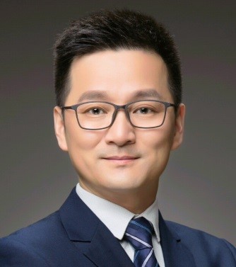 Wenguo Cui (崔文国)合作论文数Shanghai Key Laboratory for Prevention and Treatment of Bone and Joint Diseases, Ruijin Hospital, School of Medicine, Shanghai Jiao Tong University;Department of Orthopaedics, Shanghai Institute of Traumatology and Orthopaedics8
 Mingzhao Xiao (肖明朝)合作论文数8Tinghe Yu合作论文数7Wang Han合作论文数6Liyi Huang合作论文数Rehabilitation Medical Center and Institute of Rehabilitation Medicine, Sichuan University;Key Laboratory of Rehabilitation Medicine in Sichuan Province;Rehabilitation Medical Center and Institute of Rehabilitation Medicine, Sichuan University6Qin-Qin Duan合作论文数6
 Xinna Wang合作论文数Tissue Engineering Laboratory, The University of Hong Kong6XiaoHong Li (李小宏)合作论文数6
 Jing Xu (徐静)合作论文数中国人民解放军第三军医大学新桥医院内分泌科6
 Qinghua Zhao (赵庆华)合作论文数The First Affiliated Hospital of Chongqing Medical University6
 Albert Wingnang Leung合作论文数6Junyan Xiang合作论文数6Mengtong Guan合作论文数5Shuyu Chen合作论文数5
 Zhengwei Cai合作论文数5Han Wang合作论文数5DaWei Chen (陈大为)合作论文数5Qinxiang Zhou合作论文数4
 Juncai Pu合作论文数Chongqing Med Univ, Affiliated Hosp 1, NHC Key Lab Diag & Treatment Brain Funct Dis, Chongqing, Peoples R China4Juan Wang合作论文数Shanghai Inst Traumatol & Orthopaed, Shanghai Jiao Tong Univ4Lan Shao (邵岚)合作论文数4Qing Gao (高青)合作论文数4
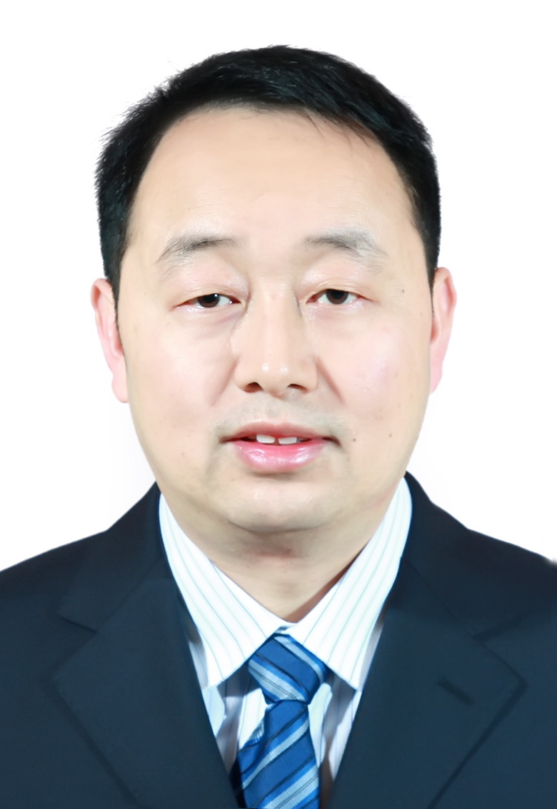 Jiang Zhu (朱江)合作论文数The First Affiliated Hospital of Chongqing Medical University4
 Yunsheng Ou (欧云生)合作论文数The First Affiliated Hospital of Chongqing Medical University4
 Ping Wang (王萍)合作论文数The First Affiliated Hospital of Chongqing Medical University4Lehua Yu (喻乐华)合作论文数华东交通大学土木工程与建筑学院4
 Yuan Jiang合作论文数4Xiya Guo合作论文数3
 Zeyu Han合作论文数3Dawu Wang合作论文数3Yong He合作论文数3Jia Qiao合作论文数3




