BACKGROUND:In recent decades, thermal ablation (TA) has gained acceptance as an effective and safe treatment for benign thyroid nodules (BTNs). However, despite its increasing popularity, the indications and techniques of TA for BTNs lack a unified standard, resulting in differences in treatment outcomes. In particular, the current guidelines and consensus statements adopt indications based on surgical criteria, which focus on larger BTNs with symptoms or cosmetic concerns. However, these indications may not adequately demonstrate the advantages of TA, as it is a fundamentally distinct therapeutic approach. To establish novel and specific indications for TA in BTNs and to standardize the use of this technique, a panel of experts issued the current expert consensus. MATERIALS AND METHODS:Based on a systematic review of the literature and clinical experience, the drafting group developed preliminary recommendations on TA for BTNs. A multidisciplinary panel of 30 experts with specific competence and expertise in TA for thyroid nodules reviewed, rated, and revised these recommendations through multiple rounds of the modified Delphi method. RESULTS:Twenty-six recommendations on TA for BTNs were proposed in the present consensus, covering indications and contraindications, physician training suggestions, preablation preparation, technical procedures, complications, efficacy assessment, follow-up strategies, and postablation management. CONCLUSION:The present consensus emphasizes the indication of TA for BTNs and outlined the technique details and periablation management. The implementation of this consensus is expected to standardize treatment practices, enhance patient outcomes, and shape future research and policy developments in the management of BTNs.
Purpose Developing a deep learning model to simultaneously evaluate lymph node status and distinguish between benign and malignant breast masses has been a challenging clinical task. This study aimed to use radio frequency (RF) signal data to create a deep learning, multimodal multitasking model, incorporating Class Activation Mapping (CAM) heatmaps to assist ultrasonographers in assessing the overall patient state based on their expertise. Research has shown a correlation between lymph node metastases and the heterogeneity of neutrophil distribution in malignancies. Therefore, the study also aimed to analyze the correlation between CAM heatmaps and neutrophil distribution, exploring the physiological mechanisms underlying the model. Methods A total of 308 eligible breast cancer patients with B-mode data, RF data and ultrasound reports were selected as the training set (n = 246) and validation set (n = 62) at the Second Affiliated Hospital of Harbin Medical University from September 2018 to October 2019. Using ResNet-18 as a feature extraction network, a multi-task loss (MTL) function to design a model for breast mass classification and axillary lymph node status assessment. The model’s CAM heatmaps were analyzed and integrated with the sonographer’s expertise to evaluate the patient’s condition. Sensitivity, specificity, accuracy and receiver operating characteristic (ROC) curve analyses were calculated. In the animal experiment, CAM heatmaps from mouse transplanted tumors were analyzed, and differences in neutrophil distribution at different heatmap color locations were examined. Results The B + RF mode data with MTL was the optimal combination for modeling. The area under the ROC curve (AUC) for the DLMMRF analysis of breast mass prediction was 0.967, for lymph node status prediction was 0.922 and for patient overall status prediction was 0.944. In CAM heatmaps, the red portion for patients with lymph node metastases focused on mass margins, while for those without metastases, it centered on the mass (p < 0.001). The AUC for sonographers with CAM assistance in breast mass prediction was 0.901, for lymph node status prediction was 0.874 and for patient overall status prediction was 0.887. Animal experiments showed that heatmap patterns correlated with neutrophil distribution in tumors, with a larger neutrophil-positive area in the red section than in the blue section in both metastatic and non-metastatic groups, and a greater neutrophil-positive area in the metastatic group’s red section compared to the non-metastatic group. Conclusion This study developed a multimodal multitasking deep learning model using RF data to generate CAM heatmaps, assisting ultrasonographers in assessing patient status, especially lymph nodes. The CAM heatmaps’ red regions displayed higher neutrophil concentrations than the blue areas, with the metastatic group showing more neutrophils in the red regions compared to the non-metastatic group, suggesting a possible association between neutrophil infiltration and the model’s attention regions.
Triple negative breast cancer (TNBC) remains a major therapeutic challenge due to its aggressive nature and immunosuppressive tumor microenvironment (TME). To overcome these barriers, we developed a multifunctional ultrasound-responsive nanobubble formulation (RLC). The RLC consists of a sulfur hexafluoride (SF6) gas core encapsulated by a lipid shell, which is co-loaded with the glycolysis inhibitor lonidamine (LND) and the sonosensitizer chlorin e6 (Ce6), and functionalized with cRGD peptides for tumor targeting. Upon ultrasound irradiation, RLC provides acoustic contrast imaging and promotes ultrasound-responsive therapeutic activation. Mechanistically, ultrasound activated Ce6 generates reactive oxygen species (ROS), resulting in mitochondrial dysfunction and impaired oxidative phosphorylation in tumor cells. Concurrently, LND inhibits glycolysis and decreases extracellular lactate accumulation, thereby mitigating acidosis associated immunosuppression. These combined strategies induce immunogenic cell death (ICD) and enhance anti-tumor immunity by stimulating dendritic cell maturation and cytotoxic T lymphocyte infiltration. Our findings suggest that the RLC nanobubbles effectively remodel the immunosuppressive TME into a more immunologically responsive state, presenting a promising theranostic strategy for TNBC management.
Accurate prediction of recurrence risk is crucial for personalizing therapy in human epidermal growth factor receptor 2-positive (HER2-positive) breast cancer. We aimed to develop an interpretable machine learning model integrating multimodal data to address this need. This retrospective study enrolled 148 patients with human epidermal growth factor receptor 2 (HER2)-positive breast cancer between 2017 and 2021. On preoperative ultrasound images, the intratumoral region (region of interest B, ROIB) was manually delineated by experienced radiologists. Based on the manually segmented ROIB, the 5 mm inward inner peritumoral region (ROIC) and 5 mm outward outer peritumoral region (ROIA) were automatically generated via the built-in adaptive tool of 3D Slicer, with non-breast tissues excluded manually. Radiomic features were extracted from each ROI using PyRadiomics. A pre-fusion strategy (i.e. feature fusion) was used to construct combined feature sets (ROIA+B and ROIA+B+C [ROI All]), and the radiomic model based on ROI All was defined as Rad All. After feature selection via Student’s t-test/Mann-Whitney U test, Pearson correlation analysis, maximum relevance minimum redundancy (mRMR) algorithm, and least absolute shrinkage and selection operator (LASSO) regression, multiple machine learning models were compared, and the support vector machine (SVM) was selected as the optimal algorithm for radiomic model construction. Independent clinical risk factors were screened by Cox regression analysis, and a post-fusion strategy integrated these factors with the radiomic signature to build a combined nomogram model. Model performance was evaluated using the area under the receiver operating characteristic (ROC) curve (AUC), calibration curves, decision curve analysis (DCA), DeLong test, net reclassification improvement (NRI), and integrated discrimination improvement (IDI). Survival analysis was performed with the Kaplan-Meier method, and SHapley Additive exPlanations (SHAP) analysis was applied to enhance model interpretability. Among the tested radiomic models, the Rad All model that integrated intratumoral, inner peritumoral, and outer peritumoral features showed the best overall performance in the internal validation set, with an AUC of 0.820 (95
PurposeTo develop a radiomics model based on ultrasound images for predicting risk of recurrence in breast cancer patients.MethodsIn this retrospective study, 420 patients with pathologically confirmed breast cancer were included, randomly divided into training (70%) and test (30%) sets, with an independent external validation cohort of 90 patients. According to St. Gallen recurrence risk criteria, patients were categorized into two groups, low-medium-risk and high-risk. Radiomics features were extracted from a radiomics analysis set using Pyradiomics. The informative radiomics features were screened using the minimum redundancy maximum relevance (mRMR) and the least absolute shrinkage and selection operator (LASSO) algorithms. Subsequently, radiomics models were constructed with eight machine learning algorithms. Three distinct nomogram models were created using the features selected through multivariate logistic regression, including the Clinic-Ultrasound (Clin-US), Clinic-Radiomics (Clin-Rad), and Clinic-Ultrasound-Radiomics (Clin-US-Rad) models. The receiver operating characteristic (ROC), calibration, and decision curve analysis (DCA) curves were used to evaluate the model’s clinical applicability and predictive performance.ResultsA total of 12 ultrasound radiomics features were screened, of which wavelet.LHL first order Mean features weighed more and tended to have a high risk of recurrence. The higher the risk of recurrence, the higher the radiomics score (Rad-score) in all three sets (training, test, and external validation set, all p < 0.05). Rad-score is equally applicable in four different subtypes of breast cancer. In the test set and external validation set, the Clin-US-Rad model achieved the highest AUC values (AUC = 0.817 and 0.851, respectively). The calibration and DCA curves also demonstrated the good clinical utility of the combined model.ConclusionThe machine learning-based ultrasound radiomics model were useful for predicting the risk of recurrence in breast cancer. The nomograms show promising potential in assessing the recurrence risk of breast cancer. This non-invasive approach offers crucial guidance for the diagnosis and treatment of the condition.
Inflammatory dilated cardiomyopathy (iDCM) represents a severe immune-related condition provoked by the progression of myocarditis. In patients suffering from myocarditis, a vicious cycle of inflammation orchestrated by CD4+ T cells, neutrophils, and fibroblasts is the culprit that drives the deterioration of myocarditis into iDCM. This study designed composite microneedles and ion solutions using calcium silicate bioceramics, which deliver SiO32- directly into myocardial tissue or indirectly via systemic circulation. These interventions modulate the cell microenvironment by regulating CD4+ T/T helper 17 (TH17) cells and their interactions with neutrophils and fibroblasts through the forkhead box O (FOXO) signaling pathway. Specifically, SiO32- inhibits the hyperdifferentiation of CD4+ T cells to TH17 cells by regulating FOXO1 and neutrophils to neutrophil extracellular traps as well as fibroblasts to myofibroblasts by regulating FOXO3, thereby ultimately disrupting the vicious cycle of myocardial inflammation and subsequent fibrotic lesions in iDCM. This discovery indicates that the biomaterial-based strategy may have great potential for the treatment of iDCM.
Transplant rejection and the side effects of immunosuppressive therapy have hindered heart transplantation development. Rejection of a heart transplant can lead to cellular and antibody-mediated immunoinflammatory responses and allograft dysfunction, thereby significantly affecting patients’ survival and prognosis. To address these challenges, many new technologies and materials, including nanomaterials, have been developed for potential applications in the heart transplantation field. Nanomaterials are most commonly used as drug delivery carriers, and the addition of specific ligands can enhance drug utilization, strengthen therapeutic effects, and reduce the occurrence of adverse reactions. In addition, nanomaterials have been developed as targeted molecular probes to support various imaging techniques and to assist in monitoring the infiltration of immune cells (such as T cells and macrophages) into cardiac tissue, thus facilitating the early diagnosis of acute rejection (AR). Continuous advances in nanotechnology have led to the development of “theranostic” and intelligent-response nanomaterials for precise disease diagnosis and simultaneous treatment. Nanomedicine primarily relies on the development of Nanomaterials and nanostructured surfaces, along with the application of nanotechnology, for molecular diagnosis, therapy, monitoring, and disease treatment. In this review, we examine the recent development of nanomaterials for the diagnosis and treatment of AR in heart transplantation, and discuss the challenges and future directions for the clinical translation of nanomaterials in heart transplantation.
AIMS:Pro-inflammatory macrophages are critical mediators of the viral myocarditis (VMC) pathological process. Methylenetetrahydrofolate dehydrogenase 2 (MTHFD2), a key enzyme involved in one-carbon metabolism, plays an essential regulatory role in macrophage function. However, the regulatory effect of MTHFD2 on macrophages in VMC remains unclear. Here, we investigated whether MTHFD2 regulates macrophage function to exert a protective effect against coxsackievirus B3 (CVB3)-induced myocarditis. METHODS AND RESULTS:To establish the VMC model, 6-week-old C57BL/6J and BALB/c mice were intraperitoneally injected with CVB3, and blood samples from the mice were examined for targeting analysis of folate metabolism-related compounds. The myeloid cell-specific MTHFD2 knockout mice Mthfd2fl/flLyz2-Cre+ (MTHFD2-KO-Mϕ) and littermate mice underwent peripheral blood proteomic analysis. We observed activation of the one-carbon metabolism folate cycle and upregulation of MTHFD2 in macrophages during myocarditis. Furthermore, CVB3-infected MTHFD2-KO-Mϕ mice exhibited higher cardiac immunocyte infiltration, especially pro-inflammatory macrophages, aggravated myocardial injury, and cardiac dysfunction. MTHFD2 knockdown also enhanced the migration of bone marrow-derived macrophages and increased their polarization towards a pro-inflammatory phenotype. Proteomic analysis identified Rap1 as a direct downstream target of MTHFD2 in VMC. Specifically, MTHFD2 modulated integrin-regulated monocyte-macrophage migration via Rap1a and reduced cellular pro-inflammatory differentiation in VMC by inhibiting Rap1/p38 MAPK signalling. Both MTHFD2 administration and high-folate diet feeding reduced cardiac inflammation and fibrosis and improved cardiac function in mice with VMC. CONCLUSION:We identified MTHFD2 as an immune regulator of monocyte-macrophage homeostasis to protect against CVB3-induced VMC. Targeted regulation of MTHFD2 is a potential therapeutic option for VMC clinically.
The assessment of Human Epidermal Growth Factor Receptor 2 (HER2) expression status is crucial for determining the eligibility of breast cancer (BC) patients for HER2-targeted therapies. This study aims to develop a nomogram model that incorporates multimodal ultrasound imaging features alongside clinicopathological characteristics to evaluate HER2 status. A retrospective analysis was conducted on 456 breast cancer patients who underwent breast ultrasound between January 2019 and December 2021. The dataset was randomly divided into a training cohort (n = 319) and a validation cohort (n = 137) in a 7:3 ratio. Independent factors predicting HER2 status in the training cohort were evaluated using univariate and multivariate logistic regression. Subsequently, a combined model was developed and validated in the validation cohort. Model performance was assessed through receiver operating characteristic (ROC) curves, decision curve analysis (DCA) and calibration curves to evaluate discrimination, net clinical benefit, and calibration, respectively. Of the 456 patients enrolled, 120 (26.32
Coronary microvascular dysfunction (CMD) refers to clinical symptoms caused by structural and functional damage to coronary microcirculation. The timely and precise diagnosis of CMD-related myocardial ischemia is essential for improving patient prognosis. This study describes a method for the multimodal (fluorescence, ultrasonic, and photoacoustic) noninvasive imaging and treatment of CMD based on ischemic myocardium-targeting peptide (IMTP)-guided nanobubbles functionalized with indocyanine green (IMTP/ICG NBs) and characterizes their basic characteristics and in vitro imaging and targeting abilities. The IMTP/ICG NBs enable the accurate location of myocardial ischemia via photoacoustic imaging, and when loaded with tannic acid (TA), can be used to effectively treat myocardial ischemia and fibrosis in CMD mice, achieving an effect superior to that of free TA. The origin of this high therapeutic efficiency is revealed by transcriptomic and proteomic analyses. This investigation lays the groundwork for visual monitoring and the drug-targeted treatment of CMD.
This study aimed to develop a new ultrasonographic dating formula to estimate gestational age (GA) based on fetal crown–rump length (CRL) in a Chinese population, evaluate model accuracy and compare its performance with established dating formulas. A prospective, multicenter study was conducted across mainland China. Participants included healthy, low-risk women with spontaneously conceived singleton pregnancies and a regular menstrual cycle in the preceding year. Ultrasonography was performed between 11 and 14 weeks of gestation, with GA determined based on the last menstrual period. Participants were randomly assigned to a development or validation cohort in a 7:3 ratio. A best-fit regression model was constructed for GA estimation based on CRL in the development cohort. For validation, mean differences between the new estimated GA and menstrual age were calculated and compared with those obtained using five established CRL-based dating formulas in the validation cohort. All participants were followed through to delivery. The study recruited 4,710 women with singleton pregnancies, with 3,297 in the development cohort and 1,413 women in the validation cohort. The mean and standard deviation values of CRL changed linearly with GA during 11–14 weeks. CRL demonstrated a linear relationship with GA between 11 and 14 weeks, yielding the regression equation GA = 59.590085 + 0.458539×CRL (R2 = 0.8042). The mean difference between estimated GA and menstrual age was 0.32 days (95
Donor heart-resident C-C chemokine receptor 2 (CCR2+) macrophages induce the recruitment of CCR2+ monocytes to a transplanted hearts through the secretion of monocyte chemoattractant protein-1 (MCP-1), which mediates the incidence of acute rejection (AR). In this study, we synthesized MCP-1 peptide-modified polyethylene glycol-poly (lactic-co-glycolic) acid (PEG-PLGA) nanoparticles loaded with the sonosensitizer dihydroporphyrin e6 (Ce6) and administered them via intramyocardial injection and used in combination with sonodynamic therapy (SDT) to selectively deplete donor cardiac-resident and infiltrating CCR2+ macrophages. In vitro experiments confirmed that Ce6-NP-MCP-1 targets and has chemotactic effects on CCR2+ macrophages, thereby enhancing the therapeutic efficacy of STD. In mouse heart grafts, the chemotactic effect of Ce6-NP-MCP-1 on CCR2+ macrophages has been used to induce donor heart-resident and infiltrating CCR2+ macrophages to aggregate and phagocytose nanoparticles in combination with SDT to induce macrophage apoptosis. This therapy inhibits the number of donor heart-resident CCR2+ macrophages and downregulates the expression of proinflammatory cytokines and inflammatory infiltration. In addition, it significantly prolongs the allograft survival time. Therefore, CCR2-targeted nanoparticles combined with SDT for the selective depletion of donor heart-resident CCR2+ macrophages provide a promising paradigm for AR target treatment.
Glioma represents a highly lethal form of malignant tumour, with RNA methylation emerging as a critical regulator of its oncogenesis and progression. As a prevalent post-translational modification, methylation influences various biological functions, particularly RNA processing, by modulating splicing, transport, and degradation of both mRNAs and noncoding RNAs. Key methylation types such as N6-methyladenosine (m6A), N5-methylcytosine (m5C), N7-methylguanosine (m7G), and N1-methyladenosine (m1A) are dynamically regulated by specific enzymes known as writers, erasers, and readers. Dysregulation of these modifications contributes to glioma pathophysiology, while offering potential biomarkers for early detection and promising therapeutic targets. This review explores the mechanistic roles of RNA methylation in glioma and highlights its translational implications, aiming to advance molecular diagnostics and targeted interventions in glioma treatment.
Objective:We aimed to establish a model to estimate the energy required for microwave ablation (MWA) to achieve the desired effect and analyze the factors influencing its therapeutic efficacy. Materials and Methods:We retrospectively analyzed 117 patients with benign thyroid nodules. A quadratic regression model was established to analyze the relationship between the technical parameters of MWA and volume reduction rate (VRR). Both univariate and multivariate logistic regression analyses were used to identify factors influencing the efficacy of MWA treatment. Results:The volume of nodules continued to decrease at 1, 3, 6, and 12 months after ablation, and the mean of VRR was 77.5 ± 15.9% at 12 months after ablation. Among these nodules, 72 (61.5%) had a VRR ≥ 75%, whereas 45 (38.5%) had a VRR < 75%. The energy volume ratio was significantly correlated with the VRR. When the VRR ≥ 75%, the energy volume ratio ranges 784-2,274 J/mL. Among all parameters, only the energy volume ratio and calcification were independent factors influencing the treatment efficacy for benign thyroid nodules (P < 0.05). Conclusions:The efficacy of the treatment was optimized when the energy volume ratio of the MWA fell within a certain range. The energy volume ratio and calcification are related to the efficacy of MWA treatment.
ABSTRACTObjectivesSupra‐normal left ventricular ejection fraction (snLVEF) represents a heterogeneous group with distinct prognoses. Left atrial (LA) strain, measured by speckle tracking echocardiography (STE), is a validated prognostic indicator. This study aimed to evaluate LA and left ventricular (LV) mechanical strains in hypertensive patients with snLVEF.MethodsThis retrospective study included 101 patients (mean age 59.7 ± 8.4 years; 61.4% men) with primary arterial hypertension and preserved LVEF (≥50%). Patients were categorized into low‐normal LVEF (lnLVEF; 50%–59%), mid‐normal LVEF (mnLVEF; 60%–69%), and snLVEF (≥70%). LV global longitudinal strain (LVGLS) and LA strains during reservoir (LASr), conduit (LAScd), and contraction (LASct) phases were measured using STE.ResultsRelative wall thickness was significantly higher in snLVEF patients compared to mnLVEF (p < 0.01), with no difference in LVGLS (p = 0.933). Compared to mnLVEF, snLVEF patients had reduced LASr and LAScd (both p < 0.01) but preserved LASct (p = 0.057). In contrast, lnLVEF patients showed greater reductions in all phasic LA strains (all p < 0.01). Loess regression revealed an inverted U‐shaped relationship between LASr and LVEF, peaking at LVEF 65%–70%. The mitral E/e′mean ratio and LVGLS correlated moderately to strongly with LASr (r = −0.39 and r = −0.65, respectively; both p < 0.001).ConclusionHypertensive patients with snLVEF exhibit impaired LA reservoir and conduit functions while maintaining pump function, suggesting snLVEF may be an intermediate stage between mnLVEF and lnLVEF as hypertension progresses. Further studies are needed to explore the prognostic potential of LA strain in this population.
Acute Myocardial Infarction (AMI) has seen rising cases, particularly in younger people, leading to public health concerns. Standard treatments, like coronary artery recanalization, often don't fully repair the heart's microvasculature, risking heart failure. Advances show that Mesenchymal Stromal Cells (MSCs) transplantation improves cardiac function after AMI, but the harsh microenvironment post-AMI impacts cell survival and therapeutic results. MSCs aid heart repair via their membrane proteins and paracrine extracellular vesicles that carry microRNA-125b, which regulates multiple targets, preventing cardiomyocyte death, limiting fibroblast growth, and combating myocardial remodeling after AMI. This study introduces ultrasound-responsive phase-change bionic nanoparticles, leveraging MSCs' natural properties. These particles contain MSC membrane and microRNA-125b, with added macrophage membrane for stability. Using Ultrasound Targeted Microbubble Destruction (UTMD), this method targets the delivery of MSC membrane proteins and microRNA-125b to AMI's inflamed areas. This aims to enhance cardiac function recovery and provide precise, targeted AMI therapy.
To investigate the role of ADIPOQ gene in gestational diabetes mellitus (GDM). We genotyped single nucleotide polymorphisms (SNPs) rs266729 and rs1501299 within the ADIPOQ gene in a cohort of 1157 pregnant women of north Chinese Han ethnicity. This cohort comprised 560 pregnant women diagnosed with GDM and 597 pregnant women who exhibited normal oral glucose tolerance test at 24–28 weeks’ gestation. All participants were recruited from the Department of Obstetrics and Gynecology at the Second Affiliated Hospital of Harbin Medical University. Additionally, we used conventional bioinformatics analysis methods to conduct multi-omics analysis (transcriptome, epigenome, and single-cell level) of ADIPOQ-regulated GDM. The systolic blood flow velocity/diastolic blood flow velocity (S/D) ratio of the umbilical artery in GDM patients with CC genotype of rs266729 and GG genotype of rs1501299 was higher than control. Single-cell analysis suggested that ADIPOQ was expressed in extravillous trophoblast (EVT), T cell, monocytes, myelocyte, NK cell and syncytiotrophoblast (SCT). Functional enrichment analysis showed ADIPOQ gene was associated with response to nutrient levels, fat cell differentiation. The findings of our study indicate a correlation between SNPs of ADIPOQ in GDM patients, and ADIPOQ is involved in the transcriptional regulation of GDM.
To develop and test a relation knowledge distillation three-dimensional residual network (RKD-R3D) model for predicting breast cancer molecular subtypes using ultrasound (US) videos to aid clinical personalized management. This multicentre study retrospectively included 882 breast cancer patients (2375 US videos and 9499 images) between January 2017 and December 2021, which was divided into training, validation, and internal test cohorts. Additionally, 86 patients was collected between May 2023 and November 2023 as the external test cohort. St. Gallen molecular subtypes (luminal A, luminal B, HER2-positive, and triple-negative) were confirmed via postoperative immunohistochemistry. The RKD-R3D based on US videos was developed and validated to predict four-classification molecular subtypes of breast cancer. The predictive performance of RKD-R3D was compared with RKD-R2D, traditional R3D, and preoperative core needle biopsy (CNB). The area under the receiver operating characteristic curve (AUC), sensitivity, specificity, accuracy, balanced accuracy, precision, recall, and F1-score were analyzed. RKD-R3D (AUC: 0.88, 0.95) outperformed RKD-R2D (AUC: 0.72, 0.85) and traditional R3D (AUC: 0.65, 0.79) in predicting four-classification breast cancer molecular subtypes in the internal and external test cohorts. RKD-R3D outperformed CNB (Accuracy: 0.87 vs. 0.79) in the external test cohort, achieved good performance in predicting triple negative from non-triple negative breast cancers (AUC: 0.98), and obtained satisfactory prediction performance for both T1 and non-T1 lesions (AUC: 0.96, 0.90). RKD-R3D when used with US videos becomes a potential supplementary tool to non-invasively assess breast cancer molecular subtypes.
PURPOSE:Mucinous breast carcinoma (MBC) tends to be misdiagnosed as fibroadenomas (FA) due to its benign imaging characteristics. We aimed to develop a deep learning (DL) model to differentiate MBC and FA based on ultrasound (US) images. The model could contribute to the diagnosis of MBC for radiologists. METHODS:In this retrospective study, 884 eligible patients (700 FA patients and 184 MBC patients) with 2257 US images were enrolled. The images were randomly divided into a training set (n = 1805 images) and a test set (n = 452 images) in a ratio of 8:2. First, we used the training set to establish DL model, DL+ age-cutoff model and DL+ age-tree model. Then, we compared the diagnostic performance of three models to get the optimal model. Finally, we evaluated the diagnostic performance of radiologists (4 junior and 4 senior radiologists) with and without the assistance of the optimal model in the test set. RESULTS:The DL+ age-tree model yielded higher areas under the receiver operating characteristic curve (AUC) than DL model and DL+ age-cutoff model (0.945 vs. 0.835, P < .001; 0.945 vs. 0.931, P < .001, respectively). With the assistance of DL+ age-tree model, both junior and senior radiologists' AUC had significant improvement (0.746-0.818, P = .010, 0.827-0.860, P = .005, respectively). CONCLUSIONS:The DL+ age-tree model based on US images and age showed excellent performance in the differentiation of MBC and FA. Moreover, it can effectively improve the performance of radiologists with different degrees of experience that may contribute to reducing the misdiagnosis of MBC.
OBJECTIVE:To develop and validate a nomogram based on ultrasound and mammographic imaging features for predicting human epidermal growth factor receptor 2-low (HER2-low) expression status in breast cancer. METHODS:Patients with HER2-negative breast cancer (n = 316) were retrospectively recruited and randomized into training (n = 221) and validation (n = 95) cohorts. Patients were categorized into HER-low and HER2-zero expression groups. Ultrasound and mammography images were collected. Univariate and multivariate analyses were used to identify independent risk factors for HER-low expression status in the training cohort. A predictive nomogram model was developed and validated in the validation cohort. The calibration, discrimination, and clinical net benefit of the nomogram model were assessed using calibration curves, receiver operating characteristic curves, and decision curve analyses, respectively. A Kaplan-Meier curve was drawn, and the log-rank test was used to compare progression-free survival of the two groups of patients. RESULTS:A longer diameter, tumor margin that was not circumscribed in ultrasound and mammography images, posterior acoustic shadowing, and a higher maximum elasticity were independent predictors of HER2-low expression status; thus, they were incorporated into the nomogram model. The area under the receiver operating characteristic curve (AUC) of the nomogram model was 0.783 in the training cohort. The nomogram also showed good discrimination in the validation cohort (AUC = 0.810), and good calibration efficiency in both cohorts. Decision curve analysis indicated that the nomogram was clinically useful. The log-rank test result revealed a significant difference in progression-free survival. HER2-low expression correlated with improved breast cancer prognosis. CONCLUSION:This nomogram may provide reference for selecting candidates for appropriate management.
 Guoqing Du (杜国庆)合作论文数The 2nd Affiliated Hospital of Harbin Medical University74
 Litao Sun (孙立涛)合作论文数The 2nd Affiliated Hospital of Harbin Medical University55
 Shuangquan Jiang (姜双全)合作论文数51
 Xiaoping Leng (冷晓萍)合作论文数The 2nd Affiliated Hospital of Harbin Medical University47Ziyao Li合作论文数37
 Yanxin Su (苏雁欣)合作论文数The 2nd Affiliated Hospital of Harbin Medical University36
 Zhenzhen Wang (王珍珍)合作论文数哈尔滨医科大学附属第二医院35Hairu Li合作论文数34
 Min Ren (任敏)合作论文数Department of Ultrasound, Second Affiliated Hospital of Harbin Medical University33Lei Zhang合作论文数State Key Lab Nat & Biomimet Drugs, Peking Univ29Yujie Liu (刘宇杰)合作论文数26Hao Cui (崔浩)合作论文数24Chunping Ning (宁春平)合作论文数24
 Heng-Da Cheng合作论文数Department of Computer Science, College of Science, Utah State University19Xiaoying Li (李晓莹)合作论文数17Ping Sun (孙平)合作论文数哈尔滨医科大学附属第二医院16
 Yanhui Guo合作论文数Department of Computer Science, University of Illinois Springfield16Xu-dong WANG (王旭东)合作论文数15Chong LIU (刘冲)合作论文数14SuMei Wang (王素梅)合作论文数14Wu Tong合作论文数14Dongmo Wang合作论文数13Shaohui Qu合作论文数13Chao Wang合作论文数13
 Li Shouqiang合作论文数13
 Yuxin Jiang (姜玉新)合作论文数Peking Union Medical College Hospital13Zihong Guo (郭子鸿)合作论文数12Xiangli Xu合作论文数12
 Jianjun Yuan (袁建军)合作论文数郑州大学人民医院12
 Yingtao Zhang (张英涛)合作论文数School of Computer Science and Technology, Faculty of Computing, Harbin Institute of Technology;Institute for Artificial Intelligence, Harbin Institute of Technology12
 Qing Zhou (周青)合作论文数Renmin Hospital, Wuhan University12Qinliang Sun合作论文数11
 Weidong Ren (任卫东)合作论文数中国医科大学附属盛京医院超声科11Ying Wang (王颖)合作论文数11
 Haitao Ran (冉海涛)合作论文数The Second Affiliated Hospital of Chongqing Medical University11Hanqing Kong合作论文数10Zhonghui Jiang合作论文数10Weidong Yu合作论文数10Yan WU (吴言)合作论文数10ZhiXin Di (邸郅欣)合作论文数10GuiXia Zheng (郑桂霞)合作论文数10
 Qi Zhou (周琦)合作论文数The Second Affiliated Hospital of Xi'an Jiaotong University10Keting Li合作论文数9
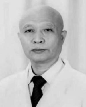 Bao-Ming Luo合作论文数9Shuai Fu合作论文数9GuoYing Che (车国英)合作论文数9HanMei Wang (王寒梅)合作论文数9DanDan Yu (于丹丹)合作论文数9




