BACKGROUND AND AIMS:Adequate bowel preparation is the cornerstone of high-quality colonoscopy. The Boston Bowel Preparation Scale (BBPS) is the gold standard for assessment, yet its application suffers from inter-observer variability and lacks a fully automated solution for entire video analysis. This study proposes EndoClean, a novel, fully automated deep learning framework designed to compute the full-segment BBPS score from colonoscopy videos, aiming to provide a standardized, objective, and near expert-level assessment. METHODS:EndoClean integrates three distinct models: frame selection, anatomical segmentation, and BBPS scoring. Its performance was rigorously evaluated against a reference standard established by senior experts and compared with junior endoscopists. We assessed assessment precision, inter-rater agreement (quadratic weighted Kappa), and consistency across all colonic segments. RESULTS:The EndoClean system demonstrated superior reliability, achieving a global accuracy of 97.8% for the total BBPS score, with satisfying agreement with senior experts (κ = 0.984; 95% CI: 0.976-0.989). Notably, EndoClean performed significantly better than junior endoscopists in overall BBPS agreements (κ: 0.984 vs. 0.949, p < 0.001) and overall accuracy (97.8% vs. 94.6%, p = 0.037). In segment-specific analysis, the EndoClean surpassed junior doctors particularly in the transverse colon (Accuracy: 97.5% vs. 90.4%, p < 0.001) and effectively reduced misclassifications in clinically ambiguous intermediate scores. For binary adequacy classification, the system achieved a sensitivity of 98.2% and a specificity of 97.3%. CONCLUSIONS:EndoClean represents a robust solution in automated quality control, demonstrating performance comparable to senior experts in bowel preparation assessment. By significantly reducing the variability seen in junior endoscopists and providing objective, full-video BBPS scoring, this framework offers a viable, standardized, and efficient solution for clinical practice and large-scale quality monitoring.
Achalasia is a rare esophageal motility disorder of poorly understood etiology. Here, we perform a large trio-based whole-genome sequencing study of achalasia and identify a recessively inherited frameshift variant in FAM129C (p.Ala454fs). A CRISPR/Cas9-engineered Fam129c-mutant mouse model recapitulating key features of achalasia was established, including growth retardation, elevated lower esophageal sphincter (LES) pressure, and selective loss of inhibitory neurons. Multi-omic analyses revealed substantial B cell expansion and activation within the LES, accompanied by enhanced humoral immune responses. Time-course experiments demonstrated that B cell accumulation preceded overt neuronal loss, while B cell depletion via anti-CD20 antibodies or intravenous immunoglobulin treatment partially rescued the phenotypes. Further protein profiling and cell-based assays suggested that the GABAA receptor may represent one potential neuronal antigen targeted by circulating autoantibodies. Together, these findings identify FAM129C as a genetic contributor to achalasia and support a neuroimmune mechanism in which B cell activation and autoantibody-mediated responses contribute to inhibitory neuronal injury. These results provide important insights into achalasia pathogenesis and highlight the potential of immunomodulatory strategies for disease intervention in the early stage.
Gas-related adverse events (GRAEs) are common after peroral endoscopic myotomy (POEM). Although air insufflation is a recognized risk factor, determinants of GRAEs in the era of carbon dioxide insufflation remain unclear. We aimed to identify independent risk factors after excluding air-insufflation cases and to develop and validate a clinically applicable prediction model. We retrospectively analyzed 3965 consecutive patients who underwent POEM between 2011 and 2024 at a tertiary referral center. GRAEs were defined as pneumoperitoneum, pneumothorax, pneumomediastinum, or subcutaneous emphysema requiring intervention. Patients were randomly allocated to training (80
Idiopathic achalasia is a rare esophageal motility disorder caused by the loss of enteric neurons leading to impaired relaxation of the lower esophageal sphincter and absent peristalsis of the esophageal body. Despite being described over three centuries ago, the precise etiology and molecular pathogenesis of idiopathic achalasia remain unclear. Recent studies indicate that an initial injury to the esophagus, potentially caused by viral infection, may trigger inflammation in genetically predisposed individuals. Ultimately, chronic inflammation causes the destruction of inhibitory myenteric neurons, resulting in clinical symptoms of achalasia-like dysphagia, regurgitation, chest pain, and weight loss. Although extensive research has been conducted on the etiology of achalasia, the majority of these studies have been limited to exploration of the infection and immune factors within the context of genetic susceptibility. In this review, we aim to provide a detailed understanding of the etiology of achalasia. In addition to the genetic factors, viral infection, and autoimmunity, we additionally propose that psychological factors, gut microbiota, and the gut-brain axis play a role in the pathogenesis of achalasia.
Vision-language models remain underused in colonoscopy despite the rich expert descriptions recorded in routine reports. These reports document lesion appearance, size and location but summarise entire procedures rather than caption individual frames, leaving clinical findings only weakly linked to the corresponding images. Here we develop EndoCLIP, a colonoscopy vision-language foundation model trained on 125,756 lesion-level image-text pairs progressively recovered from 280,476 routine colonoscopy records. Across lesion-level image-text retrieval, structured report generation and six multi-centre clinical classification tasks, EndoCLIP outperforms general-purpose and biomedical vision-language encoders in both zero-shot and linear-probe settings. On benign-versus-malignant classification, its linear probe approaches the performance of expert readers in a blinded study involving 12 endoscopists. These results suggest that recovering finding-to-frame correspondence can transform routine documentation into scalable supervision, enabling clinical targets to be specified in language rather than separately annotated for each task.
BACKGROUND:Prophylactic complete closure of mucosal defects after resection of gastrointestinal lesions is key to reducing delayed bleeding, but complete closure for large defects can be challenging with conventional through-the-scope clips (TTSC). The introduction of a TTSC with anchor prongs offers ability to approximate margins of larger defects. OBJECTIVE:The study objective was to evaluate prophylactic complete closure after polypectomy, endoscopic mucosal resection (EMR), or endoscopic submucosal dissection (ESD) in large (≥ 20 mm) nonpedunculated colorectal lesions (LNPCLs). METHODS:We conducted a multicenter, single-arm prospective cohort study of the TTSC with anchor prongs for prophylactic closure after EMR/polypectomy or ESD for LNPCLs. Patients were followed for 30 days after the index procedure. The primary outcome was the rate of complete closure of the defect. Other outcomes were the rate of delayed (postprocedural) bleeding, and rate of serious adverse events (SAEs). RESULTS:One hundred five eligible patients were enrolled. Ninety-nine (94.3%) defects had complete closure, with rates of 93.0% (80/86) for EMR/polypectomy and 100.0% (19/19) for ESD procedures. Delayed bleeding occurred in 2 (1.9%) patients by 30 days after the index procedure. Eight (7.6%) patients had ≥ 1 SAE, including bleeding (2 patients), perforation (1), microperforation (1), aspiration (1), nausea (1), and post-polypectomy syndrome (1). CONCLUSION:Prophylactic use of the TTSC with anchor prongs achieved a 94% rate of complete defect closure after EMR/polypectomy or ESD for LNPCLs. The rate of delayed bleeding after closure in this cohort was 1.9%. A prospective RCT is ongoing to further evaluate the clinical outcomes of a TTSC with anchor prongs used for prophylactic closure. TRIAL REGISTRATION:ClinicalTrials.gov number, NCT05653843.
Endoscopic papillectomy is an effective approach for selected papillary lesions. However, no consensus on the optimal choice of endoscope for the operation. Here we conducted a comparative study to investigate the effect of forward-viewing endoscopes (FVE) and oblique-viewing endoscopes (OVE) in the efficacy and safety of endoscopic papillectomy. Patients who underwent endoscopic papillectomy with OVE and FVE between February 2019 and August 2025 were reviewed retrospectively, regardless of histopathological type. Demographic characteristics, procedure-related results, adverse events, and follow-up outcomes were analyzed. Univariate and multivariate analyses were conducted to identify the risk factors of piecemeal resection and adverse events. A total of 125 patients were included, with 65 patients in the FVE group and 60 patients in the OVE group specifically. Baseline characteristics was comparable between the two groups. The OVE-based papillectomy had a longer procedure time (21 vs. 39 min; p = 0.001), and a higher en bloc resection rate (61.5
The prognosis of non-curative endoscopic submucosal dissection (ESD) for undifferentiated-type early gastric cancer (UD-EGC) remains unclear. This study aimed to identify risk factors for residual tumor, lymph node metastasis (LNM), and recurrence after non-curative ESD, and to define a potential low-risk subgroup suitable for active surveillance rather than additional surgery. This retrospective multicenter study included patients with non-curative ESD for UD-EGC from five institutions between 2017 and 2021. Patients were categorized into an additional surgery group or an observation group according to subsequent management. Clinicopathologic characteristics and survival outcomes were compared. Ninety-nine patients were analyzed, including 44 in the observation group and 55 in the additional surgery group. The median follow-up duration was 64 months. Six patients undergoing surveillance experienced recurrence within 12.0 to 37.3 months after ESD. Fourteen patients in the additional surgery group showed residual tumor or LNM. Vertical margin involvement independently predicted recurrence in the observation group, while ulceration and vertical margin involvement were independent predictors of residual tumor or LNM in surgical specimens or recurrence. Overall and disease-free survival were worse in the observation group. Disease-free survival in the observation group was associated with depth of invasion, lymphatic invasion, venous invasion, and resection margin status. Lesions with negative resection margins and no lymphovascular invasion meeting either (1) pT1a, UL0, > 2 cm, or (2) pT1a, UL1, ≤ 3 cm demonstrated excellent long-term outcomes without recurrence. Additional surgery remains standard after non-curative ESD for UD-EGC. However, a carefully selected low-risk subgroup may be appropriate candidates for close surveillance instead of further surgical intervention.
The precise monitoring of gastrointestinal mucosal damage and the standardized training of endoscopic submucosal dissection (ESD) are the research focuses in the field of digestive endoscopy. Electrochemical sensors, with their high sensitivity and real-time quantitative advantages, have become the core medical testing tools for detecting biological markers of gastrointestinal mucosal damage and for damage warning. The combination of these sensors with the ESD training model provides molecular biomedical support for improving the accuracy and scientificity of the training. This study is based on the application of electrochemical sensors in the detection of gastrointestinal mucosal damage, aiming to verify the training efficacy of the pig gastric isolated ESD training model and compare its effect with that of the pig gastric in vivo model on improving the operational skills of inexperienced ESD trainees. Ten endoscopists without ESD operational experience were selected and randomly divided into the experimental group (pig gastric isolated model training) and the control group (pig gastric in vivo model training). Using the detection principle of biomarkers related to mucosal damage based on electrochemical sensors, combined with traditional indicators such as operation time, resection speed, and perforation rate, the training effects of the two groups were comprehensively evaluated at the molecular and operational levels. The practicality of the model was analyzed through satisfaction scores, and statistical analysis was conducted using chi-square test and Mann-Whitney U test. The results showed that there was no significant difference in the expression of mucosal damage-related biomarkers detected by the electrochemical sensor in the two groups, and there were no statistical differences in various performance indicators of the trainees in simulated operations and real patient operations between the two groups. The whole resection rate and complete resection rate reached 100%. The isolated model had a more favorable applicability score for novices. The research conclusion indicates that the pig gastric isolated ESD training model and the in vivo model training have the same efficacy and can effectively improve the ESD operational skills of inexperienced trainees. The electrochemical sensor, as an important medical testing technology, can provide objective quantitative evidence for the evaluation of ESD training effects at the molecular level. The combination of it with the isolated training model can promote the development of ESD training towards a more precise molecular biomedical direction, providing new technical ideas for the optimization of the digestive endoscopy training system.
Introduction The global status of core labor force (CLF) health is underestimated, particularly the burden of patients with early-onset gastrointestinal cancers (EOGICs), aged 15-49 years old. We aim to investigate the pattern and trend of EOGICs among CLF from 1990 to 2021. Methods EOGIC burden and its attributable risk factors were estimated using data from the GBD 2021. The ASR and EAPC by age, sex and SDI were utilized for measuring incidence rate trends. Joinpoint regression analysis was utilized to explore the variation in disease burden. The Bayesian Age-Period-Cohort (BAPC) model was performed to forecast the disease burden up to 2050. Results From 1990 to 2021, the global ASIR for EOGICs among core labor force remained broadly stable, moving from 10.9 to 9.62 per 100,000(EAPC=-0.42, -1.70 to 0.87), while incident cases increased from 295,514 to 379,709. The overall incidence pattern was driven largely by declines in early-onset GC. The age-standardized DALY rate declined from 375.16 to 238.40 per 100,000(EAPC=-1.46, -2.76 to -0.15). Only early-onset CRC showed increasing DALYs. Joinpoint analysis showed a continuous downward trend in the ASIR for overall EOGICs(AAPC=-0.97%; -0.81 to -1.08). Risk-factor decomposition indicated substantial contributions from modifiable exposures, mainly led by smoking, alcohol use, and dietary risks, with marked sex- and age-specific variation. Conclusion The results of the present study are significant for global health policy and practice in core labor force. Differentiated intervention and outreach strategies based on age and gender would be necessary to reduce the impact of EOGICs.
Colonoscopy video generation delivers dynamic, information-rich data critical for diagnosing intestinal diseases, particularly in data-scarce scenarios. High-quality video generation demands temporal consistency and precise control over clinical attributes, but faces challenges from irregular intestinal structures, diverse disease representations, and various imaging modalities. To this end, we propose ColoDiff, a diffusion-based frame-work that generates dynamic-consistent and content-aware colonoscopy videos, aiming to alleviate data shortage and assist clinical analysis. At the inter-frame level, our TimeStream module decouples temporal dependency from video sequences through a cross-frame tokenization mechanism, enabling intricate dynamic modeling despite irregular intestinal structures. At the intra-frame level, our Content-Aware module incorporates noise-injected embeddings and learnable prototypes to realize precise control over clinical attributes, breaking through the coarse guidance of diffusion models. Additionally, ColoDiff employs a non-Markovian sampling strategy that cuts steps by over 90% for real-time generation. ColoDiff is evaluated across three public datasets and one hospital database, based on both generation metrics and downstream tasks including disease diagnosis, modality discrimination, bowel preparation scoring, and lesion segmentation. Extensive experiments show ColoDiff generates videos with smooth transitions and rich dynamics. ColoDiff also produces customized contents tailored for diverse tasks, e.g., colitis, polyps, and adenomas for diagnosis. Incorporating synthetic videos into training promotes discriminative representation learning and improves diagnosis accuracy by 7.1%. ColoDiff presents an effort in controllable colonoscopy video generation, revealing the potential of synthetic videos in complementing authentic representation and mitigating data scarcity in clinical settings.
Multimodal Large Language Models (MLLMs) show promise in gastroenterology, yet their performance against comprehensive clinical workflows and human benchmarks remains unverified. To systematically evaluate state-of-the-art MLLMs across a panoramic gastrointestinal endoscopy workflow and determine their clinical utility compared with human endoscopists. We constructed GI-Bench, a benchmark encompassing 20 fine-grained lesion categories. Twelve MLLMs were evaluated across a five-stage clinical workflow: anatomical localization, lesion identification, diagnosis, findings description, and management. Model performance was benchmarked against three junior endoscopists and three residency trainees using Macro-F1, mean Intersection-over-Union (mIoU), and multi-dimensional Likert scale. Gemini-3-Pro achieved state-of-the-art performance. In diagnostic reasoning, top-tier models (Macro-F1 0.641) outperformed trainees (0.492) and rivaled junior endoscopists (0.727; p>0.05). However, a critical "spatial grounding bottleneck" persisted; human lesion localization (mIoU >0.506) significantly outperformed the best model (0.345; p<0.05). Furthermore, qualitative analysis revealed a "fluency-accuracy paradox": models generated reports with superior linguistic readability compared with humans (p<0.05) but exhibited significantly lower factual correctness (p<0.05) due to "over-interpretation" and hallucination of visual features.GI-Bench maintains a dynamic leaderboard that tracks the evolving performance of MLLMs in clinical endoscopy. The current rankings and benchmark results are available at https://roterdl.github.io/GIBench/.
Background and study aims:Intramural esophageal cysts (ECs) are rare congenital malformations. This study aimed to investigate clinical characteristics of intramural ECs and evaluated safety and efficacy of endoscopic resection. Patients and methods:From August 2012 to June 2024, 67 patients with intramural ECs treated at the Endoscopy Center of Zhongshan Hospital were retrospectively analyzed. Data on baseline characteristics, clinical outcomes, and follow-up were collected. Results:Twenty-nine patients (17 males, mean age 49.8 years) underwent submucosal tunneling endoscopic resection (STER) and 38 patients (26 males, mean age 53.0 years) underwent endoscopic submucosal dissection (ESD) for intramural ECs. Mean specimen sizes were 2.8 ± 0.9 cm and 1.1 ± 0.6 cm, respectively ( P < 0.001). The STER group showed longer operative times (51.2 ± 20.6 vs. 32.6 ± 17.5 minutes, P < 0.001) and slower operation speed (0.13 ± 0.09 cm 2 /min vs. 0.21 ± 0.14 cm 2 /min, P = 0.032) compared with the ESD group. Complete resection rates for the STER and ESD groups were 82.8% and 94.7%, respectively ( P = 0.127). No major adverse events occurred in the groups. Complete resection was achieved in seven cases with extraluminal growth in the STER group without serious complications. During follow-up (median 37 months and 46 months, respectively), no local recurrence or strictures were observed in either group. Conclusions:Endoscopic resection of intramural ECs is safe and effective with fairly good long-term follow-up outcomes. The STER technique has advantages of completely resecting intramural esophageal cysts originating from the deep muscularis propria layer, particularly lesions with extraluminal growth.
 Liqing Yao (姚礼庆)合作论文数The First Affiliated Hospital of Xiamen University376
 Meidong Xu (徐美东)合作论文数School of Medicine, Tongji University;East Hospital Affiliated to Tongji University308
 Yunshi Zhong (钟芸诗)合作论文数Zhongshan Hospital, Fudan University304
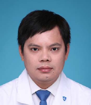 Quanlin Li (李全林)合作论文数Shanghai Geriatric Medical Center301
 Yiqun Zhang (张轶群)合作论文数Zhongshan Hospital, Fudan University207
 Ming-Yan Cai (蔡明琰)合作论文数Shanghai Geriatric Medical Center198
 Jian-Wei Hu (胡健卫)合作论文数127
 WeiFeng Chen (陈巍峰)合作论文数125Wen-Zheng Qin (秦文政)合作论文数94Zu-Qiang Liu合作论文数88LiLi Ma (马丽黎)合作论文数84
 Qiang Shi合作论文数76
 Weifeng Chen合作论文数Adobe69
 Xinyu Qin (秦新裕)合作论文数Zhongshan Hospital, Fudan University59Cai Shilun合作论文数57
 Shiyao Chen (陈世耀)合作论文数Zhongshan Hospital, Fudan University;Big Health Informatics Research Center, Fudan University55
 Zhipeng Qi合作论文数54Zhong Ren (任重)合作论文数54Xin-Yang Liu (刘歆阳)合作论文数50
 Tao Chen (陈涛)合作论文数School of Medicine, Tongji University;East Hospital Affiliated To Tongji University;Institute for Advanced Study, Tongji University49
 Yan Zhu (朱炎)合作论文数School of Life Sciences, Fudan University44
 Jiaxin Xu合作论文数43Meng-Jiang He合作论文数42Shengli Lin合作论文数40WeiDong Gao (高卫东)合作论文数40Xiao-Yue Xu合作论文数37JingZheng Liu (刘靖正)合作论文数37Bing Li合作论文数36Peiyao Fu合作论文数35Ping-Ting Gao合作论文数34GuoJie He (何国杰)合作论文数34Li Wang合作论文数31Tian-Yin Chen合作论文数31Hao Hu合作论文数27
 BoQun Zhu (朱博群)合作论文数复旦大学附属中山医院27Ping Wang (王萍)合作论文数25GENG Zi-han (耿子寒)合作论文数24Jia-Qi Xu合作论文数23Lu Yao合作论文数20
 Yun Wang (王云)合作论文数Institutes of Brain Science, Fudan University20
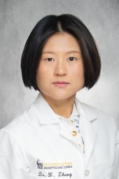 Xiaocen Zhang合作论文数Dept Med, Mt Sinai St Lukes West Hosp Ctr19
 Jianmin Xu (许剑民)合作论文数Zhong Shan Hospital, Fudan University17
 Shuo Wang (王烁)合作论文数Digital Medical Research Center, School of Basic Medical Sciences, Fudan University;Shanghai Medical College, Fudan University16Jing Cheng合作论文数16XianLi Cai (蔡贤黎)合作论文数16Li-Yun Ma合作论文数15En-Pan Xu合作论文数13Dong-Li He合作论文数13
 Zhen Zhang (章真)合作论文数Department of Radiotherapy, Shangahi Cancer Center, Fudan University12




