Abstract Background Interstitial brachytherapy is an effective and organ-preserving radiotherapeutic modality for pediatric and adolescent patients with head and neck tumors, offering favorable outcomes with minimal impact on surrounding normal tissues. However, the long-term effects of brachytherapy on dental development in young patients remain underexplored, despite clinical observations of tooth anomalies in tumor survivors. This study aimed to provide longitudinal outcomes of tooth developmental anomalies in pediatric and adolescent tumor survivors who underwent head and neck interstitial brachytherapy and to investigate the relevant factors. Methods The longitudinal panoramic radiographs of the patients at different ages before brachytherapy and during follow-up were evaluated. A modified Demirjian staging technique was used to allocate a developmental stage for each tooth, and the defect index classification criteria were applied to describe the severity of tooth damage. Logistic regression analysis was performed to identify factors associated with long-term dental abnormalities. Results A total of 210 developing permanent teeth from seven patients were included and evaluated. There were 31 teeth (14.76%) that exhibited developmental anomalies. The incidence and severity of tooth developmental abnormalities after brachytherapy were jointly determined by the radiation dose received by a given tooth and its developmental stage at the time of treatment. Multivariable analysis indicated that D2cc had the largest effect size (OR 1.029, 95% CI 1.017–1.042) and the most significant statistical association (P = 0.002). When D2cc was 0–10 Gy, the incidence of tooth developmental abnormalities was 1.90%; and when D2cc>100 Gy, the incidence reached 85.7%. The maxillary region with implanted radioactive seeds had a significantly higher risk of dental abnormalities compared to other regions. Conclusion This study suggests that interstitial brachytherapy may contribute to dental developmental alterations in pediatric and adolescent patients, with severity potentially influenced by radiation dose and tooth developmental stage. D2cc appeared to be the most informative dose-volume parameter, with lower risks observed at < 10 Gy and higher risks at > 100 Gy. Although limited by sample size and retrospective design, these findings underscore the importance of considering developing dentition when planning individualized radiotherapy for young patients.
Catheter-based pulmonary artery denervation (PADN) has achieved promising outcomes to treat pulmonary hypertension (PH). We herein present stereotactic body radiotherapy (SBRT) as a novel noninvasive approach for PADN. A single fraction of 15 Gy, 20 Gy or 25 Gy was delivered for PADN in a thromboxane A2 (TxA2) - induced acute PH swine model. We demonstrated that PADN by 20-Gy SBRT reduced mean pulmonary artery (PA) pressure during the TxA2 challenge. All SBRT dosages led to a deeper denervation area compared with radiofrequency ablation (RFA) and reduced sympathetic neural norepinephrine synthesis in the ablation zone. Probable radiation related side effects were mostly found in animals treated with 25-Gy. In subsequent monocrotaline-induced chronic PH animals, PADN by 20-Gy SBRT resulted in more significant improvement in pulmonary hemodynamics and PA remodeling in comparison to RFA. In summary, our findings suggest that appropriate SBRT scheme could balance the efficacy and safety for PADN, potentiating to be a novel strategy to treat PH. Catheter-based pulmonary artery denervation (PADN) has achieved promising outcomes to treat pulmonary hypertension (PH). Here, the authors show stereotactic body radiotherapy is a novel noninvasive approach for PADN with improved pulmonary hemodynamics in both acute and chronic PH swine models.
Tumor heterogeneity is not effortlessly detected. We present the case of a 66-year-old man with a history of rectal adenocarcinoma diagnosed four years ago, where mixed adeno-neuroendocrine carcinoma were identified via dual-tracer 18 F-FDG and 68 Ga-DOTATATE PET/CT. This case highlights the value of dual-tracer PET/CT imaging in assessing tumor heterogeneity and compensating for potential pathological sampling bias.
This multi-center analysis of data from China reviews the management and long-term outcomes of patients with bilateral Wilms tumors (BWT), and explores prognostic risk factors. We retrospectively analyzed a cohort of pediatric patients with synchronous BWT treated at 18 pediatric oncology centers in China between 2006 and 2023. The overall survival (OS) and event-free survival (EFS) rates were calculated using Kaplan–Meier methodology. Prognostic risk factors were determined using univariable and multivariable analysis. A total of 167 patients with BWT and a median age at diagnosis of 13 months (range 0–78 months) were included in the study. Neoadjuvant chemotherapy was administered to 149 index cases; tumor biopsy was performed before initiating chemotherapy in 70 patients. One hundred and three children underwent bilateral nephron-sparing surgery (NSS) and two hundred fifty-two of the three hundred one kidneys underwent NSS. The four-year OS and EFS rates in the study cohort were 86.5
BACKGROUND:This study aimed to investigate the impact of brachytherapy on dental development in pediatric and adolescent patients with head and neck tumors. Specifically, the study characterized the clinical manifestations of dental anomalies and evaluated the potential influence of patient- and treatment-related factors. PROCEDURE:A total of 35 panoramic radiographs of head and neck tumor survivors were evaluated and compared with panoramic radiographs of matched healthy controls. The mean age from the start of brachytherapy was 6.6 years (0.8-16.0 years), and the mean age for imaging examinations in the study group was 13.2 years (4.3-21.2 years). Tooth agenesis, microdontia, and root shortage were identified. RESULTS:Dental anomalies were found in 32 head and neck tumor survivors (91.43%) and 11 control subjects (28.95%). Teeth with short roots were found in 25 tumor patients (71.43%) and 8 control subjects (21.05%). Microdontic teeth were found in 17 tumor survivors (48.57%) and 2 control subjects (5.26%), whereas agenesis of teeth was found in 18 tumor patients (51.43%) and 5 control subjects (13.16%). The occurrence of dental anomalies is correlated with the age at start of brachytherapy and the treatment modalities. CONCLUSIONS:Dental development was considerably impacted by brachytherapy, especially for children under the age of 3, whereas the post-10-year age period is regarded as a relatively low-risk window. Short roots were identified as the most commonly observed type of dental developmental anomaly.
Permanent radioactive iodine-125 seed implantation (RISI), known as radioactive seed implantation, is a minimally invasive internal radiation technique. This method involves implanting 125I seeds (4.5 × 0.8 mm, encapsulated in a nickel-titanium alloy) into tumors under image guidance. The radionuclide continuously releases low energy γ-rays, effectively killing tumor cells. RISI delivers high local doses with minimal damage to surrounding normal tissues. It is performed through image-guided percutaneous puncture, accompanied by high precision, minimal trauma, and rapid recovery. In Western countries, RISI is primarily utilized for early-stage prostate cancer. In 2002, Professor Junjie Wang introduced computed tomography (CT)-guided technology for RISI, expanding its indications to head and neck, thoracic, abdominal, pelvic, and spinal tumors. In 2014, he proposed the concept of image-guided interventional brachytherapy, advancing minimally invasive brachytherapy. In 2015, he integrated three-dimensional 3D-printing template (3D-PT) with CT-guided technology, significantly enhancing the precision, quality, and efficiency of RISI, and introduced the concept of stereotactic brachytherapy. Over nearly 20 years, RISI has developed into a standardized procedure, involving preoperative planning, intraoperative optimization, and postoperative verification, highlighting its role in comprehensive cancer treatment. The main treatments for soft tissue sarcoma (STS) include surgery or surgery combined with radiotherapy and chemotherapy. However, STS is prone to local recurrence, and effective treatments are lacking after recurrence. Experts have conducted extensive trials on RISI for the treatment of recurrent STS (r-STS), accumulating significant clinical experience. This study aimed to establish standards and consensus on 3D-PT-assisted CT-guided RISI for the treatment of r-STS.
Background and purpose: Salivary gland cancers (SGCs) are hard to treat when inoperable, and sole brachytherapy appears to be a promising therapeutic strategy. This study aimed to evaluate the effectiveness, safety, and capability of pain palliation using sole brachytherapy for inoperable, recurrent, and irradiated SGCs. Materials and methods: Patients with inoperable SGCs treated using sole brachytherapy at Peking University School and Hospital of Stomatology were retrospectively included. Patients were divided into primary and recurrent groups and irradiated and non-irradiated groups. Local control (LC), overall survival (OS), radiation relevant toxicities, and Visual Analogue Scale (VAS) score for pain, were recorded and evaluated.Results: A total of 176 patients from 2006 to 2020 were included. The 5-year LC rate was 48.6 %; for the primary, recurrent, non-irradiated and irradiated groups, the rates were 72.6 %, 39.5 %, 56.8 %, and 34.5 %, respectively. The 5-year OS rates was 52.6 %; for the primary, recurrent, non-irradiated, and irradiated groups, the rates were 62.9 %, 48.6 %, 58.9 %, and 42.3 %, respectively. The mean +/- standard deviation of posttreatment VAS score of pain was 2.154 +/- 2.989, which was significantly decreased from the score of 6.923 +/- 2.280 prior to brachytherapy. Skin hyperpigmentation, mucositis, and dysphagia were the most frequently reported adverse events. Conclusions: Brachytherapy as a sole modality, was retrospectively proven effective and safe in the management of inoperable SGCs and was beneficial in multiple irradiation and pain control.
At present, selective laser melting (SLM) 3D printing technology can accurately control the internal pore structure and complex cell shape. Three types of reticulated meshes with cubic, G7 and composite structure cell shapes were fabricated by the SLM 3D printing technology using Ti-6Al-4V alloy powders. The bone stresses around the implant and the stresses in the implant were analyzed by ANSYS finite element software, which comprehensively evaluated the effect of porous dental implants with different spatial porosity characteristics on osseointegration. The results show that porous dental implants with composite structure of pore characteristics have improved mechanical and biological properties and can better promote the growth and integration of bone tissue.
Background: Desmoid tumor (DT) is rare and challenging, often affects head neck (HN) region in children, and its appropriate treatments are under discussed. This study aimed to retrospectively evaluate long-term effectiveness and safety of I seed brachytherapy for pediatric DT in HN. Procedure: Seven pediatric patients with median age of 3 years old suffered from DT in HN treated with I brachytherapy from January 2008 to June 2018 were included. Among which, 5 underwent sole brachytherapy and the others combined with surgery under a prescription doses ranging from 10 000 cGy to 12 000 cGy. The rate of local control (LC), complete response (CR) and partial response (PR) were calculated after evaluation by radiological and pathological means. The radiation-associated toxicities were also evaluated Results: The LC rate was 7/7 during the follow-up time ranging from 43 to 135 months and with a mean of 57 months. No recurrent lesion was found in the patients receiving surgery combined with brachytherapy. In patients treated with sole brachytherapy, the radiological PR rate and CR rate were 4/5 and 1/5, respectively. In those reaching radiological PR, 3/4 were pathological CR. Slight acute radiation-associated toxicities were observed in all patients, and no late or severe acute toxicity was observed. Conclusion: I brachytherapy is effective and safe in the management of pediatric DT in HN as sole modality or combined with surgery in long term.
Objective To investigate the imaging features of patients with focal cortical dysplasia(FCD) characterized by interrupting precentral gyrus and explore the surgical strategy. Methods At Beijing Tiantan-Fengtai Epilepsy Center from January 2015 to December 2021, patients with drug-refractory epilepsy, surgical treatment, whose pathological or imaging results showing FCD, and whose imaging manifestations showing interruption of the precentral gyrus were selected. The data of the patients was documented, including imaging data, medical history, physical examination, scalp EEG, magnetic resonance imaging, positron emission computed tomography, stereoelectroencephalography, intraoperative condition, pathology and postoperative follow-up, etc. Results A total of 5 patients with FCD were included. Head MRI showed that all patients had the precentral gyri transected by the malformed sulcus. Among them, there was 1 patient with blurred gray-white matter junction, 2 patients with abnormal manifestations of transmantle sign. There were 5 patients with reduced metabolism of the malformed sulcus revealed by PET-CT. 4 patients with sulcus-centered resection and 1 patient with radiofrequency-thermocoagulation. Pathological examination showed that 4 specimens were all type Ⅱ FCD, including 2 ⅡA and 2 ⅡB. Four patients who underwent resection were followed up for 1 to 4 years without epileptic seizure. Conclusion Imaging characteristics of interrupting precentral gyrus may be unique for patients with type Ⅱ FCD in the central region. Focal excision of the gray matter of the sulcus can achieve better therapeutic effect.
Rhabdomyosarcomas (RMSs) are highly malignant soft-tissue sarcomas. Head and neck RMSs often pose unique challenges to treatment because of their closeness to important structures. We here report a rare case of a 1-year-old boy with a 1-month history of right eye swelling and an eye mass. Biopsy of deep tumors in the maxillofacial region supports embryonal RMS. Postoperative positron emission computed tomography showed a 5.0 cm × 4.8 cm × 4.2 cm malignant tumor in the right maxillary region. In accordance with the international RMS study group guideline, the child was diagnosed with IIIa and TNM stage T2bN1M1 embryonal RMS. The child was treated with a combination of chemotherapy and 125I seed implantation radiotherapy and eventually achieved partial remission. This case report shows that 125I seed implantation is a safe and effective means of delivering radiotherapy to young children with head and neck RMSs. It may be an option for children with RMSs for whom surgery or external radiotherapy is unsuitable.
Brachytherapy has the advantages of being minimally invasive and highly conformal, and it achieves good results in head and neck tumors. To precisely implant the radioactive seeds according to the preplan in deep head and neck regions, the surgical navigation is applied. This study aims to explore the clinical application and accuracy of imaging-based surgical navigation-guided I-125 interstitial brachytherapy in terms of seed position. We included 41 patients with tumors in deep head and neck regions. The brachytherapy treatment plan was designed, and the preplanned data were transferred to the navigation system. Needle implantation and seed delivery were performed under surgical navigation system guidance with or without the combination of individual template. The treatment accuracy was evaluated by comparing seed cluster locations between the preoperative treatment plan and the postoperative treatment outcome. A total of 2879 seeds were delivered. The range, mean and median distances between the geometric centers of the preoperative seed point clusters and the postoperative seed point clusters were 0.8-10.5 mm, 4.5 +/- 2.3 mm and 4.1 mm, respectively. The differences between preoperative and postoperative volumes of the minimum bounding box of seed point clusters were nonsignificant. In conclusion, the imaging-based surgical navigation system is a promising clinical tool to provide the preplanned data for interstitial brachytherapy intraoperatively, and it is feasible and accurate for the real-time guidance of needle implantation and seed delivery in deep head and neck regions.
Objective:To explore the value of 18F-FDG PET/CT radiomics in predicting the cervical lymph node metastasis in salivary gland cancer. Methods:Sixty-eight patients with salivary gland carcinoma treated in the Peking University School and Hospital of Stomatology were retrospectively studied. They were randomly divided into training group ( n=40), validation group ( n=14), and test group ( n=14). The primary tumor lesions were semi-automatically delineated on PET images as regions of interest (ROIs) and the radiomic features were extracted from ROIs. After feature selection and dimension reduction, an artificial neural network (ANN) prediction model was constructed. The prediction performance of the model was assessed using receiver operating characteristic (ROC) curves, the area under ROC curves (AUC), accuracy, sensitivity, and specificity. Moreover, the performance of various models was compared using the Delong test. Results:The radiomic model yielded an AUC of 0.88 (95% CI: 0.78-0.95), a sensitivity of 75%, specificity of 92.3%, and accuracy of 88.2%. By contrast, the combined model constructed based on the clinical node status (cN) reported by PET/CT and radiomic features yielded an AUC of 0.97 (95% CI: 0.89-0.99), a sensitivity of 87.5%, specificity of 100%, and accuracy of 97.1%. The Delong test showed that there was a statistically significant difference between the combined model and cN ( Z=2.27, P<0.05), but there was no statistically significant difference between the radiomic model and cN ( P>0.05). Conclusions:The ANN model based on 18F-FDG PET/CT radiomics combined with cN reported by PET/CT can more accurately predict cervical lymph node metastasis in patients with salivary gland carcinoma.
Background The present study aimed to investigate oral cancer awareness and its related knowledge among residents in Beijing. Methods A questionnaire survey was conducted among Beijing residents concerning their knowledge of oral cancer, and its prevention and treatment. Results A total of 3055 questionnaires were completed, 45.8% by males and 54.2% by females. The ages of the respondents ranged from 15 to 93 years; 12.4% were smokers, 1.1% chewed betel nuts, and 82.5% brushed their teeth at least twice a day. Lung cancer was heard of by the most respondents, followed by gastric cancer and liver cancer; oral cancer was the least heard of. More than 60% of respondents were unaware of the risk factors and early signs of oral cancer. Conclusions This survey demonstrated a general lack of public awareness and knowledge about oral cancer. Specific measures should be taken to improve public awareness of oral cancer and its prevention and treatment.
结构磁共振(structural magnetic resonance imaging,sMRI)对癫痫的诊治至关重要,尤其是考虑行癫痫外科治疗时.但癫痫外科部分常见病理类型(如皮质发育不良、海马硬化)在常规阅片中表现隐蔽,给临床工作带来了巨大挑战.因此,结合利用神经影像后处理及统计分析方法,对影像数据进行分析、建模和解释,进一步挖掘其量化识别和分析致痫灶的价值,将有利于增加其临床指导价值.本文将针对目前癫痫外科结构磁共振常用的分析方法、计算模型和统计应用的研究进展进行综述.
Immune checkpoint inhibitors (ICIs) present significant efficacy in the treatment of malignant tumors, and they have been approved as the first-line of treatment for various cancers. Pembrolizumab monotherapy or combined with chemotherapy has been recommended by domestic and foreign guidelines for the first-line treatment of recurrent/metastatic head and neck squamous cell carcinoma. Although ICIs represent a milestone in the treatment of head and neck squamous cell carcinoma, potential problems still need to be addressed, such as the selection of the efficacy predictors for ICIs, the evaluation of the tumor response to ICIs, and the treatment of immune hyperprogression and immune-related adverse events. Therefore, to form a relatively unified understanding of ICIs treatment for head and neck squamous cell carcinoma, we integrated the clinical experience of multi-disciplinary experts of head and neck cancers on the basis of current clinical hot issues and finally developed this consensus.
Objective:To summarize and evaluate the target and dose design of 125I seed brachytherapy treatment plan of pediatric borderline tumor in head neck region. Methods:Eleven patients underwent definitive 125I brachytherapy or combined with surgery in Peking University Hospital of Stomatology from January 2010 to December 2018 were retrospective analyzed. The target region was set by extending the tumor gross region by 0.5 to 1.0 cm. The prescription dose and activity ranged from 80 to 120 Gy and 18.5 MBq, respectively. The treatments were performed according to the plan under general anesthesia. Response and toxic reaction were recorded during follow-up. The preoperative and postoperative dosimetric results were compared; and the local control rate, objective response rate, complete response rate and acute toxic reaction rate were calculated. Results:There was no statistically significant difference between preoperative and postoperative dosimetric results ( P>0.05). The follow-up time ranged from 33 to 131 months, with a median of 48 months. The local control rate, objective response rate, complete response rate and acute toxic reaction rate were 100%, 100%, 71.4% and 81.8%, respectively. Conclusions:Under well-designed target and dose, 125I brachytherapy for treatment of pediatric borderline tumor in head neck region would bring ideal therapeutic and toxic outcomes, and could be regarded as a feasible therapy.
 Jie Zhang (张杰)合作论文数Hospital of Stomatology, Peking University70
 Mingwei Huang (黄明伟)合作论文数Peking University Hospital of Stomatological;Peking University School of Stomatological64
 Lei Zheng合作论文数56Shuming Liu合作论文数39Xiao-Ming Lv合作论文数31
 Guangyan Yu (俞光岩)合作论文数School of Stomatology, Peking University25
 Fangang Meng (孟凡刚)合作论文数Beijing Tiantan Hospital, Capital Medical University;Beijing Neurological Institute23
 Kai Zhang (张凯)合作论文数Beijing Tiantan Hospital, Capital Medical University23Wen-Jie Wu合作论文数20
 Zhigang Cai (蔡志刚)合作论文数Peking University School of Stomatology15
 Wenhan Hu (胡文瀚)合作论文数Graduate School of Capital Medical University;Beijing Neurosurgical Institute, Capital Medical University15
 Chuanbin Guo (郭传瑸)合作论文数Department of Oral and Maxillofacial Surgery, School and Hospital of Stomatology, Peking University14
 Huanguang Liu合作论文数13Lei ZHENG (郑磊)合作论文数13TieLi Song (宋铁砾)合作论文数13
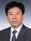 Yi Zhang (张益)合作论文数Hospital of Stomatology, Peking University13
 Anchao Yang (杨岸超)合作论文数Beijing Tiantan Hospital, Capital Medical University12FuYun Zhao (赵福运)合作论文数10
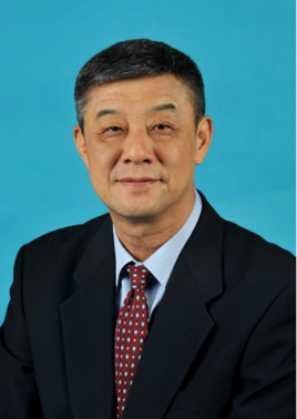 Shengde Bao (鲍圣德)合作论文数Peking University First Hospital10Ming-Hui Mao合作论文数9
 Xiu Wang合作论文数9HuiMin Wang (王慧敏)合作论文数8
 Yan Shi (时艳)合作论文数北京大学深圳研究生院8Guo-Hao Zhang合作论文数7
 Ming Ge (葛明)合作论文数Beijing Children's Hospital, Capital Medical University7JiaYong Zhang (张家涌)合作论文数6
 Xiaoming Lyu合作论文数6
 Yan Gao (高岩)合作论文数Peking University School of Stomatology6Zheng Lei (郑磊)合作论文数5Chao Duan合作论文数5Zhi-Yuan Wu合作论文数5YuCai You (尤玉才)合作论文数5
 Yunze Xuan (玄云泽)合作论文数Medical College, Yanbian University5
 Xin Zhang (张鑫)合作论文数Beijing Chao-Yang Hospital, Capital Medical University5
 Liang Li (李良)合作论文数Peking University First Hospital5Hua-Qiu Guo合作论文数4Shiying Fan合作论文数4Zhu Qiao合作论文数4Yi-Wei Zhong合作论文数4
 Yi Fan合作论文数4Lin Sang合作论文数4
 Yin Jiang合作论文数4CHEN Ning (陈宁)合作论文数4Qian Zhao (赵倩)合作论文数4Xisi Wang合作论文数4
 Yong Wang (王勇)合作论文数Hospital of Stomatology, Peking University4
 Xiaofeng Shan (单小峰)合作论文数Peking University Hospital of Stomatology4




