BACKGROUND:Neuroimaging studies frequently report aberrant spontaneous brain activity and functional connectivity within core functional networks, including the default mode network (DMN), frontoparietal network (FPN), and salience network (SN) in subclinical depression (SD). However, the dynamic coordination among these networks remains poorly understood, impeding comprehensive elucidation of the underlying neuropathology of SD. METHODS:Resting-state functional magnetic resonance imaging (fMRI) data were collected from subjects with SD (n = 26) and healthy controls (HCs, n = 33). A preclustering-based co-activation pattern method was developed to investigate the dynamic patterns of network coordination. Finally, machine learning analysis was conducted to evaluate the potential of network dynamics for clinical diagnosis. RESULTS:Subjects with SD exhibited decreased dwell time in the SN and increased transition frequency from the SN to DMN, which was positively correlated with depressive severity. Furthermore, an ensemble learning model based on SN-DMN dynamic features achieved a classification accuracy of 96.44% in distinguishing SD from HC. CONCLUSION:These findings underscore the potential of altered SN-DMN dynamics as candidates for future neuroimaging markers of SD and support a neurocognitive model whereby altered SN-DMN dynamic coordination makes subjects with SD more prone to internal directed attention biases, thereby contributing to self-related depressive symptoms like rumination.
BACKGROUND: Acute exacerbation of chronic obstructive pulmonary disease (AECOPD) is characterized by a significant worsening of respiratory symptoms. Blood eosinophil levels are a key predictor of glucocorticoid efficacy in AECOPD patients; however, their stability can present challenges. Predicting stable eosinophil levels from CT images is essential for optimal patient management. METHODS: This study utilized CT images from 482 AECOPD patients across two hospitals. Dataset 1 comprised 193 patients for model development, while Dataset 2 included 289 patients for external validation. A threshold of 2% eosinophil was used to differentiate between high and low eosinophil levels. A machine learning model was developed to predict eosinophil levels using CT radiomics and quantitative computed tomography (QCT) features. Radiomics features were extracted, and feature selection was performed using random forest (RF) algorithms. Segmentation of pulmonary lobes, airways, and blood vessels yielded 20 QCT features. A Gradient Boosting (GB) classifier was then trained on the fused features. RESULTS: The GB classifier with radiomics features demonstrated strong performance, achieving an accuracy (ACC) of 0.734 and an area under the curve (AUC) of 0.838 on the test set of Dataset 1. In external validation, the ACC and AUC were 0.624 and 0.671, respectively. After fusing QCT features, the ACC and AUC improved to 0.786 and 0.843, respectively, with external validation results of 0.673 and 0.697. CONCLUSION: The CT image-based machine learning model can predict blood eosinophil levels in AECOPD patients, providing a noninvasive and stable assessment. It has potential for future clinical application following further validation and external testing.
Myocardial pathology segmentation is crucial for quantifying myocardial danger zones in the diagnosis of acute myocardial infarction. However, effective segmentation remains challenging due to incomplete image sequences, the need to view different pathologies on different CMR sequences, difficulties in aligning pathology regions, the limited resolution of some clinical CMR sequences and the small size of myocardial lesions. To address these issues, we propose a two-stage method for segmenting myocardial lesions in CMR images. This method considers the inherent incompleteness of CMR sequences and the distinct characteristics of edema and scar tissue, enabling the segmentation of normal myocardium and two pathological regions: edema and scar tissue. We validated our method using CARE 2025 Challenge CMR images, and the results on the validation set demonstrate its effectiveness in segmenting complex myocardial pathologies. Validation showed Dice scores of 0.5302 and 0.5308 for edema and scar, respectively. These findings highlight the effectiveness and broad applicability of our segmentation approach in clinical settings.
In medical image analysis, diagnostic interpretation and downstream clinical tasks can be strongly influenced by data quality, particularly in ultrasound imaging, where speckle noise and low contrast can obscure critical anatomical details. To address these challenges, we propose SAFUS-Net, a self-supervised, attention-guided framework for ultrasound speckle suppression that learns directly from noisy data without requiring clean reference images. Unlike conventional supervised approaches, which depend on difficult-to-acquire clean targets, training pairs are generated using a multiplicative noise simulation strategy, enabling reproducible and scalable speckle-to-speckle learning. An auxiliary constant input channel is introduced to stabilize optimization by providing an input-dependent bias across receptive fields, facilitating smoother gradient propagation and improved convergence. In addition, an Adaptive Frequency-Channel Attention module is incorporated at the model bottleneck to enhance feature representation by emphasizing salient anatomical structures while suppressing irrelevant background responses. Extensive experiments are performed on both synthetic and clinical ultrasound datasets spanning multiple anatomical regions and imaging characteristics. The results demonstrate that SAFUS-Net consistently outperforms existing enhancement methods across both reference-based and no-reference evaluation metrics, achieving a more favorable balance between noise reduction, contrast enhancement, and edge preservation. These findings indicate that SAFUS-Net is a robust and practical approach for ultrasound image enhancement, with potential value for improving clinical image interpretation and supporting downstream ultrasound analysis tasks.
Deep learning-based methods for synthesizing late gadolinium-enhanced (LGE) cardiac magnetic resonance (CMR) images have attracted increasing attention at present, as they enable the generation of LGE images without the use of gadolinium-based contrast agents (GBCA). Traditional solutions such as generative adversarial networks (GANs) have demonstrated potential but are frequently limited by issues including mode collapse and training instability, which compromise their reliability in clinical applications. Diffusion models have recently gained attention as a promising alternative, owing to their stable training dynamics and superior ability to produce high resolution and realistic images. In this study, we introduce a diffusion model framework that leverages cine CMR, a noninvasive imaging modality, as a conditional input to synthesize LGE images. By incorporating the stability of diffusion modeling with cine-guided conditioning, our method generates controllable and consistent LGE images. We validate our method on the CARE2025 challenge dataset, and the experimental results demonstrate that our model outperforms previous approaches in terms of both image realism and overall visual quality. These findings highlight the potential of our cine-controllable diffusion framework as a reliable and accurate solution for LGE image synthesis, paving the way for safer and more accessible myocardial assessment without contrast administration.
Breast cancer remains a leading cause of cancer-related mortality among women worldwide. The effectiveness of early diagnosis and accurate prognosis plays a vital role in improving patient outcomes. With recent advancements in artificial intelligence (AI), automated breast cancer analysis has seen substantial improvements in both efficiency and diagnostic performance. Among the emerging trends, multi-regional feature learning, particularly the integration of tumor and peritumoral information, has demonstrated clear advantages over conventional tumor-centric approaches. In this systematic narrative review, we provide a synthesis of AI-driven methodologies that incorporate multi-regional features across various clinical tasks, including lesion differentiation, cancer staging, prognosis prediction, lymphovascular invasion (LVI) assessment, molecular subtype classification, and lymph node status prediction. Among these, prognosis assessment emerged as the most frequently studied task, followed by lesion differentiation, while cancer staging and LVI assessment were relatively underexplored, each addressed by only a single study. In addition, general paradigms in multi-regional feature learning for breast cancer screening are introduced to help bridge the knowledge gap between model developers and medical professionals. Despite encouraging progress, challenges persist, including a lack of standardization in regional feature delineation, limited understanding of their contributions to clinical outcomes, and insufficient incorporation of multimodal and multicenter datasets. To address these issues, this review highlights promising directions such as the adoption of adaptive, multimodal, and privacy-preserving learning strategies, particularly those based on federated and generative models. Finally, the integration of clinician expertise into AI workflows is emphasized as essential to improving trust and real-world applicability.
The application of deep generative models (DGMs), including generative adversarial networks (GANs), diffusion models, and variational autoencoders, is rapidly transforming the field of echocardiography. These models have proven effective in addressing key challenges in cardiovascular imaging, such as improving image quality, enhancing segmentation and classification accuracy, and mitigating data scarcity. By generating large-scale, high-quality annotated datasets, DGMs enable more reliable and efficient automated cardiac assessments, which are crucial for early and accurate diagnoses. In this study, we provide a scoping review of the use of DGMs in echocardiography, examining their role in augmenting echocardiographic analysis and supporting advanced diagnostic decision-making. This work also introduces various DGM architectures and their core implementation principles, offering fundamental knowledge to professionals from fields like medicine. DGMs have shown significant promise in generating high-quality synthetic data that enhance model performance, particularly in tasks such as cardiac structure segmentation and abnormality detection. Furthermore, our review highlights that cardiac structure segmentation is the most extensively studied task, with GANs being the most widely adopted DGM type. We also identify common challenges in DGM applications and discuss emerging research directions aimed at improving model performance, the clinical relevance of generated data, and the scalability of DGMs in clinical settings. Overall, this scoping review offers a comprehensive overview of DGMs in echocardiography, identifies gaps in current evidence, and outlines future pathways toward more reliable and clinically meaningful generative approaches for cardiovascular imaging
Annotation marks in ultrasound images can obscure diagnostically relevant features and introduce bias into automated analysis pipelines. In this study, we systematically compare two annotation suppression techniques: end-to-end image restoration frameworks and a two-stage segmentation-guided inpainting approach. Representative models from both techniques are evaluated on a curated dataset of nearly 5,000 images from 95 patients, containing both synthetically applied and physician-generated annotation marks. Experimental results demonstrate that while end-to-end enhancement methods perform favorably in removing synthetic annotations, the segmentation-guided inpainting system achieves superior performance on physician-generated marks, effectively preserving structural and diagnostic details. These findings highlight the robustness of segmentation-guided inpainting and its potential as a reliable solution for mitigating annotation artifacts, thereby improving the accuracy and usability of computer-aided ultrasound analysis.
Depression significantly contributes to global disability, yet remains underdiagnosed due to lack of awareness. Social media platforms provide rich, real-time data reflecting users' emotional and psychological states, making them promising resources for automated depression detection. However, general-purpose Large Language Models (LLMs) often perform suboptimally in clinical domains, lacking interpretability and domain specificity. To address these limitations, this study introduces a knowledge-augmented framework combining Retrieval-Augmented Generation (RAG) guided by clinical psychiatric criteria from the DSM-V with a fine-tuned DeepSeek-R1-7B model. We constructed a domain specific knowledge base comprising 329 expert-reviewed, retrieval-friendly DSM-V-derived entries. By retrieving relevant DSM-V-TR diagnostic criteria alongside user posts, our framework provides clinically grounded contextual information that may support human review by relating model predictions to established psychiatric criteria. Experiments on three balanced social media datasets showed that the DSM-V-guided RAG module, when combined with fine-tuned DeepSeek-R1-7B, achieved improved performance over the compared settings under the current experimental protocol. The retrieved DSM-V entries also provided clinically grounded contextual information that may help interpret model predictions. These findings suggest that DSM-V-guided knowledge augmentation is a promising direction for improving the transparency of benchmark-based depression detection on social media.
Precise cancer lesion analysis in medical imaging critically depends on the accurate definition of regions of interest (ROIs), which directly influence diagnostic and clinical outcomes. While peritumoral features are known to enhance lesion characterization, efficiently defining meaningful peritumoral ROIs remains a challenge. We propose an adaptive peritumoral area selection approach (APASA) that systematically identifies the most informative ROI surrounding a lesion, enabling the extraction of meaningful radiomic features for improved diagnostic performance. Unlike conventional heuristic or morphology-based methods, APASA leverages the minimum coverage graph algorithm, using the tumor ROI as a reference to construct a graph encompassing both the tumor and its peritumoral microenvironment. The effectiveness of the proposed approach was evaluated within AI-based frameworks for automated lesion differentiation in breast and thyroid cancers. Extensive experiments employing five widely used machine learning models demonstrated that APASA-selected peritumoral features consistently outperformed conventional morphological dilation. Performance improvements reached up to 30.75% in AUC and 29.00% in F1-score compared with the tumor ROI baseline. Moreover, the optimal model was found to vary depending on the ROI type, shape, and cancer type, offering new insights into the interaction between ROI selection and model choice. These results highlight APASA as a principled and efficient strategy for adaptive ROI definition in ultrasound-based cancer lesion analysis, demonstrating effectiveness across two ultrasound datasets, with potential extension to other imaging modalities and clinical settings pending further validation.
Accurate segmentation of acute ischemic stroke (AIS) lesions in multimodal magnetic resonance imaging (MRI) is essential for timely clinical intervention but remains challenging due to low lesion contrast, variability across modalities, and the need to capture both local detail and global context. In this work, we propose MHMF‑Diff, a novel diffusion‑based segmentation framework that integrates independent Mamba feature encoders for each MRI sequence (T1, FLAIR, ADC, DWI) with a Hierarchical Global Feature Fusion Block (HGFFB) for cross‑modal information aggregation at multiple decoder levels. During the reverse diffusion process, a U‑Net‑style denoising network is augmented with lightweight Mamba modules that model long‑range dependencies with linear complexity. HGFFB then injects fused, multimodal context into each decoding stage via residual projection. We evaluate MHMF‑Diff on the SOOP dataset comprising 950 stroke cases using the merged lesion label. Our method attains a Dice similarity coefficient (DSC) of 71.64
Acute ischemic stroke (AIS) is a major cause of long-term disability and mortality worldwide. Accurate segmentation of stroke lesions, particularly the infarct core and penumbra, is critical for effective treatment planning. In this study, we propose a deep learning-based multimodal segmentation network to improve the accuracy of AIS lesion delineation. The proposed framework consists of independent encoders, Multimodal Spatial and Channel Fusion (MSCF) modules, and Decoder Spatial and Channel Attention (DSCA) modules. Independent encoders are used to extract modality-specific features, while the MSCF module integrates complementary information across modalities. The DSCA module is introduced in the decoder to refine feature fusion between encoder representations and decoder features. The model was trained and evaluated on the ISLES SPES 2015, ISLES 2017, and ISLES 2018 datasets using Dice Similarity Coefficient (DSC), Hausdorff Distance (HD95), Recall, and Precision. On the ISLES SPES 2015 dataset, the proposed method achieved a DSC of 83.91
Pulmonary vascular diseases may affect arteries and veins through different physiological mechanisms, necessitating separate assessment of the two vascular trees. However, manual analysis of chest computed tomography (CT) images is time-consuming, subjective, and challenging to scale for clinical studies. To address this, we propose a novel Human-in-the-Loop (HITL) framework for annotation and model training to develop an automated pulmonary artery-vein segmentation model. Through three iterative HITL rounds, we constructed 30 gold-standard annotated datasets and trained three deep learning models. The optimal model achieved strong performance, with a Dice coefficient of 86.2%, IoU of 75.7%, sensitivity of 85.8%, and precision of 88.1%. This model was then applied to segment pulmonary arteries and veins in CT scans from patients with chronic obstructive pulmonary disease (COPD). Quantitative analysis revealed that, as COPD progresses, the volume and surface area of both pulmonary arteries and veins increase. Moreover, small vessel truncation becomes more pronounced, with small arteries showing greater structural loss than veins. These findings suggest distinct vascular remodeling patterns between arterial and venous trees across different disease stages. Our HITL-based framework not only enhances annotation efficiency but also supports the development of robust segmentation models. This approach holds promise for broader applications in medical image analysis and provides a valuable tool for characterizing vascular changes in COPD, contributing to improved diagnosis and patient management.
Integrating structural and functional connectomes remains challenging because their relationship is non-linear and organized over nested modular hierarchies. We propose a hierarchical multiscale structure-function coupling framework for connectome integration that jointly learns individualized modular organization and hierarchical coupling across structural connectivity (SC) and functional connectivity (FC). The framework includes: (i) Prototype-based Modular Pooling (PMPool), which learns modality-specific multiscale communities by selecting prototypical ROIs and optimizing a differentiable modularity-inspired objective; (ii) an Attention-based Hierarchical Coupling Module (AHCM) that models both within-hierarchy and cross-hierarchy SC-FC interactions to produce enriched hierarchical coupling representations; and (iii) a Coupling-guided Clustering loss (CgC-Loss) that regularizes SC and FC community assignments with coupling signals, allowing cross-modal interactions to shape community alignment across hierarchies. We evaluate the model's performance across four cohorts for predicting brain age, cognitive score, and disease classification. Our model consistently outperforms baselines and other state-of-the-art approaches across three tasks. Ablation and sensitivity analyses verify the contributions of key components. Finally, the visualizations of learned coupling reveal interpretable differences, suggesting that the framework captures biologically meaningful structure-function relationships.
Generative models play a pivotal role in the field of medical imaging. This paper provides an extensive and scholarly review of the application of generative models in medical image creation and translation. In the creation aspect, the goal is to generate new images based on potential conditional variables, while in translation, the aim is to map images from one or more modalities to another, preserving semantic and informational content. The review begins with a thorough exploration of a diverse spectrum of generative models, including Variational Autoencoders (VAEs), Generative Adversarial Networks (GANs), Diffusion Models (DMs), and their respective variants. The paper then delves into an insightful analysis of the merits and demerits inherent to each model type. Subsequently, a comprehensive examination of tasks related to medical image creation and translation is undertaken. For the creation aspect, papers are classified based on downstream tasks such as image classification, segmentation, and others. In the translation facet, papers are classified according to the target modality. A chord diagram depicting medical image translation across modalities, including Magnetic Resonance Imaging (MRI), Computed Tomography (CT), Cone Beam CT (CBCT), X-ray radiography, Positron Emission Tomography (PET), and ultrasound imaging, is presented to illustrate the direction and relative quantity of previous studies. Additionally, the chord diagram of MRI image translation across contrast mechanisms is also provided. The final section offers a forward-looking perspective, outlining prospective avenues and implementation guidelines for future research endeavors.
Background and objective: Knee joint lesions are common and can lead to significant pain and disability. Accurate detection is crucial for effective treatment. The YOLO series model has shown promise in automating medical image analysis but needs improvement to handle diverse clinical scenarios and complex lesion types. This study aims to enhance the YOLOv9 model to improve robustness and precision in detecting knee joint lesions. Methods: We created a knee joint lesion Magnetic Resonance Imaging (MRI) dataset (180 patients, 12 injury categories, 7,233 injury sites) and proposed an improved YOLOv9 model with multi-scale attention and structural enhancements (MASE-YOLOv9). We added extra structural layers and detection heads to handle various scales and incorporated the Efficient Multi-scale Attention (EMA) module in the Head to focus on critical image regions. Additionally, we integrated the Convolutional Block Attention Module (CBAM) in the Neck to adaptively fuse features from different levels. We also evaluated the model using the external fastMRI + dataset (1,172 patients, 21 injury categories, 16,175 injury sites). Results: On the internal dataset, MASE-YOLOv9 achieved a mAP@0.5 of 36.0%, outperforming other YOLO models. On the fastMRI + dataset, it achieved a mAP@0.5 of 37.1%, signficantly surpassing other models. After the ablation of CBAM, mAP@0.5 dropped to 36.3%, and after EMA ablation, it decreased further to 34.6%. Visual results confirmed that MASE-YOLOv9 effectively highlights important regions related to knee joint lesions. Conclusions: MASE-YOLOv9 effectively detects various knee joint lesions in MRI images, especially small ones, and can assist radiologists in providing more accurate diagnoses.
BackgroundIt is fundamental for accurate segmentation and quantification of the pulmonary vessel, particularly smaller vessels, from computed tomography (CT) images in chronic obstructive pulmonary disease (COPD) patients.ObjectiveThe aim of this study was to segment the pulmonary vasculature using a semi-supervised method.MethodsIn this study, a self-training framework is proposed by leveraging a teacher-student model for the segmentation of pulmonary vessels. First, the high-quality annotations are acquired in the in-house data by an interactive way. Then, the model is trained in the semi-supervised way. A fully supervised model is trained on a small set of labeled CT images, yielding the teacher model. Following this, the teacher model is used to generate pseudo-labels for the unlabeled CT images, from which reliable ones are selected based on a certain strategy. The training of the student model involves these reliable pseudo-labels. This training process is iteratively repeated until an optimal performance is achieved.ResultsExtensive experiments are performed on non-enhanced CT scans of 125 COPD patients. Quantitative and qualitative analyses demonstrate that the proposed method, Semi2, significantly improves the precision of vessel segmentation by 2.3%, achieving a precision of 90.3%. Further, quantitative analysis is conducted in the pulmonary vessel of COPD, providing insights into the differences in the pulmonary vessel across different severity of the disease.ConclusionThe proposed method can not only improve the performance of pulmonary vascular segmentation, but can also be applied in COPD analysis. The code will be made available at https://github.com/wuyanan513/semi-supervised-learning-for-vessel-segmentation.
Accurate breast tumor segmentation in ultrasound images is essential for cancer diagnosis and treatment planning. However, challenges such as low image contrast, irregular shapes and tumor boundary ambiguity often hinder the segmentation process. To address these issues, this study proposes a novel deep learning framework termed MOM-BUS, which utilizes a multi-tumoral area segmentation approach. It leverages shared characteristics among multiple segmentation tasks to enhance performance. Specifically, the framework delineates the intra-tumoral area (ITA), peri-tumoral area, and enlarged tumoral area (ETA) simultaneously, using their interconnected features to produce more accurate results. Furthermore, a conditional test-time ensemble approach is introduced to handle outliers and refine segmentation results by eliminating undesired elements from the network output. The effectiveness of the proposed framework has been validated through extensive experiments on two distinct datasets using five different backbone models. Experimental results consistently demonstrate that the proposed framework achieves superior segmentation performance compared to single-output counterparts, with improvements in Dice coefficient and Jaccard Index values of up to 5.35% and 5.39%, respectively. These improvement gains highlight the reliability of our framework in accurately delineating breast tumor, offering significant potential to improve subsequent malignancy assessment and clinical decision-making processes.
BACKGROUND:Automated breast lesion differentiation in ultrasound (US) imaging has advanced through deep learning (DL) techniques. However, existing 2D approaches predominantly rely on individual static images, overlooking valuable temporal information between frames, which limits their performance. While 3D models can utilize this temporal information, their high computational requirements make them impractical in resource-constrained settings. METHODS:This study introduces an improved framework that combines spatial and temporal lesion features by incorporating consecutive frames from US videos. Unlike existing 2D models, our framework utilizes a multi-channel input strategy to effectively learn lesion characteristics across frames, avoiding computational burden of 3D models. It offers potential use in resource-limited settings and real-time environments. The framework's effectiveness has been validated on multicenter data from two different regions. RESULTS:Extensive experiments demonstrate that the proposed multi-channel input technique significantly outperforms single-image approaches. Across five distinct DL backbone models, the framework consistently achieved higher precision, recall, and AUC values, highlighting the positive impact of temporal information on classification accuracy. Specifically, the multi-channel approach yielded improvements of up to 8.6% in AUC, 9.86% in precision, and 23.68% in recall compared to single-image inputs. These results highlight the potential of the proposed multi-channel framework as an effective and practical solution for breast lesion differentiation. CONCLUSIONS:By effectively utilizing temporal information without additional computational burden, our proposed approach serves as a computationally efficient alternative to 3D models, advancing DL-based breast US analysis. It shows promising potential for delivering more accurate and accessible diagnostic solutions, making it highly applicable for clinical practice.
Automated lesion segmentation in ultrasound (US) images based on deep learning (DL) approaches plays a crucial role in disease diagnosis and treatment. However, the successful implementation of these approaches is conditioned by large-scale and diverse annotated datasets whose obtention is tedious and expertise demanding. Although methods like generative adversarial networks (GANs) can help address sample scarcity, they are often associated with complex training processes and high computational demands, which can limit their practicality and feasibility, especially in resource-constrained scenarios. Therefore, this study is aimed at exploring new solutions to address the challenge of limited annotated samples in automated lesion delineation in US images. Specifically, we propose five distinct mixed sample augmentation strategies and assess their effectiveness using four deep segmentation models for the delineation of two lesion types: breast and thyroid lesions. Extensive experimental analyses indicate that the effectiveness of these augmentation strategies is strongly influenced by both the lesion type and the model architecture. When appropriately selected, these strategies result in substantial performance improvements, with the Dice and Jaccard indices increasing by up to 37.95% and 36.32% for breast lesions and 14.59% and 13.01% for thyroid lesions, respectively. These improvements highlight the potential of the proposed strategies as a reliable solution to address data scarcity in automated lesion segmentation tasks. Furthermore, the study emphasizes the critical importance of carefully selecting data augmentation approaches, offering valuable insights into how their strategic application can significantly enhance the performance of DL models.
 Wei Qian (钱唯)合作论文数College of Medicine and Biological Information Engineering, Northeastern University79
 Yueyang Teng (滕月阳)合作论文数College of Medicine and Biological Information Engineering, Northeastern University43
 Y. D. Yao (姚育东)合作论文数Wireless Information Systems Laboratory, Department of Electrical and Computer Engineering, Jr. School of Engineering and Science, Stevens Institute of Technology;College of Medicine and Biological Information Engineering, Northeastern University43
 Chen Li (李晨)合作论文数Software College, Northeastern University35
 Yan Kang (康雁)合作论文数College of Medicine and Information Engineering, Northeastern University32Yong Yue合作论文数27
 Yanan Wu (吴亚楠)合作论文数Institute of Plasma Physics, Hefei Institutes of Physical Science, Chinese Academy of Sciences17
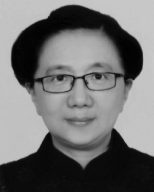 Shuyue Xia (夏书月)合作论文数15
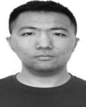 He Dianning合作论文数14
 He Ma (马贺)合作论文数College of Medicine and Biological Information Engineering, Northeastern University13
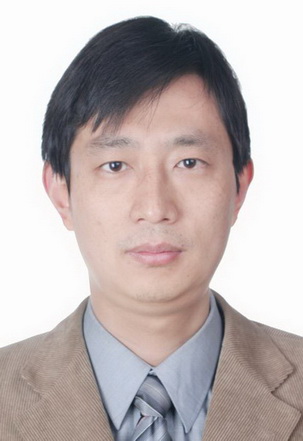 Peng Zhang (张鹏)合作论文数School of Mechanical Engineering, Shanghai Jiao Tong University12
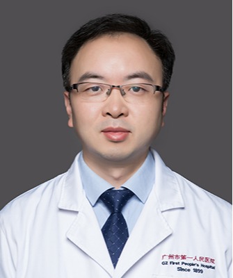 Xinhua Wei (魏新华)合作论文数Guangzhou First People's Hospital12
 Lisheng Xu (徐礼胜)合作论文数College of Medicine and Biological Information Engineering, Northeastern University11Chaoyang Jin合作论文数10
 Haowen Pang合作论文数College of Medicine and Biological Information Engineering, Northeastern University10BaiHua Zhang (张百华)合作论文数10
 Han van Triest合作论文数9Runsheng Chang合作论文数9
 Zhenyu Liang (梁振宇)合作论文数The First Affiliated Hospital of Guangzhou Medical University9
 Rongchang Chen (陈荣昌)合作论文数State Key Laboratory of Respiratory Disease, Guangzhou Medical University;The First Affiliated Hospital of Guangzhou Medical University9Fangfang Han合作论文数9Yang Duan (段阳)合作论文数9
 Ruzhu Wang (王如竹)合作论文数Institute of Refrigeration and Cryogenics, School of Mechanical Engineering, Shanghai Jiao Tong University;Engineering Research Center of Solar Power and Refrigeration of Ministry of Education, Shanghai Jiao Tong University9Jiaxuan Xu合作论文数8
 Yubao Guan (关玉宝)合作论文数The Fifth Affiliated Hospital of Guangzhou Medical University8
 Xin Zhao (赵鑫)合作论文数School of Resources and Civil Engineering, Northeastern University8
 Xuemin Xu (徐学敏)合作论文数School of Biomedical Engineering, Shanghai Jiao Tong University8
 Jie Feng合作论文数7
 Lei Yang合作论文数7Dan Zhao合作论文数7
 Bo Zhang (张波)合作论文数Medical School, Tianjin University7
 Tao Jiang合作论文数Harbin Engineering University, Harbin Engineering University7Xiuhang Ruan合作论文数6
 Frank Kulwa合作论文数College of Medicine and Biological Information Engineering, Northeastern University6
 Patrice Monkam合作论文数Shenyang Jianzhu Univ, Informat & Control Engn Fac, Shenyang 110168, Liaoning, Peoples R China6
 Shuang Liu (刘爽)合作论文数Medical School of Tianjin University6
 Jianlin Wu (伍建林)合作论文数Macau University of Science and Technology6
 Hongzan Sun合作论文数6
 Dayu Xiao (孝大宇)合作论文数College of Medicine and Biological Information Engineering, Northeastern University6Jianhua Li (李建华)合作论文数College of Medicine and Biological Information Engineering, Northeastern University6
 Md Mamunur Rahaman合作论文数5
 Can Tong合作论文数5
 Ning Xu合作论文数5
 Yu Sun合作论文数5XiaoYe Zhang (张晓晔)合作论文数5JianLin Wu (伍建林)合作论文数5
 Yu Tao合作论文数5
 Xu Mingjie合作论文数5
 Jiani Hu (胡佳妮)合作论文数School of Artificial Intelligence, Beijing University of Posts and Telecommunications5




