The binary diagnostic approach does not reflect the entire spectrum of metabolic dysfunction associated steatotic liver disease (MASLD. We used an elastography technology, dual elastography ultrasound (DEUS), to discriminate the different stages of MASLD. This prospective multicenter study was conducted from December 2020 to March 2022. All patients underwent DEUS scan, a liver biopsy, and a liver function laboratory test. The optimal model was developed (ModelDEUSC) with 10 machine learning algorithms by combining DEUS and selected clinical parameters and tested the diagnostic accuracy for distinguishing the three progression stages of MASLD: low-, intermediate-, and high-risk. The diagnostic ability of ModelDEUSC for MASH with advanced fibrosis (≥ F3) was compared with other four non-invasive tests. The study included 312 patients in the derivation cohort and 135 in the validation cohort (7:3). Combining DEUS and clinical parameters, a ternary classification of MASLD in the validation cohort achieved a macro-average AUC of 0.858 (95
Thyroid nodules are a common clinical condition of the endocrine system,and their diagnosis and differential diagnosis remain a serious challenge in the medical community.Since the first observation of acoustic enhancement in 1969,contrast-enhanced ultrasound (CEUS) has gradually matured through two major phases:the development of contrast agents (1990s) and the innovation of imaging algorithms (early 21st century).Since being introduced into clinical practice in China,2003,CEUS has undergone over two decades of localized development.Its capability for microcirculation perfusion imaging has established it as a core imaging tool for dynamic blood flow assessment in critical clinical scenarios,including the distinguishing between benign and malignant nodules,early cancer screening,and the formulation of individualized treatment plans.In recent years,with the advancements in instrument performance and the emergence of novel contrast agents,CEUS has demonstrated increasingly prominent roles in the diagnosis and treatment of thyroid diseases.This article reviews the application progress of CEUS in the diagnosis and treatment of thyroid diseases in China from 2021 to 2024.It explores the clinical application value of CEUS in thyroid nodule diagnosis,lymph node evaluation,assessment of thyroid cancer invasiveness,and guidance for thermal ablation therapy.Additionally,it makes an outlook on the future integration of CEUS with emerging technologies such as molecular ultrasound,artificial intelligence,and robotic technology,aiming to provide new insights and approaches for the precise diagnosis and treatment of thyroid diseases.
In middle-aged and older atherosclerotic renal artery stenosis (ARAS), the anatomical severity of stenosis is a poor surrogate for microvascular competence, and the renal benefit of revascularization is unpredictable. We developed Renal-Video-AI, a self-supervised deep learning framework (Video Swin Transformer with VideoMAE pretraining) that extracts spatiotemporal hemodynamic features from contrast-enhanced ultrasound, and applied it to a multi-center Discovery Cohort (N = 1,226), an independent External Validation Cohort (N = 122), a prospective Multimodal Cohort with paired 10x Visium spatial transcriptomics (N = 57), and an aged two-kidney-one-clip (2K1C) murine model. Unsupervised phenomapping identified 3 intrinsic hemodynamic phenotypes—Preserved, Delayed, and Rarefied. The Rarefied phenotype predicted major adverse renal events (MAREs) independently of anatomical stenosis [hazard ratio (HR) 4.82, 95% confidence interval (CI) 3.10 to 6.50; Fine–Gray subdistribution HR (sHR) 5.1], and adding the phenotype to a standard clinical model improved the C-statistic from 0.72 to 0.88. A significant phenotype-by-treatment interaction (P < 0.01) showed that stenting reduced events only in the Delayed phenotype (HR 0.52, 95% CI 0.35 to 0.78), not in the Preserved (HR 0.98) or Rarefied (HR 1.05) phenotypes. In absolute terms, stenting reduced the 3-year cumulative incidence of MARE in the Delayed phenotype from 25.4% to 13.2% (absolute risk reduction 12.2%; number needed to treat = 8, 95% CI 6 to 13), with no benefit in the Preserved (8.4% versus 8.0%) or Rarefied (38.6% versus 39.4%) phenotypes. Spatial transcriptomics localized a hypoxia and pyroptosis signature to rarefied tissue, and the aged 2K1C model revealed a mitochondrial reactive oxygen species (ROS)–NLRP3–pyroptosis axis whose pharmacological inhibition (MCC950) restored microvascular perfusion. AI video-phenomapping thus reframes the revascularization decision around microvascular competence rather than anatomy, identifying both therapeutic futility (Rarefied) and a treatable window (Delayed), and nominates NLRP3-driven pyroptosis as a therapeutic target.
Background:Primary breast lymphoma (PBL) is a rare extranodal lymphoma of which diagnosis is challenging due to overlapping clinical and imaging features with breast carcinoma. While the existence of PBL is known, the diagnostic significance of specific sonographic patterns like the "grid-like echo" and the critical imperative for exhaustive systemic staging to differentiate primary from systemic lymphoma warrant emphasis. This case is unique as it demonstrates a diagnostic journey from a bilateral "grid-like echo" on ultrasound to a revised diagnosis of a systemic, high-grade lymphoma, highlighting key pitfalls and the necessity of comprehensive workup. Case Description:A 38-year-old female presented with systemic symptoms. Bilateral breast ultrasound revealed Breast Imaging-Reporting and Data System (BI-RADS) 4 lesions with a distinctive "grid-like echo" pattern, well-defined hypoechoic masses, and rich vascularity. Core needle biopsy confirmed a high-grade B-cell lymphoma. However, comprehensive staging-including bone marrow biopsy, cytogenetics, and positron emission tomography-computed tomography (PET-CT)-revealed widespread disease, a t(8;14) translocation, MYC rearrangement, and multifocal extranodal involvement. The final diagnosis was revised to systemic Burkitt-like lymphoma with 11q aberration, Ann Arbor Stage IV B. The patient received systemic chemotherapy, resulting in symptomatic improvement and resolution of breast lesions on follow-up. Conclusions:While ultrasonography holds significant value in detecting and suggesting PBL, definitive diagnosis depends entirely on pathological evidence acquired through surgical biopsy. The cornerstone of treatment is systemic chemotherapy, and radical surgical resection does not improve patient survival. Therefore, the indispensable role of surgery lies in its diagnostic, not therapeutic, capacity. Enhancing awareness of PBL and establishing a multidisciplinary diagnostic model that integrates surgery, ultrasonography, pathology, and clinical practice is essential to avoid misdiagnosis and develop individualized treatment strategies.
Background Autosomal dominant polycystic kidney disease (ADPKD) is characterized by progressive cystic enlargement and an increased risk of renal cell carcinoma. Early diagnosis and treatment are often hindered by severe distortion of renal architecture and by the limitations of contrast-enhanced computed tomography or magnetic resonance imaging in patients with impaired renal function. Contrast-enhanced ultrasound offers a nephrotoxic-free alternative for lesion characterization, treatment planning, and post-ablation monitoring. Case presentation A 44-year-old woman was referred for evaluation of newly detected autosomal dominant polycystic kidney disease (ADPKD). Admission ultrasound showed markedly enlarged cystic kidneys and a 2.2-cm isoechoic lesion in the left kidney that could not be characterized due to the distorted anatomy. To avoid repeat contrast-enhanced computed tomography (CT) or magnetic resonance imaging (MRI), contrast-enhanced ultrasound demonstrated heterogeneous intralesional enhancement with relatively rapid washout, and positron emission tomography-computed tomography (PET-CT) showed focal hypermetabolism. After cessation of dual antiplatelet therapy and correction of anemia, ultrasound-guided core biopsy confirmed renal cell carcinoma. The patient underwent percutaneous radiofrequency ablation, and post-procedural contrast-enhanced ultrasound (CEUS) showed no residual enhancement. Follow-up imaging at 6 and 24 months revealed no recurrence, and renal function remained stable. Conclusions This case highlights the role of CEUS in detecting and characterizing renal tumors in ADPKD, and its utility in post-ablation surveillance. Ultrasound-guided radiofrequency ablation (RFA) is a safe, nephron-sparing treatment for localized RCC in ADPKD patients, especially when traditional contrast-enhanced imaging is limited by renal dysfunction.
Background: Whether BRAF V600E positivity should preclude radiofrequency ablation (RFA) in otherwise suitable patients with cT1N0M0 papillary thyroid carcinoma (PTC) remains unclear. This study aimed to explore the association between BRAF V600E status and structural recurrence/metastasis after RFA. Methods: We retrospectively included 433 consecutive patients with cT1N0M0 PTC who underwent initial ultrasound-guided RFA between May 25, 2021, and May 31, 2025. All patients had preablation BRAF V600E testing performed on fine-needle aspiration specimens and valid follow-up. The primary endpoint was structural recurrence/metastasis after technically complete ablation. Sparse-event, Bayesian, time-adjusted, and sensitivity analyses were performed. Results: Among 433 patients, 215 were BRAF V600E positive and 218 were negative. During a median follow-up of 14.3 months, structural recurrence/metastasis occurred in 3 patients, all in the BRAF V600E -positive group. The event rate was 1.40% versus 0.00%, and the corrected odds ratio was 7.20, with a wide 95% confidence interval of 0.37 to 140.19. Bayesian analysis showed a posterior probability of 0.968 that the risk difference was greater than zero. The signal remained directionally consistent across sensitivity analyses but was limited by the small number of events. Events were not concentrated in the high sonographic anatomic complexity stratum. No higher recorded complication burden was observed in BRAF V600E -positive patients. Conclusions: BRAF V600E positivity identified the subgroup in which structural events occurred after RFA, but it did not by itself establish procedural unsuitability. In selected cT1N0M0 PTC patients, BRAF V600E status may be better used to guide counseling and follow-up intensity than as an absolute exclusion criterion for RFA. Trial registration: Not applicable.
Anaplastic thyroid carcinoma (ATC) is a highly aggressive malignancy with limited effective therapies. Ferroptosis, a form of programmed cell death, is closely associated with thyroid cancer prognosis and represents a potential therapeutic target. This study evaluated the antitumor effects of ultrasound-targeted microbubble (MB) destruction (UTMD)-mediated RAS-selective lethal 3 (RSL3) delivery to induce ferroptosis in ATC. MBs loaded with ferroptosis inducer RSL3 were synthesized and intravenously administered in a nude mouse ATC model, followed by localized ultrasound irradiation to enable targeted intratumoral drug release. RSL3-MBs exhibited uniform particle size, good biocompatibility, and excellent contrast performance. The combination of RSL3-MBs with UTMD significantly inhibited the growth of ATC without evident systemic toxicity. This strategy was shown to be more effective than the delivery methods involving either RSL3 solution or RSL3-MBs alone. These findings support UTMD-mediated RSL3 delivery as an effective approach to enhance ferroptosis-based therapy for ATC.
Objectives: B-Raf proto-oncogene ( BRAF) and telomerase reverse transcriptase ( TERT) mutations have established roles in the management of thyroid nodules, but rat sarcoma virus oncogene homolog ( RAS) mutations’ significance is less conclusive. Integrating mutation profiles with cytological grades provides evidence to evaluate the clinical significance of molecular testing in risk stratification and diagnosis of thyroid nodules. Material and Methods: A total of 5378 nodules were subjected to fine-needle aspiration (FNA cohort) and classified according to the Bethesda system for reporting thyroid cytology. Of them, 705 were surgically removed (thyroidectomy cohort) and pathologically examined, and 53 were followed for 0.5–53.6 months (follow-up cohort) and underwent at least one follow-up FNA. Eight follow-up nodules were resected. All FNA samples were tested for mutations of the BRAF V600E , TERT , and RAS genes. Results: 640 nodules of the thyroidectomy cohort and 6 follow-up nodules were malignant. Molecular testing showed BRAF V600E had a prevalence of 41.5, 58.0, and 15.1% in FNA-, thyroidectomy-, and follow-up cohorts, respectively, but RAS mutations’ prevalence was no higher than 4% in any of them, while TERT mutations’ prevalence was <1%. Consistently, BRAF V600E had a sensitivity of 63.9% and a specificity of 100% in diagnosing malignancy in the thyroidectomy cohort, while RAS mutations had much lower sensitivity and specificity (1.88 and 7.65%, respectively). Furthermore, BRAF V600E increased the risk of malignancy of the FNA cohort to 100% regardless of cytological grades, while RAS mutations showed no increase. In the follow-up nodules, BRAF V600E was strongly associated with malignancy and elevated cytological grades, while RAS mutations showed no association. Conclusion: BRAF V600E is a robust marker for thyroid malignancy in FNA specimens, whereas RAS mutations have limited standalone diagnostic utility but may still provide adjunctive value for risk stratification when interpreted together with cytology and clinical findings.
Background:Accurate assessment of cervical lymph node metastasis (LNM) burden is crucial for formulating individualized secondary surgical strategies in patients with differentiated thyroid carcinoma (DTC) after total thyroidectomy. High-volume metastasis (defined as LNM ≥5) is significantly associated with poor patient prognosis. Preoperative identification of high-volume metastasis prior to secondary surgery is essential for determining the extent of individualized secondary lymph node dissection (LND). However, there is currently a lack of non-invasive predictive tools integrating serum biomarkers and imaging features to quantify this risk. Therefore, this study aimed to develop and validate a multimodal predictive model for preoperative identification of patients with high-volume LNM. Methods:This retrospective study included 97 patients with pathologically confirmed recurrent DTC who underwent secondary LND between May 2020 and December 2024. We analyzed preoperative serum thyroglobulin (Tg) under thyroid stimulating hormone (TSH) suppression (0.1-0.5 mU/L) and ultrasound (US) features, including quantitative metrics (total maximum diameter, total solid component long/short diameter, number of involved neck levels) and qualitative features (microcalcifications, cystic change, hyperechogenicity, abnormal blood flow, aspect ratio ≤2). A data-driven ultrasound score (USscore) was constructed from the qualitative features. Predictors were screened via logistic regression, with internal validation using bootstrap resampling (2,000 repetitions) and clinical utility assessed by decision curve analysis (DCA). Results:Multivariate analysis identified serum Tg ≥2.57 ng/mL [odds ratio (OR) =5.60, 95% confidence interval (CI): 1.31-27.70, P=0.03] and number of involved neck levels (OR =3.38, 95% CI: 1.54-8.49, P=0.003) as independent predictors of LNM ≥5. The multimodal model integrating the USscore (Model 2) achieved an optimism-corrected area under the curve (AUC) of 0.865 (95% CI: 0.783-0.934), with a mean absolute error (MAE) of 0.076, comparable to the baseline model (AUC =0.84, MAE =0.112). DCA showed marginally higher net benefit for Model 2. The USscore demonstrated a dose-dependent association with high-volume metastasis risk (incidence: 68.4% for score ≥5 vs. 23.3% for score ≤1, P=0.006). Conclusions:This study developed and validated a multimodal model integrating serum Tg and sonographic features that effectively predicts the risk of high-volume cervical LNM in postoperative recurrent DTC, providing an objective tool for preoperative assessment and surgical planning.
Three-dimensional (3D) ultrasound provides more intuitive, detailed, and comprehensive diagnostic information. The primary objective of this study was to develop affordable, lightweight, and wide-field 3D ultrasound probes which is a critical challenge for expanding the accessibility and utility of this technology. We describe an affordable freehand scanning 3D ultrasound reconstruction system. The system utilizes a QR code film attached to the skin surface for positioning. During each ultrasound scan, a lightweight camera fixed on the probe records the QR codes within the scanning area. Based on QR code features, a skin map is constructed, and ultrasound images and film image information are preprocessed. The calibration relationship between the ultrasound probe and camera field of view is determined. Finally, comprehensive algorithms such as ORB-SLAM positioning are used for three-dimensional image reconstruction. Through hand-scanning tests, the results show that the scanning speed is 15 mm/s, and when the image acquisition frame rates are 10 Fps/s, 20 Fps/s, and 30 Fps/s, the maximum cumulative error of ORB-SLAM algorithm positioning does not exceed 2
Background:The 2020 Chinese Thyroid Imaging Reporting and Data System (C-TIRADS) regards no or scant punctate-linear enhancement on contrast-enhanced ultrasound (CEUS) as a benign feature in thyroid nodules. However, emerging evidence suggests that some malignant nodules may also exhibit this enhancement pattern. Few systematic studies focusing on the differential diagnosis of such nodules have been conducted. Therefore, this study aimed to identify the ultrasound (US) and CEUS characteristics of malignant solid thyroid nodules with no or punctate-linear enhancement and to evaluate their diagnostic value in distinguishing between benign and malignant lesions. Methods:A retrospective analysis was conducted on clinical and imaging data from 107 patients with 108 pathologically confirmed solid thyroid nodules exhibiting no or punctate-linear enhancement on CEUS, with 73 benign and 35 malignant nodules ultimately being analyzed. Punctate-linear enhancement was further categorized as internal or peripheral based on the location of microbubbles. The basic characteristics, as well as US and CEUS characteristics, were compared between benign and malignant nodules. Multivariate logistic regression analysis was performed to identify independent predictors for malignancy, with odds ratio (OR) and 95% confidence interval (CI) calculated. Receiver operating characteristic (ROC) curve analysis was used to evaluate diagnostic performance, and the area under the curve was calculated with 95% CI, as well as the sensitivity and specificity. Results:Significant differences were observed between the two groups in terms of location, margin, orientation, echogenic foci, enhancement pattern, and enhancement margin (all P<0.05). Multivariate analysis identified that the independent predictors of malignancy were isthmic location (OR =28.288; 95% CI: 4.515-177.250, P<0.001), ill-defined/irregular margin (OR =10.221; 95% CI: 1.929-54.156, P=0.006), microcalcifications (OR =6.516; 95% CI: 1.392-30.497, P=0.017), and peripheral punctate-linear enhancement (OR =15.587, 95% CI: 2.932-82.855, P=0.001), which yielded areas under the curve of 0.702 (95% CI: 0.587-0.816), 0.729 (95% CI: 0.628-0.830), 0.687 (95% CI: 0.572-0.803), and 0.765 (95% CI: 0.674-0.856), respectively; meanwhile the combined model achieved an area under the curve of 0.944 (95% CI: 0.905-0.984), with a sensitivity of 0.943 and a specificity of 0.836. Conclusions:Solid thyroid nodules with no or punctate-linear enhancement on CEUS are not exclusively benign. The combination of isthmic location, ill-defined/irregular margin, microcalcifications, and peripheral punctate-linear enhancement may accurately indicate malignancy. These findings may assist in identifying malignant lesions and reducing unnecessary invasive procedures for benign nodules.
Anaplastic thyroid cancer (ATC) is the most aggressive form of thyroid malignancy and currently lacks effective treatment options. While anti-PD1 therapy has shown remarkable clinical results in some cases, only a subset of ATC patients responds to it. Eganelisib (IPI549), a highly selective PI3Kγ inhibitor, can alleviate the tumor immunosuppressive state by reducing the proportion of M2-like tumor associated macrophages, partially overcoming patient resistance to anti-PD1 therapy and synergizing with its efficacy. However, the dose-dependent hepatotoxicity of IPI549 limits its utility in combination immunotherapy. To address this challenge, we developed integrin αvβ3 targeted microbubbles loaded with IPI549 and indocyanine green, abbreviated as IPI549@αIMBs. This platform enables dual modal ultrasound-fluorescence imaging for monitoring its drug release and distribution, while its combination with ultrasound targeted microbubble destruction(UTMD) technology facilitates the precise delivery and controlled release of IPI549, thereby enhancing synergy with anti-PD1 therapy and improving treatment safety. Our results demonstrate that IPI549@αIMBs effectively targeted to subcutaneous ATC tumors in mice, significantly enhancing IPI549 delivery efficiency. UTMD mediated delivery using IPI549@αIMBs combined with anti-PD1 therapy markedly inhibited subcutaneous ATC tumor growth in mice, effectively reduced the proportion of M2-like tumor associated macrophage, and increased CD8+ T cell infiltration, which alleviated the tumor immunosuppressive state. In summary, this study explored a novel therapeutic strategy for ATC, demonstrating the efficacy of the IPI549@αIMB-based UTMD approach combined with anti-PD1 therapy and validating the underlying immunomodulatory mechanisms.
Objective To explore the research progress and hotspots of intelligent ultrasound imaging in the diagnosis of thyroid nodules and clarify the research directions via the bibliometric method.Methods The relevant research articles on intelligent ultrasound imaging in the diagnosis of thyroid nodules were retrieved from the Web of Science Core Collection,covering the period from January 2004 to August 2024.Python was used to analyze the number of annual publications.VOSviewer was used to create the co-occurrence network of authors and the keyword density map.CiteSpace was used to demonstrate the dual-map overlays of the journals,as well as the bursts and clustering of co-citations and keywords.Results A total of 1 179 articles were included.The annual number of publications increased steadily.The involved journals demonstrated high quality,and the publications showed a trend of cross-research.Chinese researchers were the core research force in this field.Haugen et al.'s study on the guidelines for thyroid nodules had the most citations.The clustering of co-citations and keywords indicated studies in multiple fields.Thyroid nodules,cancer,and deep learning were the representative keywords in this field.Conclusions The continuous enrichment of research topics promotes the rapid development of intelligent ultrasound imaging for thyroid nodules.Intelligent diagnosis methods based on deep learning can provide diagnostic suggestions,while there are still challenges such as interpretation.One of the research directions is the deep combination of intelligent diagnosis algorithms and medical knowledge.
Background and Objective:As the detection rate of thyroid nodules increases year by year, traditional ultrasonic diagnostic methods face challenges such as inefficiency and high dependence on physician experience. This paper focuses on the research status, advantages and challenges of robot automatic scanning and intelligent diagnosis system. Methods:We systematically retrieved the PubMed and Web of Science databases, screened and integrated relevant articles, and conducted a systematic analysis and summary of the existing research. Key Content and Findings:The development of robot and artificial intelligence (AI) provides a new method for efficient and accurate ultrasound diagnosis of thyroid nodules. Robot enables automated scanning of thyroid through precise robotic arm control, positioning, and trajectory planning, significantly improving the standardization and repeatability of the diagnostic process. However, its flexibility in clinical application and patient acceptance still needs to be further improved. From the early rule matching research based on manual features to the automatic feature processing of thyroid nodules using deep learning algorithms have made AI outstanding in the ultrasound diagnosis of thyroid nodules. Meanwhile, the innovative research of deep learning in the contrast-enhanced ultrasound (CEUS) video analysis has broadened the application of intelligent diagnosis systems. The interpretability of the deep learning models is solved to some extent by Gradient-weighted Class Activation Mapping (Grad-CAM) and other techniques. However, the interpretability, data dependence, and ability to generalize deep learning models in clinical practice remain key issues to be addressed. Conclusions:Robots and AI have brought revolutionary progress to the diagnosis of thyroid diseases, but their clinical translational application still faces many challenges.
The incidence of thyroid cancer keeps rising globally,with the majority being papillary thyroid carcinoma (PTC),which has a favorable prognosis.However,some aggressive PTCs exhibit different clinical behaviors and higher mortality risks,with the growth rate surpassing that of well-differentiated PTC and undifferentiated cancers.Therefore,achieving precise diagnosis and treatment of thyroid carcinoma presents a significant challenge.Photoacoustic imaging is a molecular imaging technology that integrates optical imaging and ultrasound,providing imaging information on structure,function,and molecules.Moreover,it can utilize exogenous contrast agents to realize tumor treatment,such as photothermal therapy,serving as a promising technology for precise diagnosis and treatment of thyroid carcinoma.Researchers both domestically and internationally have explored the application of photoacoustic imaging in the precise diagnosis and treatment of thyroid tumors.This article reviews the research progress,elucidates the advantages and limitations of photoacoustic imaging in the diagnosis and treatment of thyroid carcinoma,and prospects on the precise diagnosis and treatment of this disease.
Background:Bethesda IV follicular neoplasms [2-25% of fine-needle aspiration (FNA) diagnoses] pose significant diagnostic challenges due to their malignant potential. Thermal ablation (TA) has emerged as a minimally invasive alternative to surgery, though its safety for follicular neoplasms remains controversial given cytology's inability to assess capsular/vascular invasion-the diagnostic gold standard. Case Description:This case report analyzes two patients with Bethesda IV nodules treated with microwave ablation (MWA). Case 1 involved a 26-year-old female with a 5.1 cm nodule undergoing two MWA sessions, while Case 2 featured a 12-year-old female with a 2.6 cm nodule receiving single-session MWA. Both cases deviated from guideline-recommended dual preoperative FNAs. Both patients developed recurrence: Case 1: progression of thyroid carcinoma within the ablation zone and metastatic papillary carcinoma in cervical lymph nodes were surgically confirmed two years post-ablation. Case 2: a recurrent cystic-solid nodule (1.8 cm) was detected adjacent to the ablation zone during 9-month post-procedural surveillance. Notably, neither recurrence displayed typical follicular carcinoma histology, suggesting ablation-induced diagnostic obscuration. Conclusions:These first-reported recurrence cases after TA for Bethesda IV nodules underscore critical limitations: violations of pre-ablation diagnostic protocols [dual FNA/core needle biopsy (CNB)] exacerbated malignancy risks, while TA-induced obscuration of histopathological evaluation delayed cancer diagnosis. Current guidelines appropriately exclude follicular neoplasms from TA indications, with surgical resection remaining the gold standard. TA should be restricted to validated research protocols pending establishment of robust risk-stratification criteria.
Granulomatous mastitis (GM) is a rare, chronic, benign inflammatory breast disease with an unknown etiology, primarily affecting young women. It is often characterized by the presence of a painful mass, skin changes, and persistent inflammation, which can clinically resemble breast cancer. Due to the absence of specific imaging signatures and a poorly understood pathogenic mechanism, diagnosis is primarily achieved through exclusion. We present the case of a 31-year-old woman who, three years after undergoing a radical mastectomy for triple-negative breast cancer in her right breast, was diagnosed with GM in her left breast. The diagnosis was based on magnetic resonance imaging (MRI), conventional and contrast-enhanced ultrasound, and confirmed through histological analysis. The patient has experienced recurrent episodes of GM over the past three years. Notably, this case represents the first reported instance of granulomatous mastitis occurring in the contralateral breast following a radical mastectomy for breast cancer.
This study compares the success rate, diagnostic accuracy, and safety of contrast-enhanced ultrasound (CEUS)-guided biopsy versus conventional ultrasound (US)-guided biopsy for thoracic and pulmonary lesions. A systematic search of PubMed, EMBASE, Web of Science, and Cochrane Library was conducted. The primary outcomes included success rate and diagnostic accuracy, and the secondary outcome was the odds ratio of adverse effects. A random-effects meta-analysis pooled the data, with heterogeneity assessed by I² and publication bias evaluated using Egger’s test and funnel plot analysis. Sensitivity analysis was performed to confirm result robustness. Subgroup analysis and meta-regression were conducted to explore the sources of heterogeneity. Sixteen studies with 3,459 patients were included. CEUS-guided biopsy demonstrated higher success rate (99.18
Objectives: We aimed to develop and validate machine learning (ML) models that integrate clinical and laboratory data for the non-invasive prediction of metabolic dysfunction-associated steatohepatitis (MASH) in an obese population. Methods: In this retrospective study, clinical and laboratory data were collected from obese patients undergoing bariatric surgery. The cohort was divided using stratified random sampling, and optimal features were selected with SHapley Additive exPlanations (SHAP). Various ML models, including K-nearest neighbors, linear support vector machine, radial basis function support vector machine, Gaussian process, random forest, multilayer perceptron, adaptive boosting, and naïve Bayes, were developed through cross-validation and hyperparameter tuning. Diagnostic performance was assessed via the area under the curve (AUC) in both training and validation sets. Results: A total of 558 patients were analyzed, with 390 in the training set and 168 in the validation set. In the training cohort, the median age was 35 years, the median body mass index (BMI) was 39.8 kg/m2, 39.0% were male, 37.9% had diabetes mellitus, and 62.8% were diagnosed with MASH. The validation cohort had a median age of 34.1 years, a median BMI of 42.5 kg/m2, 41.7% male, 32.7% with diabetes, and 39.9% with MASH. Among the models, the random forest achieved the highest performance among the models with AUC values of 0.94 in the training set and 0.88 in the validation set. The Gaussian process model attained an AUC of 0.97 in the training cohort but 0.79 in the validation cohort, while the other models achieved AUC values ranging from 0.63 to 0.88 in the training cohort and 0.62 to 0.75 in the validation set. Conclusions: ML models, particularly the random forest, effectively predict MASH using readily available data, offering a promising non-invasive alternative to conventional serological scoring. Prospective studies and external validations are needed to further establish clinical utility.
 Yuxin Jiang (姜玉新)合作论文数Peking Union Medical College Hospital81
 Xuehua Xi合作论文数45Ruina Zhao (赵瑞娜)合作论文数45Xingjian Lai合作论文数北京协和医学院(北京协和医院、北京协和医院、北京协和医院),中国北京43
 Xiao Yang合作论文数33MA JIAOJIAO合作论文数32
 Dan Mei (梅丹)合作论文数Chinese Academy Of Medical Sciences & Peking Union Medical College30Xiaoyan Zhang合作论文数29Luying Gao合作论文数27Shenling Zhu合作论文数26
 Jianchu Li (李建初)合作论文数北京协和医院25Jiajia Tang合作论文数24Ying Wang合作论文数17Ruyu Liu合作论文数17Wei Zuo (左玮)合作论文数17
 Meng Yang (杨萌)合作论文数Peking Union Medical College Hospital16Qiong Gao (高琼)合作论文数14Dakui Li (李大魁)合作论文数Peking Union Medical College Hospital13Yan Tang (唐彦)合作论文数13
 Qing Dai (戴晴)合作论文数Department of Ultrasound, Peking Union Medical College Hospital, Chinese Academy of Medical Sciences and Peking Union Medical College13GuangHan Li (李广涵)合作论文数12Yang Hu (胡扬)合作论文数12
 Xiaoyi Li (李小毅)合作论文数Peking Union Medical College Hospital11
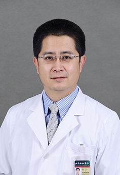 Yu Xia (夏宇)合作论文数Peking Union Medical College Hospital11WenJuan Sun (孙雯娟)合作论文数10Ying Shuai合作论文数9Wenbo Li合作论文数9LiPing Dou (都丽萍)合作论文数9
 Xiaoyan Xing (邢小燕)合作论文数China-Japan Friendship Hospital9
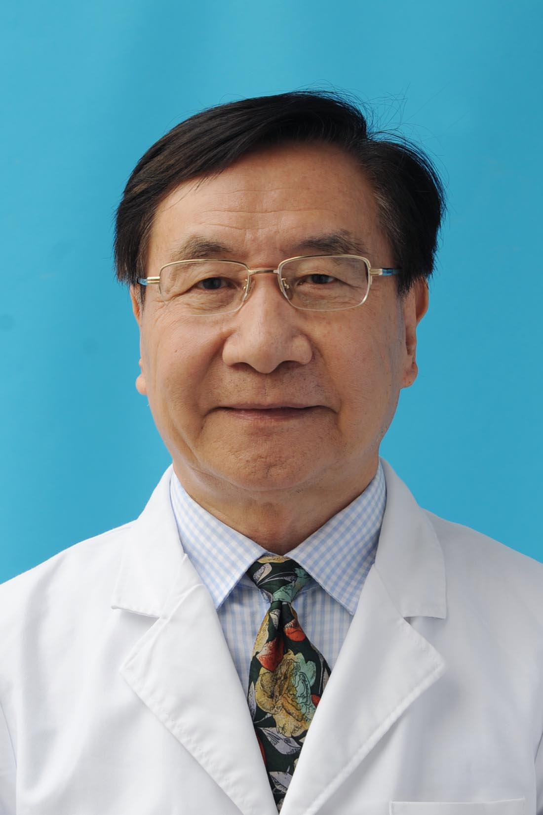 Guangwei Li (李光伟)合作论文数Department of Endocrinology, China-Japan Friendship Hospital9
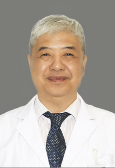 Renzhi Wang (王任直)合作论文数Peking Union Medical College Hospital;School of Medicine, The Chinese University of Hong Kong, Shenzhen9Liangkai Wang合作论文数8Huilin Li合作论文数8Tongtong Zhou合作论文数8Wu Qiong合作论文数8
 Qiuhong Gong (巩秋红)合作论文数中国医学科学院阜外医院内分泌与心血管代谢二病区7Yali An (安雅莉)合作论文数7Mai Sun合作论文数6Lin-Ping Wang合作论文数6Jinping Wang合作论文数6YuZhen Zou (邹羽真)合作论文数6JingPing Wu (武敬平)合作论文数6Sheng Cai (蔡胜)合作论文数6
 Zhiyong Liang (梁智勇)合作论文数Peking Union Medical College6
 Min Zheng (郑敏)合作论文数China-Japan Friendship Hospital6
 Qingli Zhu (朱庆莉)合作论文数Peking Union Medical College Hospital6JIA Xinying (贾欣颖)合作论文数5Li Hui合作论文数5




