目的:研究万古霉素联合美罗培南骨水泥用于骨感染患者治疗的局部释放特点及规律.方法:2020年6月至2021年3月北京积水潭医院创伤骨科收治的骨感染患者,手术感染病灶中植入万古霉素联合美罗培南骨水泥链珠,局部留置引流管,分别于术后2 h及每日清晨留取伤口引流液标本及静脉血,使用酶放大免疫法和超高效液相色谱-串联质谱法测定万古霉素和美罗培南浓度.结果:共纳入21例骨感染患者,局部释放率两药最高峰均出现在10 h,最大释放速率万古霉素和美罗培南分别为4.761,1.149 mg·h-1,累计局部释放率分别为6.38%,1.41%;术后2 h引流液中万古霉素和美罗培南药物质量浓度分别为(373.06±357.70)mg·L-1,(158.41±121.33)mg·L-1;血药浓度最大值分别为1.1,0.54 mg·L-1.结论:万古霉素联合美罗培南骨水泥自制链珠局部植入骨感染病灶可以在术后短期内获得高组织浓度,万古霉素局部药物浓度更高,两药全身药物浓度均很低安全性高.
目的:考察美罗培南骨水泥药柱的体外缓释规律,为临床使用提供参考.方法:用模具将美罗培南加入骨水泥中制成含美罗培南10%(W/W)的骨水泥植入剂,浸泡于37℃的改良模拟体液中,采用等距浸出法设定取样时间点,应用超高效液相色谱-串联质谱法对浸出液浓度进行检测,观察美罗培南骨水泥植入剂的释放规律.结果:美罗培南骨水泥药柱释药峰值出现在2 h,之后释药速率逐渐降低,19 h累计释放量达总体释放量的92.37%,在24 d内累积释放率达6.00%,体外释放曲线符合Weibull方程,r=0.9151.结论:美罗培南骨水泥具有一定的缓释特性,释放主要集中在前72 h内,后期释放少.本研究结果可以为临床使用美罗培南治疗骨感染提供参考.
开放性骨折的治疗对于任何创伤骨科医师都是一个挑战.在诸多问题上仍然需要通过研究提供更高等级证据,随着加速康复外科(ERAS)理念的引入以及循证医学的发展,可以在现有基础上做出多方面的改进,以使患者的主观体验及客观治疗效果得到改善.经过全国创伤骨科专家的讨论后达成本共识,为在急诊情况下处理开放性骨折提供参考.
目的 通过测定载万古霉素骨水泥、人工骨植入骨感染患者体内后全身及局部的药物浓度,对比万古霉素骨水泥及人工骨的局部释放规律.方法 选择2017年6月至2018年4月创伤骨科先后进行万古霉素骨水泥链珠及万古霉素人工骨植入术的16例骨感染患者,术中局部埋置引流管,分别于术后2h及此后每日清晨留取患者静脉血及伤口引流液标本,直至术后引流管拔除,使用酶放大免疫法进行静脉血及引流液万古霉素浓度测定.结果 万古霉素骨水泥、人工骨植入术后平均引流量分别为(504.50±306.52)、(278.68±136.18) ml,局部累计释放率为5.60%±3.61%、19.12%±9.86%,最大释放速度为(6.1±4.4)、(20.8±9.5) mg/h,全身血药浓度Cmax为(0.66±0.34)、(1.39±0.98)mg/L.结论 万古霉素人工骨和骨水泥植入人体后均能获得很高的局部药物治疗浓度,全身药物浓度很低,安全性较好.人工骨较骨水泥局部药物浓度更高,总体释放量、释放率、释放速度及植入后全身的药物浓度均高于万古霉素骨水泥.
目的 观察两次清创术在髋部骨折术后感染的临床疗效.方法 回顾性研究收集2012年6月至2015年6月期间在青海省人民医院创伤骨科住院的25例髋关节感染患者详细资料.其中男18例,女7例;年龄20~71岁,平均44.76岁.治疗方案:第1次先清创、取出内固定、细菌培养、抗生素骨水泥链珠植入,1周后根据细菌培养结果再次清创、敏感抗生素骨水泥间隔物植入.术后血沉、C反应蛋白恢复正常后,二期行新假体植入术.结果 25例患者中1例先后进行3次清创、抗生素骨水泥间隔物植入失败,至今感染未治愈.1例患者感染复发,准备再次手术.4例患者失访,其余21例患者随访24~80个月,平均60个月.19例患者治愈(90.48%),2例患者未治愈(9.52%).术前术后血沉、C反应蛋白结果比较,P<0.05,差异有统计学意义.术后1年Harris评分结果,优良10例(48%),较好7例(33%),尚可2例(9.5%),差2例(9.5%).术前术后Harris评分比较P<0.05,差异有统计学意义.结论 髋部骨折内固定感染术后,应选择两次清创术.术中彻底清创、5套以上送检细菌标本、针对致病菌选用敏感抗生素间隔物、合理使用敏感抗生素等措施是提高髋部骨折术后感染治愈率的有效方法.
BACKGROUND:Tibial plateau fractures remain a clinical challenge due to the complexity of the fracture patterns which have been repeatedly categorized by many researchers. However, limitations do exist in some respects. So we aimed to reclassify tibial plateau fractures based on injury mechanism and morphological characteristics.METHODS:Five hundred fourteen cases of tibial plateau fractures were enrolled. The X-rays and CT scans were analyzed.RESULTS:According to our observation and analysis, tibial plateau fractures can be categorized into the following six types: (1) Lateral condylar fractures (axial force applied while knee extending in valgus position). Two hundred fifty-one cases were included (48.83%). (2) Fracture dislocation (multiple forces especially rotational stress while knee extending). Fifty-five out of 514 cases belong to this pattern (10.70%). Correction of the subluxation remains primary and crucial during surgical procedures. (3) Simple medial condylar fractures (axial force applied while knee extending in varus position). One third of which were associated with an avulsion fracture of fibular head. Fifteen cases were included (2.92%). (4) Bicondylar fractures (axial forces applied while knee extending). One hundred twelve cases were included (21.79%). Surgical algorithm greatly depends on soft tissue conditions. (5) Posterior condylar fractures (axial stress applied while knee flexing). Sixty-five cases were seen in our study (12.65%), most of which were associated with an avulsion fracture of the intercondylar eminence (49/65, 75.38%). The fracture of posteromedial part, posterolateral part, and intercondylar eminence forms a unique pattern of injury defined as "Posterior Condylar Triad." (6) Anterior condylar compression fractures (axial, varus, or valgus forces applied while knee overextending). Posterior structural complexes, crucial ligaments, or even popliteal arteries are prone to be damaged. Sixteen cases were identified (3.11%).CONCLUSION:Our classification system has instructive significance in overall preoperative evaluation of fracture features and soft tissue problems as well as guiding clinical management for better functional outcomes.
目的:通过测定载万古霉素骨水泥链珠植入骨感染患者体内后全身及局部的药物浓度,了解万古霉素骨水泥链珠的释放规律,为临床使用万古霉素骨水泥治疗骨感染提供依据.方法:2017年1月-2018年3月收治的骨感染患者20例,手术感染灶局部植入万古霉素和骨水泥混合制成的链珠,局部埋置引流管,分别于术后2h及此后每日清晨留取患者静脉血及伤口引流液标本,直至术后引流管拔除.使用酶放大免疫法进行静脉血及引流液万古霉素浓度测定.结果:20例患者平均年龄48.35±19.37岁,男16例,女4例,引流管留置时间中位数为5d,血药峰浓度为0.99 mg·L-1,达峰时间为33.33 h,局部释放率最高峰值出现在10.5 h,最大释放速率为6.107mg·h-1.累计局部释放率为5.68%,溶出模型拟合符合Weibull方程,r=0.984 5.结论:局部植入万古霉素骨水泥可以至少在术后一周内伤口局部达到很高的药物治疗浓度,而全身药物浓度很低,可以避免全身使用万古霉素带来的不良反应,具有很高的安全性.
目的 本文旨在探讨慢性骨髓炎患者清创术中不同细菌送检套数与确定致病菌之间的关系.方法 本研究收集2007年1月至2014年1月255例慢性骨髓炎患者的临床资料,其中男性202例,女性53例;年龄15~84岁,平均年龄(45.2±16.5)岁.共送检细菌培养295次,其中送检套数分别为:63次(1套)、76次(2套)、51次(3套)、41次(4套)、64次(5套).计算出总体细菌谱、各套数检出的细菌数及检出致病菌能力,并对其各套数之间进行比较,得出结论.结果 在295次送检细菌培养中,共检出细菌42种,前5位分别是金黄葡萄球菌(35.51%)、表皮葡萄球菌(14.29%)、铜绿假单胞菌(9.80%)、阴沟肠杆菌(5.31%)、大肠埃希菌(4.49%);各套细菌检出率依次为66.7%(1套)、69.7%(2套)、74.5%(3套)、78.0%(4套)及90.6%(5套及以上);确定致病菌能力依次为42.1%(2套)、51.9%(3套)、68.3%(4套)、82.8%(5套及以上),各套数之间比较结果为2套与4套、2套与5套、3套与5套之间P<0.05,差异有统计学意义.结论 在确定致病菌方面,术中送检5套及以上细菌检出致病菌能力最强,1套送检细菌检出致病菌能力最差.因此推荐在术中至少送检5套及以上细菌标本来确定致病菌.
目的 通过测定载万古霉素人工骨微球植入骨感染患者体内后全身及局部的药物浓度,了解万古霉素人工骨的释放规律,为临床使用万古霉素人工骨治疗骨感染提供依据.方法 2017年1-7月收治的骨感染患者13例,手术时在局部感染灶植入2 9万古霉素与硫酸钙人工骨混合制成的药物微球,术中局部埋置引流管,分别于术后2h及此后每日清晨留取患者静脉血及伤口引流液标本,直至术后引流管拔除.使用酶放大免疫法进行静脉血及引流液万古霉素浓度测定.结果 13例患者中,男11例,女2例,平均年龄(41.93±14.39)岁,引流管留置时间中位数为3d,血药峰浓度为1.68 μg/mL,达峰时间为16.09 h,局部释放率最高峰值出现在9h,最大释放速率为28.72 mg/h.累计局部释放率为23.06%,溶出模型拟合符合Weibull方程,r=0.966 8.结论 局部植入万古霉素硫酸钙人工骨微球可以至少在术后1周内伤口局部达到很高的药物治疗浓度,而全身药物浓度很低,可以避免全身使用万古霉素带来的不良反应,具有很高的安全性.
目的 对比切开复位钢板内固定及非手术方法治疗成人锁骨中段1/3移位骨折的临床疗效.方法 回顾性分析我院创伤骨科2010年3月至2013年5月治疗的60例锁骨中段1/3移位的病例,30例行切开复位钢板内固定,30例行锁骨吊带固定治疗.非手术治疗组30例,男20例,女10例;年龄14~70岁,平均(43.73±4.28)岁.钢板固定组30例,男21例,女9例;年龄16~63岁,平均(36.53±4.01)岁.两组患者在人口学特征、受伤严重程度、受伤机制及骨折类型方面无差别.研究结果按照随访时间、骨折愈合时间、肩关节功能活动及术后患者对肩关节外形满意度进行评价.骨折愈合时间主要根据术后影像学来判定.肩关节功能活动主要根据Constant肩关节评分(constant shoulder score,CSS),臂、肩、手功能障碍(the disability of the arm shoulder and hand score,DASH)评分来评定.结果 两组患者均获随访,平均随访时间为(32±3.40)个月.对两组CSS及DASH评分结果进行比较,均P<0.05(P=0.000,P=0.003),且钢板固定组均值均大于非手术组(均值差分别为5.49、2.71).治疗愈合时间:钢板固定组(12.10±2.60)周,非手术治疗组(15.78±2.23)周.并发症:钢板固定组1例骨折不愈合(3%),无畸形愈合;非手术治疗组3例骨折不愈合(10%),12例畸形愈合(40%).需要二次手术取出钢板是钢板固定组主要并发症,取出率为60%.非手术治疗主要并发症是骨折畸形愈合(40%).患者对肩关节外形满意度分别为93%和77%.结论 在成人锁骨中段1/3移位骨折的治疗方法中,切开复位钢板组总体疗效优于非手术治疗组.切开复位钢板固定组在提高患者肩关节功能活动、降低骨折不愈合及畸形愈合方面优于非手术治疗.因内置物刺激症状而取出内固定是钢板固定组主要的并发症.
Objective To discuss the clinical outcomes of trochanteric digastric osteotomy and surgical hip dislocation for treatment of Pipkin Ⅳ fractures. Methods We retrospectively reviewed the 12 patients with Pipkin Ⅳ fracture who had been treated by trochanteric digastric osteotomy and surgical hip dislocation from February 2012 to June 2016. They were 10 males and 2 females, with an average age of 44. 1 years ( from 24 to 63 years ) . According to the Pipkin classification for femoral head fractures, 9 fractures were type Ⅰand 3 type Ⅱ; according to Letournel-Judet classification for acetabular fractures, 10 were posterior wall fractures ( 9 belonging to the posterior superior subgroup ) and 2 transverse plus posterior wall fractures ( one involving the posterior wall belonging to the posterior superior subgroup ) . The operation time, blood loss, complications and functional results were recorded. Results The operation time averaged 165 min ( from 150 to 195 min ); the blood loss averaged 850 mL ( from 500 to 1, 200 mL ) . A spring plate was used additionally for posterior wall fracture in 7 cases. All the 12 cases obtained an average follow-up of 19 months ( from 6 to 52 months ) . Motion function was fully recovered from 6 weeks to 4 months after operation in 2 cases of preoperative incomplete sciatic nerve injury and in one case of iatrogenic incomplete sciatic nerve injury. Anatomical reduction was achieved in all femoral head fractures. According to the Matta criteria for acetabular fractures, anatomical reduction was achieved in 9 cases, good reduction in 2 and poor reduction in one. Heterotopic ossification occurred in 4 cases ( gradeⅠin one, gradeⅡin 2 and gradeⅢin one by the Brooker grading system ) . The trochanteric osteotomy was repeated in one case. No femoral head avascular necrosis was found. According to the modified Merle d'Aubigné and Postel scoring at the final follow-ups, the functional results of the affected hip were excellent in 4 cases, good in 7 and poor in one. Conclusion Trochanteric digastric osteotomy and surgical hip dislocation is a safe and effective treatment for Pipkin Ⅳ fractures.
OBJECTIVE:To compare the difference between non-operative and operative treatment of humeral shaft fractures.METHODS:From March 2005 to October 2012, 252 cases of humeral shaft fractures were treated and were adequately followed up. According to the treatment methods, the patients were divided into 2 groups: the non-operative group and the operative group. In the non-operative group, there were 76 cases treated with plaster/small splint fixation,meanwhile there were 176 cases treated with internal fixation either by plating or by nailing in the operative group. The follow-up parameters included: fracture healing rate, fracture union time, complications rate, Constant- Murley shoulder score and Mayo elbow score.RESULTS:The mean follow-up period was (31.24±20.06) months (ranging 6 to 103 months). There were no statistical differences in age, open fracture number, fracture site and Arbeitsgemeinschaft für Osteosynthesefragen (AO) classification between the non-operative group and the operative group. The fracture healing rate: the non-operative group: 96.1%(72/76), the operative group: 97.7%(172/176), P=0.46; the fracture union time: the non-operative group: (10.24±2.93) weeks, the operative group: (10.69±2.51) weeks, P=0.22; the complication rate: the non-operative group: 5.3%(4/76),the operative group: 15.3%(27/176), P=0.03. The complications included: nonunion: the non-operative group: 3.95%(3/76), the operative group: 2.3%(4/176), P=0.434; radial nerve palsies: the non-operative group: 0%(0/76), the operative group: 5.7%(10/176), P=0.035; bone split: the non-operative group: 0%(0/76), the operative group: 1.7%(3/176), P=0.556; elbow stiffness: the non-operative group:1.3%(1/76), the operative group: 0.6%(1/176), P=1.000; shoulder pain: the non-operative group:0%(0/76), the operative group: 5.1%(9/176), P=0.061. The Constant-Murley shoulder score: the non-operative group: 97.37±4.94, the operative group: 96.34±6.88, P=0.244. The Mayo elbow score: the non-operative group: 99.80±1.72, the operative group: 99.49±2.73,P=0.923.CONCLUSION:The results of non-operative treatment of humeral shaft fractures appeared with excellent results with lower complications rate compared with that of the operative treatment.
Objective To classify tibial plateau fractures based on the analysis of the morphology and injury mechanisms of 200 cases of tibial plateau feature.Methods We collected the X-ray and CT images of 200 consecutive cases of tibial plateau fracture in i99 patients who had been treated at our department from January 2010 to April 2011.They were 134 males and 65 females,from 15 to 77 years of age (average,45.7 years).According to the Schatzker classification,9 cases were type Ⅰ (4.5%),105 type Ⅱ (52.5%),19type Ⅳ (9.5%),37type Ⅴ (18.5%),30type Ⅵ (15.0%),and none type m.The fracture morphology and injury mechanism of each case were analyzed to propose a new classification system.Results Fractures of tibial plateau can be classified into the following five types:(a) Lateral condylar fracture and valgus injury (100 cases,50%).The injury mechanism is the axial force on the valgus and extended knee joint.(b) Fracture-dislocation injury (24 cases,12.0%).This type includes typical Schatzker type Ⅳ,and some cases of Schatzker type Ⅵ associated with lateral subluxation.Its mechanism is a compound force of valgus,varus,rotational and axial stresses.The rotational force is the key factor leading to subluxation of the knee joint.(c) Double-condylar fracture (40 cases,20.0%).This type is caused by an axial force on the extended knee,including Schatzker type Ⅴ and some cases of Schatzker type Ⅵ not associated with knee subluxation.(d) Posterior condylar fracture and flexion injury (32 cases,16.0%).This type only involves the posterior condylar plateau,and is caused by an axial force on the flexed knee.Based on the morphology,posterior condylar fractures can be further divided into three subtypes:simple split of posteromedial condyle,simple collapse of posterolateral condyle,and a combination of the two.(e) Frontal plateau compression fracture and hyperextension injury (4 cases,2.0%).This type is caused by an axial force on the hyper-extended knee.It is characterized by significant compression of the anterior plateau and intact posterior plateau.Conclusion Based on the morphological features and injury mechanisms,tibial plateau fractures can be classified into 5 types:lateral condylar fracture,fracture-dislocation injury,double-condylar fracture,posterior condylar fracture,and frontal plateau compression fracture.
[目的]对比窦道细菌培养结果与术中细菌培养结果的一致率,及前者确定致病菌的能力,为临床使用敏感抗生素提供帮助.[方法]回顾性研究积水潭医院创伤骨科2003年6月~ 2013年6月53例伴有窦道的慢性骨髓炎患者,收集相关资料比较窦道细菌培养结果与术中细菌培养结果及确定致病菌之间的关系.[结果]在窦道细菌培养中,细菌检出率为75.60%,细菌未检出率24.6%,在细菌检出病例中,检出率最高的前3位分别是:金黄色葡萄球菌(32.26%)、表皮葡萄球菌(12.90%)、铜绿杆菌(8.06%).[结论]窦道细菌培养与术中细菌培养的符合率为67.30%,窦道细菌培养结果与最终确定该病致病菌的符合率为51.50%.
目的:探讨奴卡菌病的诊治要点,减少误诊误治。方法对我院近期收治的由豚鼠奴卡菌感染引起的肱骨骨髓炎1例的临床资料进行回顾性分析,并复习相关文献。结果本例因骨折术后感染窦道形成,近期窦道流脓症状加重就诊。查体左上臂见多个结节和窦道,窦道口触之疼痛,有血性、脓性分泌物及白色颗粒排出。摄左上臂X线片示:左肱骨不连续,骨质增生、硬化,骨腔不规则,可见死骨。行病灶清创引流术,术后分泌物微生物培养及药敏试验结果示:豚鼠奴卡菌生长,对万古霉素及磺胺类药物敏感。确诊为奴卡菌感染致肱骨骨髓炎,予万古霉素和复方磺胺甲噁唑治疗,并每周进行一次窦道清理术,2个月后病情好转出院。出院后继续口服复方磺胺甲噁唑,后窦道基本愈合,择期行骨髓炎病灶清除术。结论奴卡菌感染是一种少见但严重的感染,及时行微生物培养、药敏试验和组织病理学检查,尽早确诊并予有效治疗,是避免或减少误诊误治的关键。
Objective To compare biomechanical effects of augmentative plating assisted by different types and numbers of intramedullary nails in the treatment of nonunion of femoral shaft fracture after intramedullary nailing.Methods Twenty-five specimens of synthetic femur were randomly divided into an experimental group (20 bones) and a control group (5 bones).All the bones were made into models of nonunion of femoral shaft fracture after intramedullary nailing.The control group was subjected to no augmentative plating.The experimental group was further divided into 4 subgroups (n =5) which were subjected to fixation respectively by:a 7-hole plate with 3 bicortical screws on each side,a 7-hole plate with 3 unicortical screws on each side,a 5-hole plate with 2 bicortical screws on each side,and a 5-hole plate with 2 unicortical screws on each side.The torque values of all the subgroups were measured and compared when the models were subjected to torsional angles of 5°,10°,15°,20° and 25°.Results The anti-torsional strengths at torsional angles of 5°,10°,15°,20° and 25° in the 4 experimental groups were all significantly greater than in the control group(P < 0.05).At a torsion angle of 25°,the mean torque value in the 3-bicortical-screw group (46.51 ±6.66 nm) was significantly greater than in the 2-bicortical-screw group (39.03 ±8.52 nm) (P <0.05).At torsion angles of 5°,10°and 25°,the mean torque values in the 3-unicortical-screw group (13.48±1.68 nm,23.29±3.72 nm and 48.19±5.34 nm,respectively) were significantly greater than in the 3-unicortical-screw group (9.56 ± 3.62 nm,16.99 ± 5.49 nm and 38.69 ± 6.56 nm,respectively) (P < 0.05).At torsion angles of 20° and 25°,the mean torque values in the 3-unicortical-screw group (39.15 ± 5.07 nm and 48.19 ± 5.34 nm) were significantly greater than in the 2-bicortical-screwgroup (32.50 ± 5.65 nm and 39.03 ± 8.52 nm) (P <0.05).Conclnsions The anti-torsional strength of fixation by 3 screws on each side is significantly greater than by 2 screws on each side.When the number of screws is the same,there is no significant difference between fixation by unicortical locking screws and that by bicortical conventional screws.Augmentative plating with 3 unicortical screws or 3 bicortical conventional screws on each side is recommended for treatment of nonunion of femoral shaft fracture after intramedullary nailing.
Objective To provide the reference of pathogenic diagnosis and clinical medication for 1 case with chronic osteomyelitis of femur.Methods Anaerobic,aerobic and microaerobic cultures and the identification of bacteria from fester and tissue specimens obtained during operation were performed by smear and microscopy. Thein vitro drug susceptibility test was performed by disk diffusion and E-test methods.Results Facultative anaerobicStreptococcus anginosus,obligate anaerobicVeillonella parvula and microaerophilic bacillusAeromonas foam were isolated from wound tissues. Thein vitro drug susceptibility test showed that they were highly sensitive to piperacillin-tazobactam,meropenem and vancomycin. Clinical medication program was as follows:preoperative and postoperative intravenous drip of piperacillin-tazobactam,medullary cavity with bone cement implantation of imipenem and vancomycin,artificial bone implantation with meropenem and vancomycin, and repair of muscle flap. The patient recovered after the above treatments.Conclusions Special attention should be paid to the patient with chronic osteomyelitis. All pathogens should be isolated,and drug susceptibility test should be performed. Individual medication program should be laid down and performed,which is a prerequisite fora thorough treatment of infection.
Objective Compare with the rare incidence of distal tibiofibular syndesmosis diastasis without any fracture, the incidence of distal tibiofibular syndesmosis diastasis associated with medial or/and posterial malleolus but without fibular fracture may be a little bit more common but offen missed diagnosis. These two can be treated as one special kind of ankle joint injury, which can be called as ‘distal tibiofibular syndesmosis diastasis without lateral malleolus fracture’. This study focus on the patients suffered from distal tibiofibular syndesmosis diastasis without ateral malleolus fracture, discussed the mechanism, diagnosis and proper treatment methods.MethodIt was a retrospective study of nine such kind of patient, the average age is 28.2 years old. Three cases without any fracture. Five of them were associated with posterial malleolus fracture, the other four were not. Four cases were associated with medial malleolus fracture, the other five were not. Seven of them were associated with deltoid ligament rupture, the other two were not. The diagnosis depend on stress test under anesthesia.In addition to one case was treated by external fixation postoperatively,we obtained reduction under fluoroscopy in eight cases,and one to two screws were fixed the syndesmosis.ResultThe average follow up was 87.9 months (32~131 months). One case complained occasional pain. The mean degree of dorsiflexion was 14.4°(10°~20°), with 5.6°(0~20°) less than the normal side, the mean degree of plantar flexion was 56.7°, with 3.3°(0~10°) less than the normal side. The mean Philips and Schwartz score was 93.3 (86~96).ConclusionIt is a special kind of ankle joint injury that distal tibiofibular syndesmosis diastasis without lateral malleolus fracture. The stress test under anesthesia is very important to the early diagnosis.Surgical treatment is always suggested. The correct diagnosis, porper treatment and rehabilitation could benefit to the patients’ functional recovery.
目的 研究依替米星骨水泥植入剂在家兔体内的局部释放及其全身浓度并评价其肾毒性.方法 将自制定量的依替米星骨水泥药柱,手术植入兔股骨髓腔,测定植入依替米星骨水泥后不同时间的骨及周围组织浓度和全身血药浓度,应用Phoenix WinNonlin 6.3软件进行药物动力学参数计算.应用全自动生化分析仪对给药前后兔的血肌酐和尿素氮进行测定,评价其肾毒性.结果 依替米星骨水泥植入剂,t1/2(13.2±4.71)h,tmax (4.44±0.88)h,ρmax(7 163.15±1 673.62)ng·mL-1,术后1,2,4周骨髓局部浓度分别为(226.14±60.20),(16.00±11.40),(5.98±1.97)μg· g-1,骨皮质浓度(54.68±36.98),(3.95±1.73),(1.39±0.77)μg·g-1.结论 依替米星骨水泥植入剂较全身注射给药可以达到较好的缓释作用.植入1个月后局部组织浓度仍能达到多数细菌MIC以上,而且肾毒性较全身用药低,可以为临床使用提供参考.
目的 探讨后内和后外侧入路治疗胫骨平台后髁骨折的疗效、适应证及术中要点. 方法 回顾性分析2005年1月至2010年12月收治的24例胫骨平台后髁骨折患者资料,男17例,女7例;年龄23~ 63岁,平均39.2岁.根据骨折类型选用相应体位和入路;单纯后内髁劈裂骨折2例,取仰卧位,行后内侧入路;单纯后外髁骨折11例,取侧卧或俯卧位,行后外侧入路;后内髁劈裂合并后外髁塌陷11例,取俯卧位,行后内、后外双侧入路.20例合并髁间前嵴撕脱骨折患者中有4例加用前侧髌腱旁入路进行处理.末次随访时根据美国特种外科医院(HSS)评分评定膝关节功能. 结果 本组患者手术时间为1.5~3.6h,平均2.3h.术后1例患者出现腓总神经麻痹,1例患者后内侧切口浅层感染.24例患者术后获10 ~63个月(平均28.9个月)随访.骨折愈合时间为3~5个月,平均3.8个月.膝关节活动度为110°~ 150°,平均142.2°.末次随访时HSS评分为86~100分,平均94.3分.5例患者发生膝关节伸直受限,无明显膝关节不稳定患者. 结论 后内及后外侧入路可直视下完成骨折的复位、固定,安全性良好,对膝关节稳定性亦无明显影响.采用后外侧入路须注意其解剖下限,即腘动脉分叉,术后应特别强调膝关节伸直功能练习.
 Manyi Wang (王满宜)合作论文数Beijing Jishuitan Hospital28
 Xieyuan Jiang (蒋协远)合作论文数Department of Orthopaedic Trauma, Beijing Jishuitan Hospital, Capital Medical University7
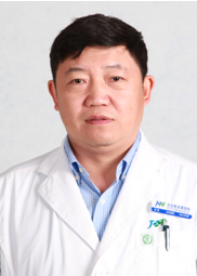 Lei Huang (黄雷)合作论文数 Department of Orthopaedic Trauma,Beijing Jishuitan Hospital,Beijing ,China6
 Lu Mao (毛璐)合作论文数北京大学6Xianzhi Ma合作论文数5GuoWei Rong (荣国威)合作论文数5HongBo Liu (刘洪波)合作论文数5
 Xinbao Wu (吴新宝)合作论文数Beijing Jishuitan Hospital5Wenyuan Qi合作论文数4YuJiang Mao (毛玉江)合作论文数4DeQuan Liu (刘德全)合作论文数4
 Lidan Zhang (张力丹)合作论文数Beijing Jishuitan Hospital4
 Liang He (贺良)合作论文数Beijing Jishuitan Hospital4
 Maoqi Gong (公茂琪)合作论文数Beijing Jishuitan Hospital4guihua di (翟桂华)合作论文数3Zhendong Wang (王振栋)合作论文数3Di Dong (董迪)合作论文数3
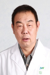 Jie Wei (危杰)合作论文数Beijing Jishuitan Hospital3
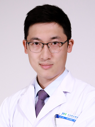 Xu Sun (孙旭)合作论文数Beijing Jishuitan Hospital3
 Yabo Liu (刘亚波)合作论文数Beijing Jishuitan Hospital,Beijing ,P.R.China3
 Ting Li (李庭)合作论文数Beijing Jishuitan Hospital/Zhongshan Jishuitan Orthopaedic Hospital3Minghui Yang合作论文数2Lili Huo合作论文数2yalian zhang (张亚莲)合作论文数2Lu JIANG (姜璐)合作论文数2GuangYun Qu (曲广运)合作论文数2ShuHui Zheng (郑淑慧)合作论文数2Jing Bian (卞婧)合作论文数2Long Zhang (张隆)合作论文数2ShuMao Wang (王树茂)合作论文数2WenYi Li (李文毅)合作论文数2XiaoFeng Gong (龚晓峰)合作论文数2XingHua Liu (刘兴华)合作论文数2Quan Zhang (张权)合作论文数2Jing Li (李静)合作论文数2Kai Wang (王凯)合作论文数2Ying Liu (刘颖)合作论文数2Gang Zhao (赵刚)合作论文数2
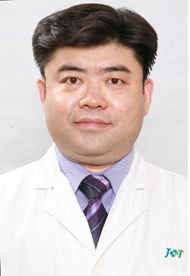 Shiwen Zhu (朱仕文)合作论文数Nanjing Cigarette Factory,Nanjing ,China2
 Junqiang Wang (王军强)合作论文数Beijing Jishuitan Hospital;Capital Medical University2
 Lin Sun (孙林)合作论文数Department of Orthopaedic Trauma Surgery, Beijing Jishuitan Hospital2
 Linong Ji (纪立农)合作论文数Peking University People's Hospital2Ming-jiang Yi (伊明江)合作论文数1bai qiu en *gu ke jia su kang fu lian meng (白求恩·骨科加速康复联盟)合作论文数1bai qiu en gong yi ji jin hui guan jie wai ke zhuan ye wei yuan hui (白求恩公益基金会关节外科专业委员会)合作论文数1Jing-ming DONG (东靖明)合作论文数1Yejun Zha合作论文数1LI Bing (李兵)合作论文数1Yuneng Li合作论文数1




