目的:探讨CXCR4在宫颈癌细胞株Hela细胞中的表达情况,及CXCL12-CXCR4生物轴对Hela的增殖、迁移和侵袭能力的影响,并研究CXCR4的特异性拮抗剂AMD3100对CXCR4的特异性阻断效果.方法:将宫颈癌Hela细胞株细胞进行体外培养.免疫组化检测Hela细胞中CXCR4的表达情况.采用MTT法研究不同浓度CXCL12对Hela细胞增殖的影响;Transwell法进行实验,判断在无血清培养液的生长条件下,不同浓度的CXCL12和CXCR4拮抗剂AMD3100对Hela细胞迁移、侵袭的影响.结果:CXCR4在Hela细胞的细胞膜、细胞质中有表达.1组Hela细胞的增殖数与2组及3组两两比较,差异有统计学意义(P<0.05),4组Hela细胞的增殖数较2组、3组低,差异有统计学意义(P<0.05).与A组Hela细胞的迁移数和侵袭数相比,B组明显更高(P<0.05);而C组Hela细胞的迁移数和侵袭数明显高于A组和B组;D组Hela细胞的迁移数和侵袭数均低于A、B、C3组,差异有统计学意义(P<0.05).结论:CXCR4在Hela细胞的细胞膜、细胞质中均表达.CXCL12-CXCR4生物轴可促进宫颈癌Hela细胞株的迁移及侵袭,而CXCR4拮抗剂AMD3100能抑制这一过程,CXCL12-CXCR4生物轴在宫颈癌Hela细胞生长和转移过程中可能起着重要作用.
Objective:To investigate the potential value of chemokine CXCL12-CXCR4 in the growth of ovarian cancer cells.Methods:SKOV3 cells transfected with CRCX4 vector or plasmid,as well as untransfected SKOV3 cells,were cultured in vitro,and assigned into 4 groups (A,B,C,and D)cell proliferation was measured using MTT Assay.Results :①In SKOV3/CXCR4 group,the cell proliferation in A and B groups is higher than that in C and D groups(P<0.05);And the cell proliferation in B group is higher than A group(P<0.05);②In SKOV3/CXCR4 group,the number and weight of metastatic tumors in experimental group are lower than those in control group. The live time of nude mice is longer than that in control group(P<0.05);③In SKOV3/neg and SKOV3 group,no significantly differences of the experimental data are observed.Conclusions:Chemokine CXCL12-CXCR4 mightimprove the growth of ovarian cancer cells.The CXCR4 antibody and antagonist AMD3100 might inhibit the growth of ovarian cancer cells.
目的:研究子痫前期患者外周血协同刺激分子CD28、CTLA-4、CD28/CTLA-4在CD3+CD4 +T细胞和CD3+ CD8+T细胞上的表达,探讨CD28/CTLA-4在子痫前期发病中的作用.方法:采集22例正常妊娠妇女(正常妊娠组),45例子痫前期患者(子痫前期组)外周血,用流式细胞技术分别检测外周血T淋巴细胞CD3+、CD4+、CD8+的表达和CD28、CTLA-4、CD28/CTLA-4在CD3+ CD4+T细胞和CD3+ CD8+T细胞上的表达.结果:子痫前期组外周血T淋巴细胞中CD3+及CD8+的表达低于正常妊娠组(P<0.05),而CD4+及CD4 +/CD8+的表达高于正常妊娠组(P<0.05).与正常妊娠组相比,子痫前期组CD28在CD3+ CD4+T细胞和CD3+ CD8+T细胞上的表达低于正常妊娠组,但差异无统计学意义(P>0.05);CTLA-4在CD3+ CD4+T细胞和CD3+ CD8+T细胞上的表达高于正常妊娠组,且重度子痫前期组高于轻度子痫前期组,差异均有统计学意义(P<0.05);CD28/CTLA-4比率在CD3+ CD4+T细胞和CD3+ CD8+T细胞上的表达低于正常妊娠组,且重度子痫前期组低于轻度子痫前期组,差异均有统计学意义(P<0.05).结论:子痫前期患者外周血T细胞亚群明显偏移,且协同刺激分子CD28/CTLA-4表达异常,高表达的CTLA-4导致T淋巴细胞亚群失衡,这可能是促使子痫前期发生的重要原因之一.
AIM:To explore the effect of human peritoneal mesothelial cells (HPMC) on the adhesion,migration and invasion of ovarian carcinoma SKOV3 cell line and the potential mechanism. METHODS:The primary culture method for HPMC was established. Isolated HPMC were identified and some cell factors expression were examined by immunohistochemical analysis. Adhesion assay,migration assay and invasion assay were used to evaluate the metastatic ability of SKOV3. The role of axis of CXCL12-CXCR4 in SKOV3 metastatic ability was also studied. RESULTS:HPMCs were successfully cultured and CXCL12 and MSLN were expressed in HPMC,but CXCR4 was not expressed. In the group of the co-cultured HPMC and SKOV3 cells,the absorbance,the number of migration and invasion cells were significantly higher than those in the group of single culture SKOV3,with statistically significant difference (P<0.05). In the group of co-cultured HPMC and stable and high CXCR4 expression of SKOV3 cells(SKOV3-CXCR4-cDNA),the absorbance,the number of migration and invasion cells were significantly higher than those in the other three groups,with statistically significant difference(P<0.05). CONCLUSION:Interactions of ovarian carcinoma SKOV3 cells and HPMC contribute to the adhesion,migration and invasion of SKOV3 and the mechanism may relate to the axis of CXCL12-CXCR4.
AIM:To construct the eukaryotic expression vector of human CXCR4 and transfect SKOV3 cells with it.METHODS:The cDNA of CXCR4 was amplified by RT-PCR.After purification,the gene fragment was cloned into a vector pReceiver-M02.The sequence of inserted CXCR4 gene fragment was identified by enzyme digestion of BamH I/EcoR I and sequencing,and then the recombinant plasmid was transfected into SKOV3 cells which did not express CXCR4 protein by lipofectamine-mediated gene transfection method.The SKOV3 cell transfected with CXCR4 were examined by RT-PCR,Western blot and immmunocytochemistry methods.RESULTS:Cell clones which were screened by G418 were obtained.The size of RT-PCR product of the positive cell clones was about 304 bp,and the positive signals of protein were obtained.The results of immunocytochemistry indicated that transfected SKOV3 cells were stained tan on its cytoplasm and membrane.CONCLUSION:SKOV3 cells that can express CXCR4 protein stably has been established successfully,which facilitates the researches of epithelial ovarian cancer.
护理服务质量是医院核心竞争力的重要组成部分。建立科学、规范、有效的护理质量管理体系是医院实施ISO9001质量体系认证过程中的重要环节。护理部在建立护理质量管理体系过程中应充分发挥组织、培训、沟通和协调的职能作用,确保体系符合科学质量管理的要求,实现护理质量的持续改进。
<正>复拇指畸形是一种较为多见的手部先天性畸形之一,其发生率约为0.08~0.18‰。该畸形分类方法较多,1969年,Wassel将复拇指畸形分为七型,龙虾钳样畸形常出现于Ⅲ、Ⅳ型中。2000年以来,我科共收治了6例龙虾钳样复拇畸形,术后疗效满意。
Objective To provide the morphological basis for the reconstruction of old central tendon.Methods Morphological observation of finger extensor tendon apparatus was carried out in 12 adults cadaveric hands.The distance from the fasciculus medialis of lateral extensor tendon to central tendon and from lateral extensor tendon to central tendon,and the transverse movement of laterphalangeal joint of band were measured when the fingers were in the extension position.Results The extensor tendon apparatus was similar among all fingers.The distance from the central extensor tendon to medial band was different,but the distance from central extensor tendon toleteral extensor band was in the range of 9~10 mm.Conclusion The finger extensor tendon apparatus is a complicated structure,so it is improtant in clinical treatment to protect the integrity of the whole extensor apparatus,especially the length between central extensortendon and lateral tendon.
 YingPu Lv (吕英璞)合作论文数3
 Xiaohua Wu (吴小华)合作论文数Shijiazhuang Obstetrics and Gynecology Hospital3FAN Hong-Wei (范红伟)合作论文数2
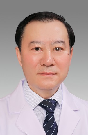 Xinzhong Shao (邵新中)合作论文数Department of Hand Surgery, The Third Affiliated Hospital, Hebei Medical University2Yadong Yu (于亚东)合作论文数2Xiangli Ji (吉向丽)合作论文数2weican zhang (张玮璨)合作论文数1Yu-xia SONG (宋玉霞)合作论文数1HongWei Wang (王宏卫)合作论文数1KeLiang Zhang (张克亮)合作论文数1XiaoGuang Zhu (朱晓光)合作论文数1Hui Han (韩辉)合作论文数1Ping Xu (许萍)合作论文数1Feng Xu (徐锋)合作论文数1Jun Ge (葛军)合作论文数1GuiSheng Zhang (张桂生)合作论文数1Bo Yang (杨波)合作论文数1Xiaohua Wang (王晓华)合作论文数1




