目的 研究基于纤维内镜吞咽功能评估(FEES)指导下的摄食训练对改善脑卒中患者吞咽功能的康复效果.方法 选择60例入住南京医科大学附属无锡市同仁康复医院神经康复科因吞咽障碍留置胃管的脑卒中患者,分别纳入观察组和对照组进行回顾性病例对照研究.给予两组患者常规的神经康复训练和吞咽功能训练.根据改良容积-黏度测试结果,选择能够安全吞咽时液体的体积和稠度带管对对照组患者进行摄食训练;在FEES指导下通过选择安全进食的食物性状和一口量、指导吞咽技巧和代偿方法等对观察组患者进行直接摄食训练.分别于干预前和干预4周后评价两组患者的洼田饮水试验分级和功能性经口摄食量表评分,记录两组拔除胃管时间和胃管拔除率、肺炎发生率等指标变化.结果 与对照组比较,干预后,观察组患者功能性经口摄食量表评分均值显著改善〔(5.33±0.48)分vs.(3.67±0.51)分,P<0.001)〕,干预期间肺炎发生率显著降低(6.7%vs.36.7%,P=0.005),拔除胃管时间减少〔(15.24±3.21)天vs.(19.36±3.58)天,P=0.019)〕,差异均有统计学意义.洼田饮水试验评级治愈率和显效率总和较对照组提高(66.7%vs.53.33%,P=0.329)、胃管拔除率升高(86.7%vs.73.3%,P=0.197),但差异无统计学意义.结论 FEES指导下的直接摄食训练可有效保证患者进食安全,并改善其吞咽功能.
肝脏淋巴管瘤是一种极为罕见的良性肿瘤,笔者对3例经病理证实的肝脏淋巴管瘤患者的临床、影像及病理资料进行整理分析,并复习相关文献,现报道如下,旨在提高对本病影像表现的认识.一、资料与方法 3例患者均经手术病理证实为肝脏淋巴管瘤,术前均行MRI平扫及增强扫描.男2例、女1例,年龄分别为43、35、10岁.3例均因上腹部不适或体检偶然发现肝脏占位性病变,发现肿物到手术时间分别为1个月、5个月、2年,其间随访病灶无明显变化.
Hepatocellular carcinoma (HCC) has a high fatality rate and most patients are in advanced stage when diagnosed with routine methods. As a molecular imaging method, positron emission tomography/computed tomography (PET/CT) has become more and more important. However, it also has certain limitations, since it always has false-negative results for well-differentiated HCC and small HCC. Therefore, a combination of various tracers can improve the sensitivity and specificity of PET/CT in the diagnosis of HCC. This article reviews the role of PET/CT in the therapeutic effect evaluation, recurrence monitoring, and prognosis of HCC.
患者女,43岁,主因"上腹部不适,伴轻微腹痛1月余"入院。查体:右上腹可触及巨大肿块,质地中等,活动度差。实验室检查:肝肾功能正常,肿瘤指标阴性。超声检查:肝右叶可见一大小约19.8cm×21.2cm高回声区,边界可辨,内部回声不均匀,呈多结节融合,内见数个无回声区(图1A)。CT检查:肝右叶见一巨块状低不均匀密度影,内见数个片状更低密度区,边界尚清,增强后病灶实性及分隔呈渐进性不均匀强化,囊性
Purpose To assess the accuracy of magnetic resonance (MR) imaging-based differential diagnosis of hepatocellular carcinoma (HCC) and intrahepatic cholangiocarcinoma (ICC) in a cohort of patients with focal liver lesions and cirrhosis. Materials and Methods This study was institutional review board approved, and the requirement for informed consent was waived. Between January 2009 and December 2014, a cohort of cirrhotic patients, including 71 with ICC and 612 with HCC, underwent unenhanced T1- and T2-weighted MR imaging, gadolinium-based contrast material-enhanced dynamic phase imaging, and partial hepatectomy. Results of pathologic examinations were obtained for all patients. Clinical, radiologic, and pathologic results in these patients were analyzed and compared comprehensively. Differences in signal intensity on baseline and contrast material-enhanced images and dynamic enhancement patterns were evaluated and compared by using the χ(2) test or the Fisher exact test. Results The preoperative diagnoses of 517 of 683 lesions were confirmed pathologically. Five ICCs and 481 HCCs displayed contrast material washout in delayed phases (P < .001). Thirty-six ICCs and 23 HCCs displayed a progressive enhancement pattern (P < .001). Twenty-six ICCs and 63 HCCs displayed a stable enhancement pattern (P < .001). ICCs displayed significantly different enhancement patterns according to size (P = .022). Conclusion The accurate discrimination of small ICCs from HCCs in the setting of cirrhosis at MR imaging is difficult, as substantial proportions of ICCs and HCCs have similar enhancement patterns. Absence of contrast material washout at MR imaging is not a factor that prevents the misdiagnosis of HCC. (©) RSNA, 2016 Online supplemental material is available for this article.
Hepatocellular carcinoma (HCC) complicated by portal vein tumor thrombus (PVTT) is associated with poor prognosis, early recurrence of HCC, and limited treatment options. Current guidelines do not have standardized diagnostic and treatment modalities, thus creating a need for a multidisciplinary treatment model for standardization of the treatment. Eastern Hepatobiliary Surgical Hospital (China) convened two working parties of experts from all the departments, to consolidate the current evidence, prevailing vision for the future, and experience of the practicing clinicians engaged in HCC management, so as to develop a consensus for PVTT diagnosis and treatment according to the GRADE system. Based on the quality of the existing evidence and the strength of recommendations, the consensus statements were categorized into 3 evidence levels (A/B/C) and 5 classes (I/II/IIa/IIb/III).The panel discussed and provided clarity on the management and research options in the field of HCC with PVTT. In addition, the panel also assessed the quality of the cited studies and assigned grades to the recommendation statements. Among the group of experts, there was excellent agreement with regard to effective diagnosis and treatment of HCC with PVTT. The recommendations of this consensus will provide guidance to physicians and clinical researchers on the effective management of HCC with PVTT.
Objective To compare the MRI manifestations and characteristics of ≤ 3 cm nonepithelial hepatic angiomyolipomas (HAML)and small hepatocellular carcinoma (SHCC),then improve the preoperative diagnostic accuracy.Methods A retrospective analysis of 20 patients ≤3 cm nonepithelial HAML and 26 cases of SHCC,confirmed by clinical pathology,with both in clinical data and MRI characteristics.Results ≤3 cm nonepithelial HAML commonly occurs in women;The enhancement patterns“wash in and wash out”in 1 1 cases,6 cases “wash in but slow out”,3 cases with delayed enhancement;Mature adipose tissue found in 5 cases, 3 cases has pseudocapsule enhancemen in delayed phase,13 cases can see central vessels;While SHCC often occurs in men,with“wash in and wash out”enhancement pattern in 23 cases,1 7 cases with pseudocapsule enhancemen in delayed phase,12 cases can see central vessels in lesions.ADC values for SHCC was significantly lower than that for ≤3 cm nonepithelial HAML,the ADC values of SHCC significantly lower than the surrounding liver parenchyma;The ADC values between ≤3 cm nonepithelial and liver parenchyma around has no significant differences.ADC values of liver parenchyma with liver cirrhosis was lower than that without cirrhosis.Conclusion The existence of mature adipose tissue,the MRI enhancement pattern and the value of ADC can help to distinguish between ≤3 cm nonepithelial HAML and SHCC,then improve the preoperative diagnostic accuracy.
Interventional treatment has become the first-line therapeutic method for intermediate-stage hepatocellular carcinoma (HCC). With the development of related techniques,drug-loaded microparticles and radioactive microspheres have been applied in clinical prac-tice,with good therapeutic effect achieved.This paper analyzes and summarizes the existing interventional treatment methods and problems, and points out that HCC patients can achieve great benefits from combination therapy,and that standardized interventional therapy is a direc-tion for future development.
Background: Accurate assessment of characteristics of tumor and portal vein tumor thrombus is crucial in the management of hepatocellular carcinoma. Aims: Comparison of the three-dimensional imaging with multiple-slice computed tomography in the diagnosis and treatment of hepatocellular carcinoma with portal vein tumor thrombus.Method: Patients eligible for surgical resection were divided into the three-dimensional imaging group or the multiple-slice computed tomography group according to the type of preoperative assessment. The clinical data were collected and compared.Results: 74 patients were enrolled into this study. The weighted kappa values for comparison between the thrombus type based on preoperative evaluation and intraoperative findings were 0.87 for the three-dimensional reconstruction group (n = 31) and 0.78 for the control group (n = 43). Three-dimensional reconstruction was significantly associated with a higher rate of en-bloc resection of tumor and thrombus (P = 0.025). Using three-dimensional reconstruction, significant correlation existed between the predicted and actual volumes of the resected specimens (r = 0.82, P < 0.01), as well as the predicted and actual resection margins (r = 0.97, P < 0.01). Preoperative three-dimensional reconstruction significantly decreased tumor recurrence and tumor-related death, with hazard ratios of 0.49 (95% confidential interval, 0.27-0.90) and 0.41 (95% confidential interval, 0.21-0.78), respectively.Conclusion: For hepatocellular carcinoma with portal vein tumor thrombus, three-dimensional imaging was efficient in facilitating surgical treatment and benefiting postoperative survivals.
Based on ontology and combined with international standards, this paper firstly introduces a process of developing a set of imaging reporting terminology of primary liver cancer through analysis of 1093 samples. The second, realizing the structured imaging reporting by mapping the terminology structure and clinical document architecture.
Objective:To explore the MR and CT appearances and imaging differential diagnosis of solitary necrotic nodule(SNN)of the liver.Methods:The clinical and imaging features of 47patients(29men and 18women;age range,32~71years;median age,49years)with pathologically confirmed SNN were retrospectively reviewed.All the cases underwent plain and dynamic contrast-enhanced MRI and/or CT scan of the abdomen(38cases with MRI scan,7cases with CT scan,and 2cases with both MRI and CT scan).Results:Thirty-nine lesions(39/47,83.0%)were located in the right lobe of the liver,and 8(8/47,17.0%)in the left lobe.Forty-two lesions(42/47,89.4%)were found in the sub-capsular region,meanwhile 5lesions(5/47,10.6%)were located within the deep hepatic parenchyma.The maximum diameter of 25masses(25/47,53.2%)were less than or equal to 2.5cm,and 22masses(22/47,46.8%)were larger than 2.5cm.Twenty-nine masses(29/47,61.7%)were irregular,meanwhile 18cases(18/47,38.3%)were round or oval.SNN presented hypo-intense or iso-intense on T1 WI in all cases.Thirty-two lesions were hyper-intense or iso-intense on T2 WI,and 8lesions were hypo-intense on T2 WI.On the plain CT,all cases showed slightly low to low density within the lesions,of which only one case showed the annular calcification.After contrast administration,the central part of the lesions showed no enhancement,and the peripheral part showed thin ring-like delayed enhancement in all cases.In addition,18 lesions showed slightly delayed enhancement of the septa within the lesions.Conclusion:The MRI and CT appearances of solitary necrotic nodules of the liver are characteristic and helpful in the diagnosis of the diseases.
目的:探讨肝脏孤立性坏死结节(SNN)的MRI表现.方法:收集我院2009年11月至2014年9月行MRI检查并经手术病理证实的SNN共40例,回顾性分析该病影像特征.结果:单结节病灶26例、多结节病灶14例.病灶最大径0.7~5.6cm(2.42 ±0.98cm),30例≤3cm、10例>3cm;分别位于肝右叶34例、肝左叶4例、中肝叶2例,病灶分布于肝缘下35例、肝脏深部5例;病灶呈圆形或椭圆形18例、葫芦状14例、蚯蚓状8例.T1WI示所有病灶均为低/等信号,T2WI示病灶呈稍高/等信号32例、稍低信号8例,增强后病灶实质均无强化,病灶周缘均可见薄环状强化,环壁厚0.87~3.05mm(2.09±0.52mm),14例病灶内见间隔线强化.结论:肝脏孤立性坏死结节多见于肝右叶肝缘下,通常最大径≤3cm,MRI增强表现为周缘薄环状强化,典型表现可以提示SNN的诊断.
目的 探讨磁共振单体素MRS技术结合LC-Model绝对定量慢乙肝后代谢物在预测慢乙肝转归的可行性.方法 对11例健康志愿者和17例慢乙肝稳定及15例慢乙肝进展的患者行常规MRI和MRS检查,利用LC-Model波谱软件分别对代谢物Cho、Lip及Cho/Lip定量测量并进行比较.结果 Cho(a.u.)分别为7.58±5.05,11.26±7.21,17.67±11.72 3组之间有统计学意义;Lip(a.u.)分别为625.32±694.12,329.28±412.57,422.65±471.45,慢乙肝稳定组与进展组之间无明显统计学意义,与对照组有统计学意义;Cho/Lip的比值在慢乙肝稳定组与进展组无意义,与对照组比较有统计学意义.结论 单体素MRS结合LC-Model技术能够分析慢乙肝后病理改变的代谢物的变化;在预测慢乙肝后进展方面有一定的意义.
Objective: To investigate the clinical features of undifferentiated embryonal sarcoma of the liver (UESL) to improve its preoperative diagnostic accuracy.Methods: Retrospectively analysis the clinical, imaging and histopathologic ifndings of 16 cases preoperatively misdiagnosed UESL which were all pathologically conifrmed. Results: Among these 16 cases, clinically diagnosed primary liver carcinoma 9 cases, hepatoblastoma 3 cases, malignant hepatic tumor 4 cases. Twelve patients mainly presented abdominal discomfort. Sonograms revealed predominant solid lesions; CT and MRI demonstrated predominant cystic masses within irregular soft tissue. Contrast-enhanced images showed intralesional foci, multiple internal septations andedges enhancement. hTe postoperative pathology showed the cut surface of tumors was variegated, with solid and cystic gelatinous areas, hemorrhage and necrosis. Intracytoplasmic hyaline globules were commonly present among cancer cells. Conclusion: UESL is a rare malignancy of the liver with no specific clinical manifestation, imaging finding inconsistent between ultrasound, CT and MRI which might be helpful to improve the preoperative diagnosis accuracy.
To establish an effective quality control mechanisms imaging reports, and explore the effect of knowledge in clinical applications, the paper describes an implementation method based on ontology knowledge base for guiding imaging reporting quality control. We obtain 152 common terms and mine quality control rules by analyzing 1093 reports.
目的:观察及分析原发性肝脏血管肉瘤(PHA)的MRI表现,结合其病理学基础及相关病理特点,以提高诊断准确性.方法:回顾性分析13例经手术病理证实行上腹部MRI三期增强扫描的PHA患者图像,分析其影像学及病理表现.结果:13例中8例为单发肿块型,2例为多发结节型,3例为巨块结节混合型.各病灶T1WI呈低信号,T2WI呈高信号,增强后早期病灶内呈斑片状及不规则片状不均匀强化,晚期呈进行性填充式强化.延迟期病灶内见不规则斑片状无强化区域.少数病灶延迟期与周围肝实质分界清楚,边缘可见“刀切征”和“假包膜征”.结论:PHA的MRI表现为块状及结节状T1WI低信号,T2WI高信号影,增强后病灶呈渐进性充填,与周围肝实质分界清楚,较大病灶内伴出血及坏死,通过上述特征性表现,有助于提高诊断率.
Objective To explore the clinical value of spectral CT Iodine-based material decomposition imaging in differentiating intrahepatic cholangiocarcinoma ( ICC ) from atypical hepatocellular carcinoma ( AHCC ) . Methods Retrospectively analyzed 52 patients of our hospital during April 2013-February 2014 who underwent tri-phase enhanced spectral CT imaging including arterial phase, portal venous phase and equilibrium phase. Primary hepatocellular carcinoma (ICC patients 24 cases, AHCC patients 28 cases)was pathologically confirmed in each case. The Iodine concentrations of ICC, AHCC, and normal hepatic tissue in tri-phase were measured. The iodine concentration differences between equilibrium phase and arterial phase (EAD), lesion and normal hepatic tissue iodine concentration difference during equilibrium phase (LNDE) were calculated. The group differences of ICC and AHCC were evaluated by two samples t test and receiver operating characteristic curve (ROC) analysis. Results During arterial phase, portal venous phase, and equilibrium phase, the mean iodine concentrations of ICC were significantly higher than those of AHCC (P<0.01). The EAD and LNDE of ICC were significantly higher than those of AHCC (P<0.01). ROC analysis showed that the sensitivity and specificity of the iodine concentration during equilibrium phase, EAD and LNDE in differentiating between ICC and AHCC were 95.8%and 92.9%, 62.5%and 89.2%, 95.8%and 82.1%, respectively. Conclusion Spectral CT iodine-based material decomposition image is a suitable tool to quantitatively evaluate contrast enhancement characteristics of ICC, the iodine concentration measurement during equilibrium phase performs best in differentiating AHCC from ICC.
Background: To investigate the clinical features of undifferentiated embryonal sarcoma of the liver (UESL) to improve its preoperative diagnostic accuracy.Methods: The clinical, imaging, and histopathologic findings of 16 UESL patients whose disease was pathologically confirmed but preoperatively misdiagnosed were retrospectively analyzed.Results: Among these 16 patients, 9 were clinically misdiagnosed as primary liver cancer, 3 as hepatoblastoma, and 4 as malignant hepatic mass. In 12 patients who were presented due to abdominal discomfort, ultrasound showed that predominantly solid lesions, whereas computed tomography (CT) and magnetic resonance imaging (MRI) demonstrated predominantly cystic masses within irregular soft tissue. Contrast-enhanced imaging showed enhancement intralesional foci, multiple internal septations, and edges. The postoperative pathology showed the cutting surface of tumors was variegated, with solid and cystic gelatinous areas, hemorrhage, and necrosis. Intracytoplasmic hyaline globules were commonly present among cancer cells.Conclusions: UESL is a rare clinical condition without specific clinical manifestations. The inconsistencies between ultrasound and CT/MRI findings may be helpful to improve the preoperative diagnosis accuracy.
Objective To investigate the MRI findings of primary hepatic angiosarcoma ( HAS). Methods Three cases of HAS confirmed by pathologically were retrospectively analyzed between 2009 and 2011 in Shanghai Eastern Hepatobiliary Surgery Hospital of the Second Military Medical University . Results OnMRI, all cases (two females and one male, age from 35 years to 71 years) appeared as hypo mixed hyper signal-intensity both in axial T1WI and T2WI.All lesions had intratumoral hemorrhage and necrosis. After GD-DTPA injected on arterial phase , three cases demonstrated heterogeneously mild enhanced and continued progressive enhancement on delay phase without enhancement of tumor center . Conclusion When the MRI representations of intrahepatic tumor show intratumoral hemorrhage and necrosis and continued progressive enhancement on delayed phase , HAS should be considered .
Objective To investigate the imaging features of hepatic epithelioid hemangioendothelioma (EHE) and to improve the level of awareness of this disease.Methods A retrospective analysis was conducted on the CT and MRI findings and the clinical data of 16 patients with liver EHE.The diagnosis was made on specimens obtained by liver biopsy or with operation.Six patients received only CT,6 only MRI,4 both CT and MRI,and 4 with DWI sequence.Results 6 of 16 patients with hepatic EHE had a solitary tumor while the remaining 10 patients had multiple tumors.CT scanning on 10 patients showed a heterogeneous mass with low density.In 4 patients,there were small calcifications in the lesions.The lesions on MRI showed a heterogeneous mass with low signal in T1WI and high signal in T2WI and DWI.In 8 patients,during enhanced scanning,the intrahepatic vein and its branches terminated at the edge of the tumor to form the "lollipop sign." On the arterial phase lesions with a diameter > 3 cm showed a centripetal enhancement pattern,which is similar to that of a haemangioma.Lesions with a diameter < 3 cm showed ring enhancement and continuous annular enhancement in the portal venous phase and in the delayed phase.The best observation period for the number of lesions was in the arterial phase.Conclusion Hepatic EHE had certain distinct characteristics on CT and MRI,e.g.the "lollipop" sign,lesions with small calcification,lesions with a diameter > 3 cm with a centripetal enhancement pattern,and lesions with a diameter < 3 cm with continuous ring enhancement on CT scanning.These radiological features help in the diagnosis of the disease. Key words: Liver; Hemangioendothelioma, neoplasms ; Magnetic resonance imaging; Tomography, X-ray computed
 Dong Chen (陈栋)合作论文数39
 Aimin Xu (徐愛民)合作论文数Department of Medicine, School of Clinical Medicine, The University of Hong Kong;Department of Pharmacology and Pharmacy, Li Ka Shing Faculty of Medicine, The University of Hong Kong39
 Mengchao Wu (吴孟超)合作论文数Naval Medical University34YuChen Jia (贾雨辰)合作论文数30XingAn Long (龙行安)合作论文数22Lun Lu (陆伦)合作论文数16
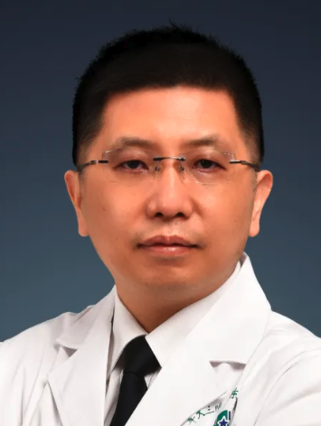 Ningyang Jia (贾宁阳)合作论文数Eastern Hepatobiliary Surgery Hospital13AiMin Xu (徐爱民)合作论文数11ZhiGang Han (韩志刚)合作论文数9Dandan SHAO (邵丹丹)合作论文数8Han Chen (陈汉)合作论文数8XueXue Wang (王雪雪)合作论文数7Bin Huang (黄彬)合作论文数7Quanyu Cai合作论文数6Hua Yu (俞花)合作论文数6XiaoYan Wang (王晓琰)合作论文数6YuFang Chen (陈玉芳)合作论文数6YanZhuo Li (李妍卓)合作论文数5GuoLi Zhao (赵国礼)合作论文数5YongBo Yang (杨永波)合作论文数5GuiMin Zhang (张桂敏)合作论文数5Liang Zhang (张亮)合作论文数5
 Shuqun Cheng (程树群)合作论文数School of Health Science and Engineering, University of Shanghai for Science and Technology5Wang Xiao-yan合作论文数4Wen XU (徐雯)合作论文数4
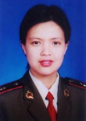 Yimin Liu (刘逸敏)合作论文数东方肝胆外科医院信息科4
 Feng Shen (沈锋)合作论文数The Third Affiliated Hospital of Naval Medical University4
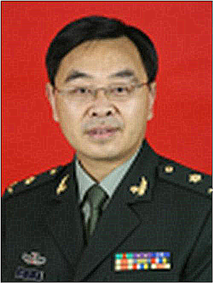 Yefa Yang (杨业发)合作论文数上海东方肝胆医院4
 Yi Shou (寿毅)合作论文数School of Medicine, Tongji University4Ping YE (叶萍)合作论文数3wenbin jiang (姜文斌)合作论文数3ZengQiang Qu (曲增强)合作论文数3Gaoqun Cao (曹高群)合作论文数3
 Wan-yee Joseph Lau (刘允怡)合作论文数Department of Surgery, Faculty of Medicine, The Chinese University of Hong Kong3
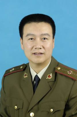 Guojun Qian (钱国军)合作论文数上海东方肝胆外科医院微创一科3Fei-Guo Zhou合作论文数3Yong-Bo Yang合作论文数2WANG Hong-zhi合作论文数2Xiang WANG (王翔)合作论文数2hong liu (刘红)合作论文数2meijuan qiu (裘美娟)合作论文数2xueting lu (吕学婷)合作论文数2Daoling ZHUANG (庄道玲)合作论文数2He Wang合作论文数2ZhenTang Wang (王振堂)合作论文数2LiSong Huang (黄立嵩)合作论文数2JiCheng Duan (段纪成)合作论文数2




