BACKGROUND:Eye movements intuitively reflect visual information processing and motor planning and execution. Studies confirm that Wilson's disease (WD) patients have abnormal eye movements, such as prolonged saccade latency and increased anti-saccade error rates, but the underlying mechanism remains unclear, a key gap in understanding copper-induced neural damage in WD. This study used non-invasive high-precision eye-tracking combined with fMRI to analyze abnormal eye movement characteristics and cognitive-sensorimotor integration disorder networks in WD patients with and without potential cognitive impairment risk. METHOD:Montreal Cognitive Assessment (MoCA) was performed on 44 WD patients: 32 scored < 26, 12 scored ≥ 26, with no differences in age, disease duration, or UWDRS scores between groups. Eye movement data from the EyeKnow™ system showed MoCA scores negatively correlated with interval saccade latency and fastest completion time, and positively correlated with maximum interval saccade speed, anti-saccade accuracy, and average/maximum saccade speeds. RESULT:1.5 T fMRI revealed 10 connectivity abnormalities across 7 brain regions among patients with MoCA < 26, with the right superior parietal lobule-right lateral occipital cortex abnormality being particularly prominent. Correlation studies found sLOC.L - AG.L and sLOC.r - AG.r positively correlated with interval saccade completion time; SPL.r - aSMG.r positively correlated with interval saccade latency. CONCLUSION:The study suggests interval saccades can serve as an important marker of cognitive-sensorimotor integration disorders in WD patients, involving collaborative participation of multiple frontal, parietal, and occipital brain regions.
Disease progression or physical decline varies considerably across patients with Wilson's disease (WD), limiting the understanding of pathological and biological processes underlying heterogeneity in the brain and clinical phenotypes of WD. Herein, using large-scale data (N = 1749), we developed a gray matter volume (GMV)-based 'brain age model' to calculate the deviation between predicted brain age and chronological age, known as the brain-predicted age difference (brainPAD) and brain structure deviation (BSD), across one hepatic subtype (HWD, N = 32) and two neurological subtypes, namely, parkinsonism (PWD, N = 78) and dystonia (DWD, N = 46). BrainPAD is associated with clinical severity and is further related to GMV, revealing heterogeneous patterns of BSD across the two neurological subtypes. On the basis of support vector machine (SVM) analysis, the GMV-based BSD pattern robustly discriminates PWD from DWD. The SVM-derived fingerprint is significantly related to brainPAD in both DWD and PWD patients, suggesting that heterogeneous BSD has a stronger effect on neurological WD. In addition, differences in cortical transcriptomic signatures between the two neurological subtypes suggest that heterogeneous cortical BSD patterns may be preferentially related to subtype-specific pathology, involving biological processes related to synaptic plasticity, dendritic development, and neuronal projection, as well as cell type-specific signatures associated with excitatory and inhibitory neurons, oligodendrocytes, and microglia. In contrast, BSD in subcortical regions exhibited largely shared transcriptomic signatures between PWD and DWD, suggesting that subcortical biological processes may predominantly reflect disease-level pathology in neurological WD. Moreover, BSD-related genes in both cortical and subcortical regions are enriched for genetic risks associated with multiple neurological disorders, suggesting a potential complex pathology underlying heterogeneous clinical manifestations in patients with neurological WD. Together, these findings highlight convergent subcortical and divergent cortical transcriptomic signatures underlying subtype-specific BSD patterns, potentially providing novel biological insights into heterogeneous neurodegenerative mechanisms across neurological WD subtypes.
To assess oral health status and identify determinants associated with elevated DMFT scores among individuals diagnosed with Wilson’s disease (WD). Between January and September 2025, a questionnaire-based survey was administered to 395 WD patients hospitalized in the neurology department. Sociodemographic characteristics (sex, education, residence, income) and behavioral variables (frequency of dental check-ups, use of mouthwash, and dental floss) were recorded through self-reported responses to the WHO Oral Health Questionnaire for Adults (Chinese version). Participants were categorized into three groups based on DMFT values: low (0–1), moderate (2–3), and high (> 3). The distribution of these categories was determined, and multivariate ordinal logistic regression was performed to identify risk factors influencing DMFT levels. The findings indicated that 52.40
Wilson disease (WD) and familial hypertriglyceridemia (FHTG) are both genetic metabolic diseases, and their comorbidity is extremely rare. This article reports a case of WD with FHTG in a 12-year-old Chinese boy. The patient was diagnosed due to elevated transaminase levels, combined with clinical manifestations, copper metabolism indexes, lipid profile analysis, and genetic testing results (pathogenic mutations of ATP7B and APOA5). The patient was treated using a copper chelating agent to lower copper levels and fibrate drugs to lower lipid levels, which resulted in improvements in his liver function and blood lipid indices. This case serves as a source of reference for the diagnosis and treatment of other similar cases. It not only reveals the potential interaction between copper metabolism disorders and lipid abnormalities, but also highlights the importance of systematic genetic testing to identify comorbid inheritance.
BACKGROUND:This study aims to build a machine learning (ML) model to predict the deterioration of neurological symptoms in Wilson's disease (WD) patients during short-term anti-copper therapy. The model combines brain T1WI MRI radiomics with clinical features and employs SHapley Additive exPlanations (SHAP) to interpret the contributions of the features. METHODS:Automated segmentation techniques were used to delineate regions of interest (ROI) in routine brain T1WI MRI scans from 107 WD cases. Radiomics features were extracted and screened using LASSO regression. Six ML models were trained to develop a predictive model for symptom deterioration during short-term anti-copper therapy. The SHAP method was applied to identify and explain the importance of the features in the ML models. RESULTS:Significant correlations were observed between UWDRS-N scores, Course of disease, age, and radiomics features of the brainstem, caudate, and corpus callosum (p < 0.05). The ML models incorporated 18 radiomics and 9 clinical features. Six ML models were used in the training and test set, the best performing model was XGBoost, with AUC values of 0.96 and 0.94, respectively. SHAP analysis revealed that the five most important features were the UWDRS-N score, age, right putamen GrayLevelNonUniformityNormalized, right caudate ZoneEntropy, and central corpus callosum SmallDependenceEmphasis. The SHAP force plot illustrated how the XGBoost model predicted neurological symptom deterioration at the patient level. CONCLUSION:An explainable XGBoost model was successfully developed using brain T1WI MRI radiomics and clinical features. This model identifies WD patients at risk of neurological symptom deterioration during anti-copper therapy and provides insight into the contributions of individual features.
Wilson's disease (WD) and Duchenne muscular dystrophy (DMD) are rare genetic diseases, and their co-occurrence is even rarer. Here, we describe our experience diagnosing a 6-year-old male Chinese patient presenting with an atypical phenotype and two genetic causative factors who was ultimately diagnosed with coexisting WD and DMD. We used a comprehensive and systematic evaluation of the patient's history, physical examinations, laboratory tests, and genetic testing to make the diagnosis. The patient was treated for one year with therapy to inhibit copper absorption and an anti-inflammatory treatment, and their condition remained stable. This case suggests that the inflammatory response could be a common pathogenesis between these two diseases. It also demonstrates the clinical efficacy of anti-inflammatory therapy for WD with DMD. Furthermore, this case illustrates the importance of taking a detailed history and performing thorough physical examinations to diagnose coexisting hereditary diseases.
We presented a case of a 34-year-old male with postoperative brainstem cavernous malformations complicated with LGI1 encephalitis and secondary hypertrophic olivary degeneration (HOD). Due to recurrent dizziness and headache, the patient was diagnosed as brainstem cavernous malformations with recurrent hemorrhage and underwent resection. He subsequently developed unexplained abnormal mental behavior 1 month after the surgery, and diagnosed with LGI1 encephalitis. Six months later, cranial MRI showed HOD. This condition is rare in clinical practice,and a complex mechanism underlies the occurrence.
Objective To share a case of anti-glutamate decarboxylase 65 antibody stiff-person syndrome and autoimmune polyendocrinopathy syndrome type Ⅱ,to improve clinicians’ understanding of this disease. Methods The clinical data of a 51-year-old woman who was admitted to the affiliated hospital of a Neurology Institute in Anhui Province in 2022 was retrospectively analyzed, and the relevant literature was reviewed. Results Patients’ clinical to repeatedly lumbago, lumbar abdomen and the stiffness of lower limbs with unable to characteristics of early misdiagnosed as separate conversion disorder, after the inspection found that serum and cerebrospinal fluid resistance to glutamic acid decarboxylase antibody positive 65,thyroglobulin antibody and higher peroxidase antibody degrees, fasting and postprandial 2 hours blood sugar, glycosylated hemoglobin, Neuroelectrophysiology showed that the continuous motor unit potential was mainly distributed by body axis muscles in the resting state. The patient was diagnosed as anti-glutamic acid decarboxylase 65 antibody SPS and APS-Ⅱ(Hashimoto’s thyroiditis, type 1 diabetes mellitus),and the condition improved after immunotherapy and symptomatic treatment. Conclusion Although anti-glutamate decarboxylase 65 antibody SPS complicated with APS-Ⅱ has certain clinical specificity, it is easy to be misdiagnosed and missed because of its rarity, especially in the early course of disease.
目的 探究急性脑出血(ICH)患者短期(90 d)预后情况及其相关危险因素.方法 回顾性分析2021年9月至2022年12月安徽医科大学第二附属医院收治的55例发病24 h内的急性ICH患者全部临床资料,在患者出院90 d后,通过电话随访行改良Rankin量表(mRS)评估其预后,并据此将患者分为预后良好组(mRS<2分)与预后不良组(mRS≥2分),比较两组临床资料差异,采用多因素logistic回归分析影响急性ICH患者预后的危险因素.结果 预后良好组23例,预后不良组32例,两组在24 h内CD3+T和CD4+T细胞、中性粒细胞与淋巴细胞比值(NLR)、肺部感染、入院时出血量、舒张压及高胆固醇血症方面比较,差异均有统计学意义(P<0.05).多因素logistic回归分析显示,24 h内低CD4+T细胞值(OR=0.980,95%CI:0.961~1.000)、高NLR(OR=1.394,95%CI:1.063~1.827)、入院时出血量大(OR=1.164,95%CI:1.032~1.313)及舒张压高(OR=1.104,95%CI:1.028~1.186)均是急性ICH患者预后的独立危险因素(P均<0.05).结论 24 h内CD4+T细胞、NLR、入院时出血量及舒张压是急性ICH患者预后的预测指标.
目的 分析3 种新型抗癫痫药物[奥卡西平(Oxcarbazepine,OXC)、左乙拉西坦(Levetiracetam,LEV)及拉莫三嗪(Lamotrigine,LTG)]和 2 种传统型抗癫痫药物[丙戊酸钠(Valproic acid,VPA)与卡马西平(Carbamazepine,CBZ)]临床使用情况及常规剂量用药后血清浓度水平,为新发癫痫患者进行药物治疗提供依据.方法 回顾性分析2018 年1 月至2021 年12 月在安徽中医药大学神经病学研究所附属医院进行抗癫痫药物治疗的1261 例癫痫患者治疗信息.按照用药模式分成 4 组,分别为单一用药、两药联合、三药联合及四药联合组;按性别分为男性、女性组;按年龄分成6 组,分别为≤10 岁、11~20 岁、21~30 岁、31~40 岁、41~50 岁、≥51 岁组.分析并比较各组间的血清药物浓度水平及血药浓度在治疗有效范围的患者例数.结果 癫痫患者单一用药多于联合用药,单一服用 OXC 最多,联合用药 VPA 最多.血清药物浓度在有效范围内比率最高的是CBZ、低于有效范围下限最多的是 LTG、高于有效范围上限最多的是 LEV.单一用药组血清 LEV 水平[7.87(5.37,9.83)μg/ml]显著高于两药及三药联合用药组[7.05(5.26,9.60)、6.23(4.07,8.94)μg/ml](P<0.05);LTG水平显著低于两药联合组[3.47(1.94,4.73)μg/ml vs.4.37(2.58,7.28)μg/ml,P<0.05],CBZ水平显著高于两药联合组[6.02(4.68,7.48)μg/ml vs.5.25(4.29,6.57)μg/ml,P<0.05].结论 本研究中,青少年患者首选新型抗癫痫药物、中老年患者首选传统型抗癫痫药物;联合用药首选VPA联合.使用LTG常规剂量进行抗癫痫治疗时,可能需增加剂量;LEV、LTG及CBZ在与其他药物两药联合抗癫痫时需依据血清药物浓度水平及病情对常规使用剂量进行调整.
目的:使用血氨等实验室指标,筛选出对肝豆状核变性(Wilson's Disease,WD)肝硬化并发肝性脑病辅助诊断效能较高的指标组合.方法:选取2017年1月~2020年3月在安徽中医药大学神经病学研究所附属医院住院的WD肝硬化135例为研究对象,其中63例肝性脑病为病例组,72例非肝性脑病为对照组;检测并比较两组的血氨、血清学指标[总胆红素(TBIL)、天冬氨酸氨基转移酶(AST)、白蛋白(ALB)、铜蓝蛋白(CP)及肌酐(Cr)];血小板(PLT)计数;凝血酶原时间(PT);计算国际标准化比值(INR)、凝血酶原活动度(PTA)、白蛋白-胆红素指数(ALBI)、AST-PLT比率指数(APRI)、MELD评分及Child-Pugh分级.根据Spearman相关系数分析观察指标与MELD评分的相关性、受试者工作特征(ROC)曲线下面积(AUC)值筛选出敏感指标;分析血氨联合其他敏感指标对辅助诊断WD肝硬化并发肝性脑病的价值.结果:病例组血氨、APRI、ALBI及MELD均高于对照组,PTA低于对照组,差异具有统计学意义(P<0.05);Child分级可见病例组以C级为主(57.2%),对照组则为A级(69.5%).病例组PTA、ALBI、APRI及血氨与MELD的相关系数分别为-0.794、0.701、0.564及0.350(P<0.01).病例组血氨、ALBI、PTA及APRI的AUC面积分别为0.915、0.827、0.804及0.736;血氨、ALBI、PTA及APRI对病例组的诊断比值比分别为49.2、11.9、8.8及6.9;血氨在辅助诊断病例组时最佳诊断界值是45μmol/L;血氨联合APRI辅助诊断的灵敏度最高(96.8%);血氨联合PTA的特异度(91.7%)、诊断准确度(89.6%)最高.结论:血氨联合PTA或APRI在辅助诊断肝豆状核变性肝硬化并发肝性脑病诊断效能高,具有临床应用价值.
目的 研究在辅助诊断Wilson病(WD)患者肝纤维化中肝脏B超单点剪切波弹性成像(pSWE)值与肝纤维化血清学指标及肝纤维化无创诊断评分模型得分的相关性.方法 选择2020年8月—2021年5月在本院住院的WD患者127例为研究对象,按照仪器厂家提供的pSWE值评判肝纤维化的标准将WD患者分为肝纤维化(0-1.34 m/s)无/轻度组、肝纤维化(1.34-2.20 m/s)中度组和肝纤维化(>2.20 m/s)重度组三组,无/轻度组42例、中度组42例、重度组43例.检测三组患者肝脏B超pSWE值、血清肝功能指标(TBA、TBIL、AST、ALT、GGT、ALB)、肝纤维化血清学指标[透明质酸(HA)、层黏蛋白(LN)、Ⅳ型胶原(CⅣ)、Ⅲ型前胶原N端肽(PⅢNP)]、PT、PLT并计算无创诊断评分模型得分[天冬氨酸氨基转移酶和血小板比率指数(APRI)、肝纤维化4因子指数(FIB-4)、γ-谷氨酰转肽酶和血小板比值(GPR)及Sheth指数],与pSWE值作对照研究和统计学分析.结果 肝纤维化重度组WD患者与无/轻度组及中度组比较,指标TBA、TBIL、AST、GGT、PT、HA、LN、CⅣ、PⅢNP、APRI、FIB-4及GPR均显著增加,ALB显著减少,差异有统计学意义(P<0.05);PLT在肝纤维化无/轻度组与中度组、重度组比较,差异有统计学意义(P<0.05);但中、重度组之间差异无统计学意义(P>0.05);ALT在肝纤维化无/轻度组与中度组之间差异无统计学意义(P>0.05),但中、重度组之间差异有统计学意义(P<0.05);三组WD患者的Sheth指数差异无统计学意义(P>0.05);pSWE值与TBA、TBIL、AST、ALT、GGT、PT、HA、LN、CⅣ、PⅢNP、APRI、FIB-4、GPR呈显著正相关(P<0.01),与ALB、PLT呈显著负相关(P<0.01);pSWE值与CⅣ(r=0.730)相关性最高,其次为GPR(r=0.710).结论 WD患者肝脏B超pSWE值与肝纤维化血清学指标及肝纤维化无创诊断评分模型(APRI、FIB-4、GPR)均具有显著相关性;三者联合用于评价WD患者肝纤维化过程具有较高价值,有利于WD患者肝纤维化筛查,避免因患者拒绝肝活检而漏诊,防止WD患者严重肝病的发生.
O'Sullivan-Mcleod syndrome is a very rare variant of MND with a good prognosis. Its clinical feature is distal lower motor neuron syndrome of both upper limbs, and there is no effective treatment at present. We reported a case of O'Sullivan-Mcleod syndrome in this paper.The patient exhibited with middle-aged progressive distal muscle weakness and atrophy of both upper limbs, without sensory, cognitive or behavioral impairment and without pyramidal tract sign. Laboratory examination, imaging and genetic tests showed no obvious abnormalities. EMG revealed neurogenic damage to the small muscles of both hands. Now we retrospectively analyzed the clinical features of a patient with O'Sullivan-McLeod syndrome, and data from 18 cases for comparative analysis, in order to improve its understanding by clinicians.
目的 探讨扩大的血管周围间隙(enlarged perivascular spaces,EPVS)与急性缺血性脑卒中(acute ischemic stroke,AIS)患者阿替普酶静脉溶栓后颅内出血转化的关系.方法 回顾性收集2019年1月 ~2021年3月就诊于安徽医科大学第二附属医院神经内科的150例AIS患者的临床资料,均在发病4.5h内接受标准剂量重组组织型纤溶酶原激活剂(rt-PA)静脉溶栓治疗.根据溶栓后24h内头颅CT检查结果,分为出血转化组和非出血转化组,比较两组患者的基线资料,实验室指标及影像学特征.采用多变量Logistic回归法分析EPVS与溶栓后出血转化的关系.结果 150例患者中共有17例(11.33%)发生溶栓后出血转化.两组患者在年龄、溶栓前NIHSS评分、溶栓后即刻NIHSS评分、溶栓前mRS评分、入院时收缩压、血尿酸、基底节区EPVS评分、半卵圆中心EPVS评分、脑室旁白质Fazekas评分、深部脑白质Fazekas评分方面比较,差异有统计学意义(P<0.05).多变量Logistic回归分析提示,在校正年龄、溶栓前及溶栓后即刻NIHSS评分等因素后,基底节区EPVS评分(OR=3.623,95%CI:1.029~12.755,P=0.045)、入院时收缩压(OR=1.062,95%CI:1.014~1.112,P=0.011)、血尿酸(OR=1.009,95%CI:1.001~1.016,P=0.022)和脑室旁白质Fazekas评分(OR=4.645,95%CI:1.099~19.633,P=0.037)是溶栓后出血转化的独立相关危险因素.结论 基底节区EPVS评分与AIS患者rt-PA静脉溶栓后发生出血转化密切相关,基底节区EPVS程度越重,发生出血转化风险越高.
肌张力障碍持续状态是一种罕见的运动障碍疾病急症,其临床特征包括高热、自主神经功能紊乱、吞咽障碍和呼吸衰竭,多预后不良.需要紧急评估,并根据患者的临床特点和并发症进行干预.目前临床上对肌张力障碍持续状态的认识不足,文中针对肌张力障碍持续状态的诊治研究进行综述.
肝豆状核变性(hepatolenticular degeneration)又称威尔逊病(Wilson disease, WD),是一种常染色体隐性遗传铜代谢障碍疾病[1].1912年金尼尔·威尔逊(Kinnier Wilson)系统描述该病是一种"进行性豆状核变性(progressive lenticular degeneration)伴有肝硬化的家族性神经病变".Ciarla(1916年)在意大利神经病理学杂志首次使用"Wilson disease"的名称,Hall (1921年)指出WD是隐性遗传病,并使用了"肝豆状核变性"(Dégénérescence Hépato-lenticulaire)的术语.1985年Frydman等将WD基因定位于13 号染色体, 1993 年Petrukhin、Bull 和Tanzi 等确定为13 号染色体长臂上( 13q14 . 3 )的ATP7B 基因,编码140 kD 铜转运 P 型 ATP 酶[2].现已明确 WD 由于铜转运 ATP7B 酶功能缺陷,导致铜蓝蛋白合成减少、胆道排铜障碍,肝脏、脑、肾和角膜等组织器官过量铜蓄积,出现进行性肝损害、锥体外系症状、精神症状、角膜色素环( Kayser-Fleischer ring , K-F 环)等.WD 通常在青少年期发病,是迄今少数几种可治疗的神经遗传病之一[3].
Wilson's disease (WD) is an inherited disorder characterized by excessive accumulation of copper in the body, particularly in the liver and brain. In the central nervous system (CNS), extracellular copper accumulation triggers pathological microglial activation and subsequent neurotoxicity. Growing evidence suggests that levels of inflammatory cytokines are elevated in the brain of murine WD models. However, the mechanisms associated with copper deposition to neuroinflammation have not been completely elucidated. In this study, we investigated how the activation of NLR family pyrin domain containing 3 (NLRP3) inflammasome contributes to copper-mediated neuroinflammation in an animal model of WD. Elevated levels of interleukin-1β, interleukin-18, interleukin-6, and tumor necrosis factor-α were observed in the sera of WD patients and toxic milk (TX) mice. The protein levels of inflammasome adaptor molecule apoptosis-associated speck-like protein containing a C-terminal caspase recruitment domain (ASC), cleaved caspase-1, and interleukin-1β were upregulated in the brain regions of the TX mice. The NLRP3 inflammasome was activated in the TX mice brains. Furthermore, the activation of NLRP3 inflammasome was noted in primary microglia treated with CuCl2, accompanied by the increased levels of cleaved caspase-1, ASC, and interleukin-1β. Blocking NLRP3 inflammasome activation with siNlrp3 or MCC950 reduced interleukin-1β and interleukin-18 production, thereby effectively mitigating cognitive decline, locomotor behavior impairment, and neurodegeneration in TX mice. Overall, our study demonstrates the contribution of copper overload-mediated activation of NLRP3 inflammasome to progressive neuropathology in the CNS of a murine model of WD. Therefore, blockade of the NLRP3 inflammasome activation could be a potential therapeutic strategy for WD.
目的 探讨良性家族性婴儿癫痫(BFIE)的临床特点.方法 回顾性分析1例PRRT2基因突变和双侧额区放电的BFIE患者的临床资料,并进行文献复习.结果 BFIE主要临床表现为局灶性癫痫发作,PRRT2为主要致病基因.发作间歇期EEG多无异常,局灶性放电部位多为额区.本病预后良好,多数抗癫痫药物对本病有效.结论 婴儿期以局灶性癫痫起病,如丛集性发作,且智力、运动发育正常,需结合基因和EEG检查,考虑BFIE可能.
历时近1年,汇聚了全国十多位知名神经病学、内科专家临床诊疗实践、学术研究成果的《内科理论与实践》:肝豆状核变性专刊终于如约面世. 专刊首次尝试以一种累及全身多系统(神经系统、消化系统、眼、皮肤等)的疾病——肝豆状核变性[又称威尔逊病(Wilson disease,WD)]为核心,从学科交叉(内科-神经内科相互联系)和中西医结合两个维度对WD的发病机制、诊治实践及最新进展进行了系统总结和介绍.内容充分体现了WD诊治的复杂性、多样性、可逆性特点以及我国学者在这一领域的诸多原创性贡献,并适时融入神经影像、超声诊断以及基因检测等技术,全方位介绍了WD在临床诊疗上的最新进展和流程,同时结合医学人文,将WD的中外历史典故和中西医研究历史也悉数呈现,颇具教育意义.
 YongZhu Han (韩咏竹)合作论文数82Nan Cheng (程楠)合作论文数59
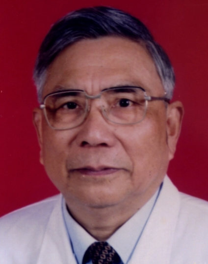 Renmin Yang (杨任民)合作论文数Anhui University of Chinese Medicine58
 Jiyuan Hu (胡纪源)合作论文数47
 Yongsheng Han (韩永升)合作论文数Anhui University of Chinese Medicine33XuEn Yu (喻绪恩)合作论文数24
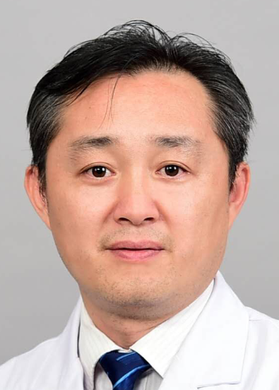 Zhihua Zhou (周志华)合作论文数The First Affiliated Hospital of Guangdong Pharmaceutical University;Department of Neurology, Guangdong Pharmaceutical University19WenBin Hu (胡文彬)合作论文数18Kai Li (李凯)合作论文数16JunXia Wu (吴君霞)合作论文数15QunRong Ye (叶群荣)合作论文数12
 Jianjian Dong合作论文数Affiliated Hosp, Anhui Univ Chinese Med11GongQiang Wang (王共强)合作论文数11YongGuang Shi (石永光)合作论文数11ChenChen Xu (徐陈陈)合作论文数10JiaWei Wang (王佳炜)合作论文数10ShiJing Wang (汪世靖)合作论文数8Jing Zhao (赵静)合作论文数8
 Mingfan Hong (洪铭范)合作论文数First Affiliated Hospital, Guangdong College of Pharmacy7Yin Xu (徐银)合作论文数6WeiMing Gao (高伟明)合作论文数6Yan Yan (严彦)合作论文数6Hai-Hong Zhou (周海虹)合作论文数5FU Xiaoming (付晓明)合作论文数5
 Zongliang Gao (高宗良)合作论文数Guangzhou Medical University5XiaoSan Wu (吴小三)合作论文数5Ju Qiu (邱菊)合作论文数5Yunfan Wu (吴筠凡)合作论文数5Dandan Sun (孙丹丹)合作论文数5Xueyun Liu合作论文数4Cong-bo SHA (沙从波)合作论文数4XianJun Yang (杨先军)合作论文数4MingShan Ren (任明山)合作论文数4
 Rao Rao (饶娆)合作论文数4
 Kai Wang (汪凯)合作论文数The First Affiliated Hospital of Anhui Medical University4Faming Pan合作论文数3Dao-yin SUN (孙道银)合作论文数3Liu Lijun (刘利君)合作论文数3YANG Guang (杨光)合作论文数3Na Nian (年娜)合作论文数3Wu Tong (吴桐)合作论文数3Jiasheng WANG (汪加胜)合作论文数3ShengQiang Xu (许胜强)合作论文数3GuangE Yang (杨广娥)合作论文数3BenChun Xue (薛本春)合作论文数3HuanXi Hou (侯焕喜)合作论文数3ZhiChao He (何志超)合作论文数3YongLiang Cai (蔡永亮)合作论文数3




