Background Definitive concurrent chemoradiotherapy (dCCRT) is the gold standard for the treatment of locally advanced esophageal squamous cell carcinoma (ESCC). However, the potential benefits of consolidation chemotherapy after dCCRT in patients with esophageal cancer remain debatable. Prospective randomized controlled trials comparing the outcomes of dCCRT with or without consolidation chemotherapy in patients with ESCC are lacking. In this study, we aim to generate evidence regarding consolidation chemotherapy efficacy in patients with locally advanced, inoperable ESCC. Methods This is a multicenter, prospective, open-label, phase-III randomized controlled trial comparing non-inferiority of dCCRT alone to consolidation chemotherapy following dCCRT. In total, 600 patients will be enrolled and randomly assigned in a 1:1 ratio to receive either consolidation chemotherapy after dCCRT (Arm A) or dCCRT alone (Arm B). Overall survival will be the primary endpoint, whereas progression-free survival, locoregional progression-free survival, distant metastasis-free survival, and treatment-related toxicity will be the secondary endpoints. Discussion This study aid in further understanding the effects of consolidation chemotherapy after dCCRT in patients with locally advanced, inoperable ESCC. Trial registration ChiCTR1800017646.
Gastric cancer is the third leading cause of cancer-related death worldwide, with relapse and metastasis being major contributors to the mortality. Circular RNAs (circRNAs) have been at the center of several researches and some circRNAs have been indicated to be involved in gastric cancer as sponges. Nevertheless, the mechanism underlying the function of circRNA remains largely unclear. Therefore, this study was conducted with the main objective of screening the associated circRNA in gastric cancer and exploring its mechanism. Expression of hsa_circRNA_0009172 was validated in gastric cancer tissues and cell lines after the correlation between hsa_circRNA_0009172 and prognosis was determined. Moreover, the binding site between miR-485-3p and hsa_circRNA_0009172 or NTRK3 was verified using dual luciferase assay and RNA pull down. Function-gain and -loss experiments were performed for the purpose of detecting the effect of hsa_circRNA_0009172 in vivo and in vitro as well as its mechanism with microRNA (miRNA)-485-3p and NTRK3 in gastric cancer. The hsa_circRNA_0009172 expression was downregulated in gastric cancer tissues and cell lines, indicating a positive association with patient prognosis. Functionally, hsa_circ_0009172 overexpression inhibited proliferative, invasive and migrative potential of gastric cancer cells as well as epithelial-mesenchymal transition (EMT)-related proteins by sponging miR-485-3p to inhibit NTRK3, while miR-485-3p overexpression could reverse the inhibitory effect of hsa_circ_0009172 on gastric cancer. Furthermore, either up-regulation of hsa_circ_0009172 or down-regulation of miR-485-3p led to the suppression of xenograft tumor growth in nude mice. In conclusion, hsa_circ_0009172 serves as a tumor suppressor in gastric cancer by targeting miR-485-3p/NTRK3 axis.
目的 分析初诊非小细胞肺癌(NSCLC)脑转移(BM)患者一线免疫检查点抑制剂联合放疗的疗效.方法 回顾性分析河南省肿瘤医院2017年12月至2021年6月首诊确诊为NSCLC BM的患者66例的资料.患者一线应用程序性死亡受体1(PD-1)/程序性死亡受体配体1(PD-L1)抑制剂联合或不联合脑部放疗.采用Kaplan-Meier法计算生存率,log-rank检验进行单因素分析,Cox模型行多因素分析.结果 全组患者中位随访13(范围4~49)个月,中位生存期24个月,1、2、3年总生存(OS)率分别是75.8%、44.7%、33.6%.1年脑内无进展生存(iPFS)分别是68.8%.放疗免疫组和免疫组脑内客观应答率icORR分别是71.4%、52.6%,脑内疾病控制率(icDCR)分别是96.4%、89.4%(P=0.122、0.291).单因素分析显示:预后评估评分(GPA)高的患者1、2、3年OS率分别是100%、85.7%、35.7%,GPA低的患者则分别是68.3%、33.6%、33.6%(P=0.115);放疗免疫组患者1、2、3年OS率分别是 88.7%、58.7%、44.0%,免疫组患者则分别是 63.6%、20.9%、20.9%(P=0.022);BM 有症状患者 1、2、3年OS率分别是60.9%、33.8%、33.8%,无症状患者则分别是84.6%、53.1%、39.8%(P=0.021).多因素分析显示,无BM症状、脑部放疗是OS率的独立影响因素(P=0.038、0.037).结论 一线免疫检查点抑制剂联合脑部放疗治疗NSCLC BM可以提高患者的生存率.建议BM患者尽早开始放疗,尤其是有脑部症状的患者.
Objective:To investigate the relationship between lung immune prognostic index (LIPI) and the prognosis of locally advanced non-small cell lung cancer (LA-NSCLC) treated with radiochemotherapy.Methods:A retrospective analysis was conducted for the clinical data of LA-NSCLC patients who received radiochemotherapy in the Affiliated Cancer Hospital of Zhengzhou University from 2013 to 2019. According to the hematologic test result of the derived neutrophil-to-lymphocyte ratio (dNLR) and the lactate dehydrogenase (LDH), the patients were divided into three groups according to their LIPI scores, namely the good-LIPI group with dNLR ≤ 3 and LDH ≤ upper limit of normal (ULN), moderate-LIPI group with dNLR >3 or LDH > ULN, and poor-LIPI group with dNLR >3 and LDH > ULN. Moreover, the overall survival (OS) and the progression-free survival (PFS) were calculated using the Kaplan-Meier method, the Log-rank test, and the Cox regression model.Results:A total of 238 patients were enrolled, and their median follow-up time was 37.1 months, median PFS 16.1 months, and median OS 30.6 months. The OS and PFS of the poor-LIPI group were significantly worse than those of the good- and moderate-LIPI groups ( χ2= 9.04, 2.88, P<0.05). The univariate analysis showed that the factors influencing OS included gender, pathological type, epidermal growth factor receptor (EGFR) mutations, and LIPI ( χ2=6.10, 13.66, 10.58, 9.04, P<0.05), and the PFS was only affected by the LIPI ( χ2=2.88, P = 0.03). Multivariate analysis suggested that EGFR mutations and LIPI were independent prognostic markers for OS ( HR = 1.31, 1.36; 95% CI: 1.03-1.67, 1.05-1.76; P<0.05). Conclusions:The LIPI is a potential prognostic indicator of radiochemotherapy in LA-NSCLC, and this result should be further confirmed by prospective studies.
Objective:To analyze the clinical efficacy of different treatment modalities and prognostic factors of patients with Masaoka-Koga stage Ⅲ thymoma.Methods:Clinical data of patients diagnosed with Masaoka-Koga stage Ⅲ thymoma admitted to Affiliated Cancer Hospital of Zhengzhou University from January 2000 to December 2018 were analyzed retrospectively. A total of 133 patients had complete treatment and follow-up data. Kaplan-Meier method was used to calculate the cumulative survival rate, log-rank method was used to compare the survival between two groups, and Cox regression model was used for multivariate analysis.Results:The median follow-up time was 50 months (3-221 months). The median overall survival (OS) was 51 (3-221) months, and the median disease-free survival (DFS) was 45 (2-221) months. The survival rate in the radical surgery group was better than that in the palliative surgery group. The 5- and 10-year OS rates in radical surgery group were 88.2% and 74.4% respectively, while in palliative surgery group were 51.8% and 32.4% respectively ( P<0.001). The 5- and 10-year DFS rates in radical surgery group were 72.2% and 45.5%, respectively, while in palliative surgery group were 32.3% and 16.1% respectively ( P=0.001). The OS in the surgery combined with radiotherapy group was better than that in the surgery alone group. The 5- and 10-year OS rates in the radical surgery group were 82.8% and 64.2% respectively, while in the palliative surgery group were 55.8% and 50.2% ( P=0.033). There was no significant difference in DFS between two groups ( P=0.176). Multivariate analysis showed that age < 50 years old ( HR=0.264, P=0.001), radical resection ( HR=0.134, P<0.001), surgery combined with radiotherapy ( HR=2.778, P=0.009) were independently associated with better OS. Age < 50 years old ( HR=0.550, P=0.046), radical resection ( HR=0.555, P=0.042), and invasion of single organ ( HR=0.111, P=0.003) were independently associated with better DFS. Conclusions:OS and DFS in patients undergoing radical surgery are significantly better than those in their counterparts treated with palliative surgery, which is the most important factor affecting prognosis. Surgery combined with radiotherapy yields better OS. It is necessary to design a rigorous and reasonable multicenter prospective study to evaluate the efficacy of various treatment modalities and prognostic factors.
Objective:To analyze the failure patterns and influencing factors of stereotactic ablative radiotherapy (SABR) for early-stage non-small cell lung cancer (ES-NSCLC).Methods:113 cases of ES-NSCLC treated with SABR from 2012 to 2020 in our hospital were retrospectively analyzed. The failure patterns, recurrence time, recurrence site and influencing factors were analyzed. Kaplan-Meier method was used to calculate the local recurrence rate, regional lymph node recurrence rate and distant metastasis rate. Univariate analysis was performed by Log-rank test, and multivariate analysis was performed by Cox model.Results:The median follow-up time was 58 months (range: 6-108 months), and a total of 45 patients (39.8%) recurred. The median recurrence time was 36 months. Distant metastasis (DM) occurred in 31 patients (27.4%) and DM alone in 24 patients (21.2%). Local recurrence (LR) was developed in 12 patients (10.6%) and LR alone in 7(6.2%). Regional lymph node recurrence (RR) occurred in 11 patients (9.7%) and RR alone in 6 patients (5.3%). LR combined with RR was observed in 1 case (0.9%), LR combined with DM in 3(2.7%), LR combined with RR and DM in 1(0.9%), and RR combined with DM in 3(2.7%). The 1-, 2-, 3-, 4-and 5-year recurrence rates were 5.4%, 16.6%, 27.5%, 44% and 51.2%, respectively. Univariate and multivariate analyses suggested that EGFR mutation was an influencing factor of high recurrence rate.Conclusion:ES-NSCLC patients treated with SABR alone have a high recurrence rate, and DM is the most common mode of failure. Follow-up consolidation therapy is recommended, especially for EGFR mutation-positive NSCLC patients.
Objective:To analyze the invasion characteristics and prognostic factors of patients with Masaoka-Koga stage Ⅲ thymoma.Methods:The tumor invasion characteristics of 179 patients who were diagnosed with Masaoka-Koga stage Ⅲ thymoma and treated in Affiliated Cancer Hospital of Zhengzhou University from January 2000 to June 2018 were analyzed retrospectively. According to the treatment methods, all patients were divided into the radical operation group ( n=94), palliative operation group ( n=39) and simple biopsy group ( n=46). The χ2 test was used to compare the classified variables, Kaplan- Meier method was utilized to calculate the cumulative survival rate, log-rank method was used for group comparison and univariate analysis, and Cox’s regression model was used for multivariate analysis. Results:Mediastinal pleural invasion (86.0%) was the most common site, followed by pericardium (50.8%), great vessel (40.8%) and lung (36.3%). The proportion of macrovascular invasion in the radical operation group was 14.9%, significantly lower than 79.5% and 60.9% in the palliative surgery group and biopsy group (both P<0.001). Multivariate analysis showed that the nature of operation ( P<0.001), age ( P=0.011), radiotherapy ( P=0.020) were the independent factors affecting overall survival (OS), while nature of operation ( P<0.001), age ( P=0.004), radiotherapy ( P=0.020), number of invasive organs ( P=0.023) and pathological type ( P=0.016) were the independent factors affecting progress-free survival (PFS). Conclusions:For patients with Masaoka-Koga stage Ⅲ thymoma, mediastinal pleura is the most common site of invasion, pericardium, lung and great vessels are also commonly invaded. The invasion of mediastinal pleura, pericardium and lung exerts slight effect on surgical resectability, whereas great vessel involvement can significantly affect surgical resectability. OS and PFS in patients undergoing radical resection are significantly better than those in patients treated with palliative resection and biopsy. Radical resection is the most important factor affecting prognosis.
Objective:To evaluate the efficacy and safety of stereotactic ablative radiotherapy (SABR) in patients with pulmonary oligometastases.Methods:Clinical data of patients with pulmonary metastases treated with SABR from 2011 to 2018 were retrospectively analyzed. The local control rate (LCR) and overall survival (OS) were calculated by Kaplan- Meier method. log-rank test was used for univariate analysis and Cox’s regression model for multivariate analysis. Results:A total of 214 lung metastases were detected in 159 patients, and the median follow-up time was 43 months. The 1-, 3-and 5-year LCR were 90.1%, 73.9% and 65.8%, respectively. The 1-, 3-and 5-year OS were 73.8%, 43.6% and 11.9%, respectively. Univariate analysis showed that biological effective dose (BED)≥100 Gy was significantly correlated with LCR ( P=0.033). Cox’s multivariate analysis showed that BED and primary tumor source were the independent prognostic factors of LCR ( P=0.023, P=0.043). No>grade 3 adverse events were observed in all patients during treatment. Conclusions:SABR is a safe and effective treatment of lung oligometastases. SABR should be actively aD ministered for pulmonary oligometastases, especially for those with lesions from lung cancer and the radiation dose should be selected as BED ≥100 Gy.
Abstract Background Stereotactic ablative radiotherapy (SABR) is the current standard‐of‐care in cases of inoperable early stage non‐small cell lung cancer (ES‐NSCLC). This study aimed to assess the survival outcomes and recurrence patterns after SABR for ES‐NSCLC in a hospital setting. Methods A single‐institution retrospective study was performed which included 109 patients who had undergone SABR. The main study endpoints were overall survival (OS), cancer specific survival (CSS), local recurrence‐free survival (LRFS), regional recurrence free survival (RRFS) and distant metastasis‐free survival (DMFS). Univariate and multivariate analysis were conducted to explore the potential factors which might be related to patient survival. Results A total of 109 patients were enrolled into the study. Median follow‐up was 44 months (range: 2–93 months). (i) Recurrence results: Among 45 patients with recurrence, 30 patients (28%) had distant metastasis (DM), 17 patients (16%) had local recurrence (LR), 10 patients (9%) had regional recurrence (RR) of lymph nodes and two patients (2%) had second primary lung cancer (SPLC). (ii) Survival results: Median OS, CSS, PFS was 78 months, 78 and 40 months. Two‐year OS, CSS, PFS, LRFS, RRFS and DMFS was 84.7%, 87.1%, 69.2%, 86.8%, 92.7% and 78.0%, respectively. Four‐year OS, CSS, PFS, LRFS, RRFS and DMFS was 55.6%, 60.7%, 37.3%, 76.3%, 88.4% and 59.4%, respectively. (iii) Univariate and multivariate analyses indicated that age was a prognostic factor of CSS in patients aged <75 years (P = 0.04 HR 2.12 95% confidence interval [CI]: 1.04–4.33). Conclusions Although high survival rates can be achieved in ES‐NSCLC patients treated with SABR, using SABR on its own may not be enough. Prolonged surveillance and adjuvant therapy is therefore needed.
Esophageal cancer represents the eighth most frequently occurring cancer, as well as the sixth most widespread cause of cancer-related deaths. In recent years, accumulating evidence has implicated long non-coding RNAs in the progression of esophageal squamous cell carcinoma (ESCC). The aim of the present study was to investigate the potential involvement and underlying mechanisms of LINC00337 in ESCC. Expression patterns of LINC00337 and targeting protein for Xenopus kinesin-like protein 2 (TPX2) in ESCC tissues and cells were detected using RT-qPCR and immunohistochemical staining. Next, the interactions among LINC00337, E2F4, and TPX2 were identified using chromatin immunoprecipitation, dual-luciferase reporter, and RNA-binding protein immunoprecipitation assays, suggesting that LINC00337 could recruit E2F4 to enhance the transcription of TPX2. Thereafter, the regulatory roles of LINC00337 and TPX2 in ESCC were analyzed by altering the expression of LINC00337 or TPX2 in ESCC cells following treatment with cisplatin (DDP). The levels of autophagy-related proteins Beclin1 and LC3II/LC3I, viability, autophagy, apoptosis, and chemoresistance of ESCC cells to DDP were measured following transfection treatment with different plasmids. Additionally, the role of the LINC00337/E2F4/TPX2 axis was assessed in vivo by measuring tumor formation in nude mice. The results demonstrated that LINC00337 upregulated TPX2, consequently leading to elevated levels of Beclin1 and LC3II/LC3I, promoted cell viability and autophagy, while inhibiting apoptosis and chemosensitivity to DDP in ESCC. In sum, the current study evidenced that the overexpression of LINC00337 could potentially enhance ESCC cell autophagy and chemoresistance to DDP via the upregulation of TPX2 by recruiting E2F4. Thus, LINC00337 may serve as a potential candidate for the treatment of ESCC.
Objective:To evaluate the role of salvage radiotherapy in the re-treatment of patients with regional lymph node oligo-recurrence after radical surgery for esophageal squamous cell carcinoma.Methods:Clinical data of patients diagnosed with thoracic esophageal squamous cell carcinoma treated with radical surgery and developed regional lymph node oligo-recurrence ( n=1-3) from January 2013 to January 2016 were retrospectively analyzed. A total of 74 cases with intact clinical data were extracted for analysis. The survival analysis was performed by Kaplan-Meier method. Group comparison was conducted by Log-rank method. Results:The median overall survival (OS) after recurrence was 9(2.5-43) months, and the median progression-free survival time (PFS) was 4(1-33) months. There were 47 cases in the salvage radiotherapy group and 27 cases in the non-radiotherapy group, and the objective response rates were 77%(36/47) and 30%(8/27), respectively. Patients in the salvage radiotherapy group had better OS ( P=0.042) and PFS ( P=0.01) compared with their counterparts in the non-radiotherapy group. Among the patients who received salvage radiotherapy, involved field irradiation and elective nodal irradiation yielded similar OS ( P=0.963) and PFS ( P=0.599), and patients treated an irradiation dose ≥ 60Gy had better OS ( P=0.001) and PFS ( P=0.001) compared with those with dose< 60Gy. Conclusions:Local salvage radiotherapy is an effective treatment of esophageal squamous cell carcinoma with regional lymph node oligo-recurrence after radical surgery. Salvage radiotherapy has better OS and PFS compared with non-radiotherapy. Prospective clinical studies should be carried out to standardize the target and dose of radiotherapy, and to further clarify the effect of radiotherapy.
Objective To evaluate the recurrence pattern and identify the risk factors of esophageal squamous cell carcinoma after neoadjuvant therapy combined with surgery.Methods Clinical data of 275 patients with thoracic esophageal squamous cell carcinoma treated with neoadjuvant therapy combined with surgery from December 2011 to December 2015 were retrospectively analyzed.The follow-up data of the enrolled patients were complete and analyzable.The recurrence pattern,recurrence time,recurrence location and influencing factors after neoadjuvant therapy in combination with surgery were analyzed.The recurrence rate was calculated by Kaplan-Meier method.The multivariate analysis was performed by Cox regression model.Results The median follow-up time was 32 (3-84) months,and the median time of the first recurrence was 10.6(2.0-69.1) months.The 1-,2-and 3-year recurrence rates were 32.0%,45.1% and 52.3%,respectively.A total of 152 cases (55.3%) had recurrence.Among them,77 cases (50.6%) had local-regional recurrence (LRR),34 cases (23.4%) had distant metastasis (DM),33 cases (21.7%) had LRR+DM and 8 cases (6.0%) had recurrence in unknown site.Among the patients with LRR,lymph node recurrence was the most common (n =98,89.1%).For DM patients,lung metastasis (n =33,49.3%),liver metastasis (n=16,23.9%),bone metastasis (n=14,20.9%) and non-regional lymph node metastasis (n=14,20.9%) were commonly observed.The multivariate analysis showed that postoperative T stage (P=0.008),N stage (P<0.001) and the number of lymph node dissection (P<0.001) were the independent risk factors for recurrence after treatment.Conclusions The recurrence rate after neoadjuvant therapy remains relatively high for esophageal squamous cell carcinoma,and the regional lymph node is the most common site of recurrence.Postoperative pathological T staging,N staging and the number of lymph node dissection are the independent risk factors for recurrence after treatment.
Objective:To evaluate the long-term survival and identify prognostic factors of patients diagnosed with early-stage non-small cell lung cancer (ES-NSCLC) receiving stereotactic ablation radiotherapy (SABR).Methods:Clinical data of 109 ES-NSCLC patients treated with SABR in Henan Cancer Hospital from 2011 to 2018 were retrospectively analyzed. The overall survival (OS), cancer-specific survival (CSS) and progression-free survival (PFS) were calculated by Kaplan- Meier method and log-rank test. Multivariate prognostic analysis was performed by Cox regression model. Results:The median follow-up time was 44 months (2-93 months). The median OS, CSS and PFS were 78 months, 78 months and 44 months, respectively. The 1-year OS, CSS and PFS were 95.4%, 97.2% and 84.1%, and 75.6%, 79.1% and 56.6% for the 3-year OS, CSS and PFS, and 55.6%, 60.7% and 37.3% for the 5-year OS, CSS and PFS, respectively. Univariate analysis showed that ECOG score, age, smoking history and derived-neutrophil/lymphocyte ratio (dNLR) were the influencing factors of OS ( P=0.03, 0.02, 0.04, 0.001). Age, smoking history and dNLR were the influencing factors of CSS ( P=0.02, 0.03, 0.001). Multivariate analysis demonstrated that dNLR was an independent prognostic factor for OS and CSS ( P=0.001, 0.001). Conclusions:ES-NSCLC patients treated with SABR can achieve favorable survival. The dNLR is an independent prognostic factor of OS and CSS, which can be considered in clinical application.
The role of definitive radiotherapy (dRT) and debulking surgery (DS) for patients with locally advanced, unresectable, Masaoka-Koga stage III thymomas was not well studied. Unresectable tumor refers to tumor that could not be completely resected because of invasion of surrounding organs. Consecutive patients with unresectable stage III thymomas between 2000 and 2017 were reviewed. According to the treatment intent and radiation dose, patients were categorized into a dRT group and a non-dRT group. The former group included patients who received radiotherapy at doses ≥ 54 Gy after DS or biopsy. The latter group included patients who did not receive radiotherapy and those who received a radiation dose < 54 Gy. A total of 82 patients were included. Compared with non-dRT, dRT significantly improved 5-year overall survival (OS, P = 0.003), progression-free survival (PFS, P = 0.008), and freedom from locoregional failure (FFLF, P < 0.001). Compared with biopsy alone, DS did not improve OS, PFS, FFLF. On multivariate analysis, dRT was an independent prognostic factor for OS (hazard ratio [HR]: 2.37, P = 0.024), PFS (HR: 2.40, P = 0.004), and FFLF (HR: 3.83, P = 0.001). In conclusion, dRT was an effective and beneficial treatment for patients with unresectable Masaoka-Koga stage III thymoma.
Objective To evaluate the clinical efficacy and prognostic factors of recurrent esophageal squamous cell carcinoma after neoadjuvant therapy combined with surgery.Methods From December 2011 to December 2015,152 cases of recurrent thoracic esophageal squamous cell carcinoma after neoadjuvant therapy combined with surgery were retrospectively analyzed.The overall survival (OS) after treatment failure,clinical efficacy and prognostic factors of different salvage treatments were analyzed.OS was calculated by Kaplan-Meier method.Prognostic analysis was performed by using multivariate Cox regression model.Results The median interval of the first recurrence was 10.6(2.0 to 69.1) months.The median OS after recurrence was 8.0(0.8 to 43.3) months.The 1-,2-and 3-year OS rates after recurrence were 36.0%,15.1% and 5.2%,respectively.The median OS of patients with locoregional recurrence alone,distant metastasis alone and locoregional recurrence combined with distant metastasis was 11.3(1.8 to 43.3) months,6.7(1.2 to 28.6) months and 5.1 (0.8 to 22.9) months,respectively.Multivariate analysis demonstrated that neoadjuvant chemotherapy (P=0.009),ypTNM stage (P=0.012),comprehensive treatment after recurrence (P=0.000) and locoregional recurrence (P=0.026) were independently correlated with the OS of patients with recurrent esophageal squamous cell carcinoma.Conclusions Neoadjuvant therapy,ypTNM stage,recurrence pattern and postrecurrence treatment are the independent risk factors for clinical prognosis of patients with recurrent esophageal squamous cell carcinoma after neoadjuvant therapy combined with surgery.Clinical prognosis of patients with recurrent esophageal squamous cell carcinoma after neoadjuvant therapy is not satisfactory.After recurrence,combined treatment mode should be adopted according to the site of recurrence and neoadjuvant treatment mode to maximize the benefits of salvage treatment.
Background Growing evidence indicates that several inflammatory biomarkers may predict survival in patients with malignant tumors. The aim of this study was to evaluate the prognostic value of pretreatment biomarkers in patients with primary small-cell carcinoma of the esophagus (PSCCE). Methods A total of 73 PSCCE patients enrolled between January 2009 and December 2017 at the Affiliated Cancer Hospital of Zhengzhou University. The total lymphocyte counts (TLC), neutrophil-to-lymphocyte ratio (NLR) and platelet-to-lymphocyte ratio (PLR) prior to anticancer therapy were collected as inflammation biomarkers. The cutoff value was determined by Receiver operating characteristic (ROC). The Kaplan-Meier method was utilized to analyze overall survival (OS). Cox proportional hazards regression was used to identify univariate and multivariate prognostic factors. Results Univariate analysis showed that high NLR group (hazard ratio [HR] = 1.685; 95% CI: 1.001-2.838; P = 0.047) and high PLR group (hazard ratio [HR] = 1.716; 95% CI: 1.039-2.834; P = 0.033) were associated with poor OS, and TLC was not correlated with OS. On multivariate analysis, high PLR (hazard ratio [HR] = 1.751; 95% CI: 1.042-2.945; P = 0.035) was an independent prognostic factor of unfavorable OS. Conclusions Pretreatment PLR and NLR are correlated with OS. These biomarkers are easily accessible, cost effective, and can serve as a marker to identify high-risk patients for further designing personalized treatment and predicting treatment outcomes.
Objective To explore the role of radiotherapy in the treatment of primary central nervous system lymphoma. Methods Clinical data of 60 patients diagnosed with primary central nervous system lymphoma from September 2010 to December 2017 were retrospectively analyzed. Among them, 50 cases were diagnosed by histopathological examination after stereotactic biopsy or tumor resection and 10 patients were diagnosed by gadolinium enhanced magnetic resonance imaging ( MRI) . Fifty-two patients underwent chemotherapy, and 45 of them received methotrexate-based chemotherapy, 25 received rituximab-based regimen. Twenty-seven patients were given with planned whole brain radiotherapy, while 33 patients were not. Salvage radiotherapy was delivered in 9 patients after treatment failure. Results The median follow-up time was 28 months ( 5-70 months) . The median overall survival time and median progression-free survival time of the whole patients was 22 months ( 5-65 months) and 13 months ( 5-55 months) , respectively. The 4-year overall survival rate and progression-free survival rate were 61% and 33%, respectively. The 4-year overall survival rates between patients with and without planned whole brain radiotherapy were 68% and 54% ( P=0.083) . The 4-year progression-free survival rates between patients with and without planned whole brain radiotherapy were 47% and 20% ( P=0.014) , respectively. Patients with and without salvage whole brain radiotherapy had a 4-year overall survival of 49% and 68%, respectively ( P=0.398) . Among patients who received whole brain radiotherapy, patients with a lower dose of ≤36 Gy had a similar overall survival compared with those with a higher dose of>36 Gy ( 80% vs. 45%, P=0.136) . Conclusions Radiotherapy is part of the comprehensive treatment of primary central nervous system lymphoma. Planned radiotherapy may bring clinical benefits to patients during the comprehensive therapy. However, the irradiation dose to the whole brain should not be too high because of neurotoxicity.
Objective To compare the effect of neoadjuvant chemoradiotherapy ( NCRT ) and neoadjuvant chemotherapy ( NCT) on the survival of patients with esophageal cancer. Methods Clinical data of 275 cases of thoracic esophageal squamous cell carcinoma treated with neoadjuvant therapy combined with surgery from December 2011 to December 2015 were analyzed retrospectively. The data of treatment and follow-up were complete and analyzable. There were 70 cases in the NCRT group and 205 cases in the NCT group. The survival rate was calculated by Kaplan-Meier method and statistically compared by log-rank test, and multivariate analysis was performed by Cox regression model. Results The median follow-up time was 32( 3-84) months. The median survival time and recurrence-free survival time was 42( 3-84) months and 30 ( 3-84) months, respectively. The overall 3-and 5-year survival rates were 56. 8% and 45. 9%, respectively, and the 3-and 5-year recurrence-free survival rates were 45. 1% and 38. 9%, respectively. The median survival time in the NCRT and NCT groups was 46( 7-84) and 40( 4-74) months, and the median recurrence-free survival time was 31( 3-84) and 28( 3-69) months, respectively. The 3-and 5-year overall survival of the two groups were 59. 1%, 47. 1% and 56. 3%, 47. 5%( P=0. 515) , and the 3-and 5-year recurrence-free survival were 44. 5%, 40. 1% and 47%, 39%, respectively. There was no significant difference in the survival between two neoadjuvant therapy modes ( P= 0. 554 ) . Multivariate analysis showed that postoperative pathological TNM staging was an independent factor affecting the prognosis of patients with esophageal cancer ( P=0. 001) . Conclusions The survival results of NCRT are similar to those of NCT. Postoperative pathological staging is an independent survival factor.
Cancer stem cells (CSCs) have been reported to be involved in esophageal cancer (EC) development. Hence, we aim to explore whether microRNA-135a (miR-135a) affects EC and its associated mechanism. Cancerous and adjacent tissues from 138 EC patients were collected. The dual-luciferase reporter gene assay and bioinformatics analysis were used to confirm the interaction between nucleotides. A series of mimics or inhibitors of miR-135a or small interfering RNA (siRNA) against Smo were introduced into EC cells. After that, the expression of miR-135a and Hedgehog (Hh) signaling pathway-related genes (Smo, Gli1, Shh, and Gli2) in tissues and cells was measured, accompanied by evaluation of cell viability, apoptosis, invasion, and migration. High expression of Smo, Gli1, Shh, and Gli2 and low expression of miR-135a were observed in EC. Smo was verified to be a target gene of miR-135a. In addition, overexpression of miR-135a or silencing of Smo decreased the expression of Gli1, Gli2, and Shh, thus inhibiting EC cell proliferation, migration, and invasion and promoting apoptosis. Silencing of miR-135a was observed to reverse the inhibitory role of miR-135a in EC. These results suggest that miR-135a inhibited the migration and invasion of EC cells through inhibition of the Smo/Hh axis.
 Hong Ge合作论文数50Chengliang Yang (杨成梁)合作论文数21Chengcheng Fan合作论文数17YaNan Sun (孙亚楠)合作论文数16YuFei Lu (陆寓非)合作论文数14Sun Yanan合作论文数11Yue Jiang (蒋月)合作论文数9Hui Luo (罗辉)合作论文数7HaiLong Liu (刘海龙)合作论文数7Yougai Zhang合作论文数6Zhuo Feng合作论文数6ChongYa Di (翟冲亚)合作论文数5Yang Yang (杨洋)合作论文数The second Affiliated Hospital of Zhengzhou University5SONG Shuai (宋帅)合作论文数4HaiLiang Li (黎海亮)合作论文数4ChenYang Guo (郭晨阳)合作论文数4Dingjie Li (李定杰)合作论文数4Xiaohui Wang (王晓辉)合作论文数4
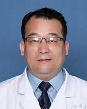 Hongtao Hu (胡鸿涛)合作论文数Henan Cancer Hospital4Peizan Ni合作论文数3Xiao Liu合作论文数3RongLiang Qiu (邱荣良)合作论文数3SiNing Shen (申思宁)合作论文数3
 Xiaoli Zheng (郑晓莉)合作论文数School of Materials Science and Engineering, Zhengzhou University3
 Yingying Cui合作论文数3
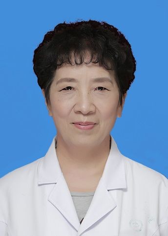 Qingxia Fan (樊青霞)合作论文数The First Affiliated Hospital of Zhengzhou University3Ruidi Jiao合作论文数2meiling liu (刘美玲)合作论文数2chen cheng (程宸)合作论文数2Ruiyun Zhang合作论文数2Shujuan Wang合作论文数2Xiao FENG (冯潇)合作论文数2LiJie Jia (贾丽洁)合作论文数2HuiYun Zhao (赵慧云)合作论文数2JianWei Zhang (张建伟)合作论文数2Zhao Huiyun合作论文数2
 Jianhua Wang (王建华)合作论文数Institute of Chemical Materials, China Academy of Engineering Physics2
 Siguo Cheng (程四国)合作论文数Henan Provincial People's Hospital2
 Yanan Sun (孙亚男)合作论文数The 2nd Affiliated Hospital of Harbin Medical University2Hao Wang (王浩)合作论文数2
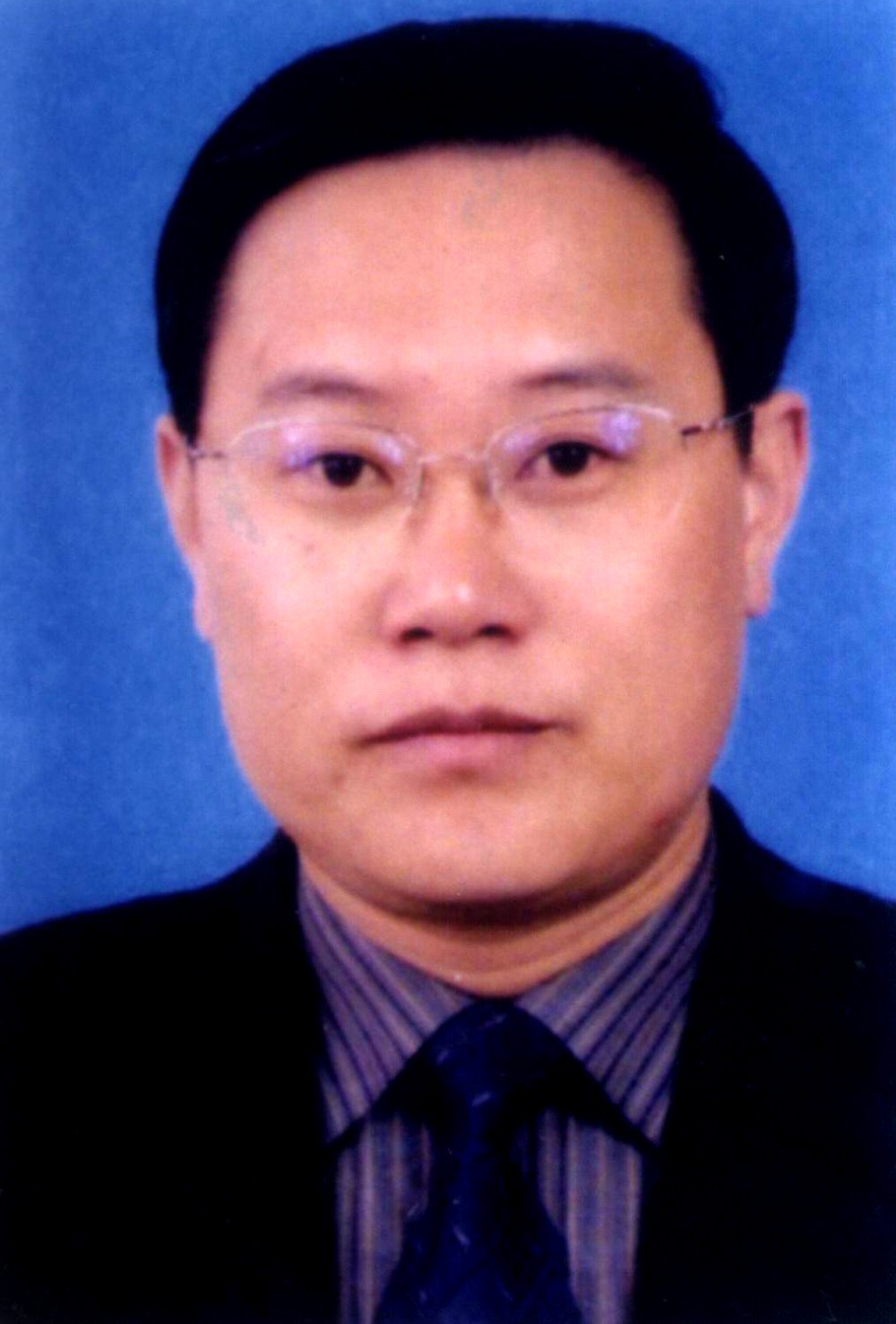 Kuisheng Chen (陈奎生)合作论文数The First Affiliated Hospital of Zhengzhou University2Ronghu Mao (毛荣虎)合作论文数郑州大学附属肿瘤医院2LIU Xin-ju (刘新菊)合作论文数2
 Ruozheng Wang (王若峥)合作论文数Radiotherapy Center, Affiliated Tumor Hospital of Xinjiang Medical University2
 Jinming Yu (于金明)合作论文数Shandong First Medical University;Shandong Cancer Hospital2Jie Ma合作论文数1Qinfu Dan合作论文数1Xu Wang合作论文数1




