BACKGROUND CONTEXT Patients with degenerative lumbar spinal stenosis (LSS) often experience limited long-term relief from conservative therapies, with many progressing to surgery. Limaprost, a prostaglandin E1 analog, may improve symptoms by enhancing microcirculation in compressed neural tissues. This multicenter prospective study evaluates its real-world effectiveness and safety. PURPOSE To evaluate the effectiveness and safety of limaprost in degenerative LSS and to identify predictors of subsequent surgical intervention. STUDY DESIGN/SETTING Multicenter prospective observational cohort study. PATIENT SAMPLE A total of 1,139 patients with degenerative lumbar spinal stenosis. OUTCOME MEASURES Primary outcomes included changes in Oswestry Disability Index (ODI) and Japanese Orthopaedic Association (JOA) subjective symptom scores. Secondary outcomes included visual analog scale (VAS) scores for pain and numbness, EuroQol-5 Dimension (EQ-5D), and walking capacity. METHODS Patients were categorized into four groups: limaprost monotherapy (n=308), standard combination (n=234), other combination (n=531), and non-limaprost control (n=66). Outcomes were assessed at 2 weeks and at 1, 3, and 6 months. Changes from baseline were compared across groups. Odds ratios (OR) with 95% confidence intervals (CI) were used to assess risk of subsequent surgical intervention. RESULTS Compared with controls, the limaprost monotherapy group demonstrated significantly greater improvement in ODI at 3 months (p=0.0123). Improvements in JOA subjective scores were greater in the monotherapy group beginning at 1 month (all p<0.05). At 6 months, all limaprost groups showed significantly greater improvements than controls in ODI and JOA scores (all p<0.05), as well as superior outcomes in pain and numbness VAS, EQ-5D, and walking capacity. Among patients without prior LSS surgery, limaprost monotherapy was associated with a significantly reduced risk of subsequent surgery (OR 0.07, 95% CI 0.01–0.39; p=0.003). Adverse event rates were low across all groups (0.9%–2.2%). CONCLUSIONS Limaprost-containing regimens are associated with improved symptoms, functional outcomes, and quality of life in patients with degenerative LSS compared with non-limaprost therapy. Limaprost monotherapy may also reduce the likelihood of subsequent surgical intervention in patients without prior surgery. FDA Device/Drug Status Limaprost (limaprost alfadex) (Investigational/Not Approved).
Background Multilevel thoracic ossification of the posterior longitudinal ligament (m-TOPLL) is frequently associated with severe neurological impairment and poor postoperative recovery. Posterior decompression and fusion (PDF) is the traditional operation strategy. Thoracic posterior dekyphosis and fusion (TPDF) has recently been introduced as an alternative surgical strategy for severe m-TOPLL. However, the advantages and disadvantages of between TPDF and PDF have not yet been compared. This study aimed to compare the clinical outcomes of TPDF and PDF in non-ambulatory patients with m-TOPLL. Methods A retrospective study was conducted involving patients who underwent surgical treatment for m-TOPLL at a single institution between 2015 and 2023. Patients were matched according to preoperative neurological status and thoracic kyphosis angle (TK). Demographic characteristics, radiographic parameters and surgical outcomes were compared between the TPDF and PDF groups. Results No significant differences were observed between the two groups regarding demographic characteristics and radiographic parameters. Compared with the PDF group, the TPDF group demonstrated significantly greater postoperative improvement in lower-extremity motor function (3 (2) vs. 1 (3), P = 0.017) and a superior mJOA recovery rate (72.5% ± 18.5% vs. 59.6% ± 23.2%, P = 0.034). However, postoperative cerebrospinal fluid leakage (CSFL) occurred more frequently in the PTDF group (20/25 vs. 13/25, P = 0.037). Conclusion TPDF provides superior neurological recovery compared with conventional PDF in non-ambulatory patients with severe m-TOPLL. Although TPDF is associated with a higher incidence of CSFL, operative duration and blood loss can be maintained within acceptable ranges when performed by experienced surgical teams.
Purpose We found that a lot of patients with thoracolumbar ossification of the ligamentum flavum (TLOLF) had Scheuermann’s disease simultaneously. To investigate the clinical characteristics and postoperative prognosis of TLOLF patients with thoracolumbar Scheuermann’s disease (TLSD), we conducted this research. Methods Patients who underwent surgery for ossification of the ligamentum flavum located in the thoracolumbar region at a single center from 2012 to 2022 were retrospectively reviewed. Patients were divided into a TLSD group and a control group. Baseline characteristics, sagittal spinal parameters, TOLF classification, spinal canal occupying rate, and postoperative recovery rate of neurological function were compared between the two groups. Results Patients in the TLSD group had a higher proportion of males (38/50 vs. 16/50, P < 0.001), greater thoracolumbar kyphosis (TLK) (14.69 ± 9.66° vs. 7.91 ± 5.43°, P < 0.001), a shorter duration of symptoms (7 (22) vs. 21 (51) months, P = 0.003), and a higher recovery rate (66.7% (54.2%0 vs. 41.4% (77.1%), P = 0.003). No significant differences were observed between the two groups in TOLF classification or spinal canal occupying rate. Conclusion TLOLF patients with TLSD had a greater TLK, which might be associated with the acceleration of ossification of the ligamentum flavum, resulting in a shorter disease course and better postoperative prognosis. TLSD does not affect the morphology of TOLF or the spinal canal occupying rate.
ABSTRACT Objective Operation for thoracic spinal stenosis (TSS) is considered a high‐risk surgery. Because of the frailty of elderly patients, the prediction for postoperative complications is crucial. This study investigated the relationship between frailty, as measured by the modified frailty index‐11 (mFI‐11), and postoperative complications in elderly patients with thoracic myelopathy secondary to TSS. Methods A retrospective review was conducted of 391 patients aged 65 years or older, with 209 males and 182 females, who underwent surgery for TSS at Peking University Third Hospital from 2012 to 2023. Patients were stratified into subgroups based on mFI‐11 score. Data on perioperative complications, including systemic and local events, were collected. Univariate and multivariate analyses were performed to determine the association between frailty and perioperative complications and to identify independent risk factors. Results A total of 391 elderly patients undergoing decompression and fusion for TSS were included and categorized by mFI‐11 score: 0 ( n = 73), 0.09 ( n = 159), 0.18 ( n = 98), and ≥ 0.27 ( n = 61). Multivariate analysis identified the mFI‐11 as an independent risk factor for surgical site infection (SSI) (OR = 7.250, p = 0.022), gastrointestinal complications (OR = 2.461, p = 0.029), urologic complications (OR = 4.855, p = 0.001), respiratory complications (OR = 13.968, p = 0.033), postoperative fever (OR = 2.256, p < 0.001), and postoperative transfusion (OR = 1.962, p = 0.014). Moreover, mFI ≥ 0.27 is a threshold for severe complications (OR = 15.886, p = 0.017), and mFI ≥ 0.18 is a threshold for any postoperative complications (OR = 6.338, p < 0.001) and minor complications (OR = 5.915, p < 0.001). Conclusions The mFI‐11 score is an effective predictor of the risk of surgical site infection, gastrointestinal complications, urologic complications, respiratory complications, postoperative fever, and postoperative transfusion in elderly patients undergoing TSS surgery. Patients with mFI scores ≥ 0.18 are at a significantly higher risk of any postoperative complications or minor complications, with mFI scores ≥ 0.27 indicating severe complications. Frailty, as assessed by mFI‐11, and non‐neurological complications did not significantly impact the long‐term recovery rate.
ABSTRACTObjectiveHigh‐grade dysplastic spondylolisthesis (HGDS) is a relatively rare condition mainly involving the L5/S1 segment of the spine and occurring in children and adolescents. Whether surgical fixation should be L5–S1 monosegmental or extended up to L4 remains controversial. This study aimed to compare clinical outcomes and the risk of adjacent segment spondylolisthesis between L5–S1 monosegmental fixation and L4–S1 double‐segmental fixation for pediatric HGDS.MethodsThis is a retrospective control study Pediatric patients diagnosed with HGDS between March 2007 and October 2022 at our hospital were followed up, and their data were analyzed. The study involved 39 HGDS patients (average slip, 70.0%) divided into two groups: the L5–S1 monosegmental reduction and fusion group (L5 group, 16 patients) and the L4–S1 double‐segmental reduction and fusion group (L4 group, 23 patients). Radiographic parameters and patient‐reported outcomes were collected before surgery and at follow‐up and compared between the two groups. Additionally, the prevalence of adjacent segment spondylolisthesis and instability (ASS/ASI) was compared between the groups at the last follow‐up. Independent sample t‐tests were employed to compare normally distributed data between the two groups. Paired sample t‐tests were employed to compare preoperative data with postoperative data. The chi‐square test was utilized to compare rates.ResultsThe average age was (12.5 ± 2.3) years. The L4 group had a greater slip percentage (77.6% vs. 59.2%; p < 0.001), a smaller Dub‐LSA (60.3° vs. 69.7°; p = 0.022), and a higher slip grade (p = 0.002) than the L5 group, indicating more severe spondylolisthesis in the L4 group. Clinical parameters, namely the visual analog scale, Oswestry disability index, and Japanese Orthopedic Association‐29 score, did not significantly differ between the two groups. The L5 group had a higher rate of ASS/ASI than the L4 group (6/16, 37.5% vs. 0/23, 0%; p = 0.002). Of all the ASS/ASI patients, one underwent a second surgery due to L4 spondylolisthesis 2 years after the primary surgery. The remaining five exhibited no symptoms or mild symptoms.ConclusionsFor pediatric HGDS, both L5–S1 monosegmental fixation and L4–S1 fixation can achieve satisfactory outcomes. However, there is a higher risk of ASS/ASI following L5–S1 fixation than following L4–S1 fixation.
Scheuermann’s disease always occurs in adolescent, and it is uncommon for elderly individuals. The aim of this research is to investigate the different potential pathogenic mechanisms and clinical characteristics of thoracolumbar disc herniation (TLDH) in Scheuermann’s disease between elderly and adolescent patients. We retrospectively analyzed data from patients with Scheuermann’s disease who underwent surgical treatment for TLDH from a single center between June 2013 and June 2023. Patients were divided into two groups: elderly (≥ 65 years) and adolescent (≤ 20 years), with 24 patients in each group. Data, including medical records and imaging parameters, were independently collected and analyzed by two doctors. The elderly group exhibited significantly lower preoperative modified Japanese Orthopaedic Association (mJOA) scores (5.58 ± 2.38 vs. 7.25 ± 2.19, P = 0.009) and longer symptom duration (48.29 ± 62.32 vs. 8.37 ± 13.19 months, P < 0.001) compared to the adolescent group. The elderly group also had a higher prevalence of diabetes mellitus (6/24 vs. 0/24, P = 0.029), fewer Schmorl’s nodes, more severe intervertebral disc degeneration, and a higher frequency of intervertebral disc vacuum phenomena. Additionally, elderly patients exhibited more frequent localized ossification or hypertrophy of the ligamentum flavum in the thoracolumbar region. The later onset of TLDH in Scheuermann’s disease among elderly patients is likely due to a combination of genetically determined higher endplate strength and age-related degenerative factors. Elderly patients also commonly show localized ossification or hypertrophy of the ligamentum flavum, which may contribute to more severe symptoms.
ABSTRACTObjectiveDuring percutaneous endoscopic interlaminar discectomy (PEID), a range of technologies including medical robotics, visual navigation, and spatial registration have been proposed to expand the application scope and success rate of minimally invasive surgery. The use of robotic technology in surgery is conducive to improving accuracy and reducing risk. This study aims to introduce a precise and efficient targeting method tailored for robot‐assisted positioning under C‐arm fluoroscopy inPEID.MethodsThis study conducted a retrospective analysis of 107 patients with lumbar disc herniation (LDH) who underwent surgical treatment at our hospital from February 2023 to February 2024 (average age: 43.3 ± 13.3 years; 61 males and 43 females). The method entails constructing a specialized end‐effector capable of simultaneous fluoroscopy calibration and robot‐to‐image‐space registration. The average time of the surgical procedure and the average number of fluoroscopy exposures were collected. Preoperative and postoperative follow‐up data were collected, including assessment scores from the Japanese Orthopedic Association (JOA) and Visual Analog Scale (VAS). Paired t‐tests were employed to compare differences in each clinical outcome between the preoperative and follow‐up time.ResultsThese techniques effectively reduce both radiation exposure and operation time. Clinical data reveals that the average time for robot‐assisted positioning stands at 2.5 ± 0.7 min, with anteroposterior and lateral radiographs demonstrating accuracies of 2.4 ± 2.8 mm and 3.1 ± 3.7 mm, respectively, during robot‐assisted positioning. Postoperative VAS scores for back pain and leg pain were significantly lower than preoperative scores (1.2 ± 1.8 vs. 4.1 ± 2.3, 0.9 ± 2.0 vs. 5.9 ± 1.8; p < 0.05, p < 0.05, respectively). Postoperative JOA scores were significantly higher than preoperative scores (26.1 ± 3.5 vs. 14.5 ± 4.9; p < 0.05).ConclusionsThrough the evaluation of the system in robot‐assisted positioning during PEID, this study substantiates the accuracy and reliability of the proposed method in clinical applications.
OBJECTIVES:Cage retropulsion (CR) is a common complication following posterior lumbar interbody fusion (PLIF). Symptomatic patients with CR often require revision surgery. However, there is a lack of literature supporting the effectiveness of conservative treatment for CR. This study compares clinical and radiographic outcomes between conservative treatment and revision surgery in patients with CR after PLIF. METHODS:A total of 55 patients with CR after PLIF treated at our institution between 2016 and 2023 were retrospectively reviewed; postoperative radiographic data of follow-up were used to diagnose CR. Clinical outcomes were assessed before therapy and at the final follow-up using the visual analog scale (VAS) for lower back pain and leg pain, Oswestry Disability Index (ODI) scores, and Japanese Orthopedic Association 29 (JOA-29) scores. The treatment effectiveness was evaluated based on whether the score change reached the minimally clinically important difference (MCID). Radiographic indicators included the fusion rates, the extent of CR into the spinal canal, and the total displacement distance. Continuous variables were compared using independent samples t-tests or Mann-Whitney U tests, while categorical variables were analyzed using Chi-square or Fisher's exact tests, as appropriate. A p-value < 0.05 was considered statistically significant. RESULTS:The fusion rates at the final follow-up for the conservative treatment group and the revision surgery group were 87.5% and 84.6%, respectively. There were no significant differences in final follow-up fusion rates, lower back pain VAS scores, leg pain VAS scores, JOA scores, or ODI scores between the two groups (all p > 0.05). Additionally, there was no difference in the proportion of patients whose lower back pain VAS, ODI, and JOA scores achieved MCID between groups (all p > 0.05). However, in the revision surgery group, the proportion of patients whose leg VAS scores reached MCID was significantly higher than in the conservative group (p = 0.001). In the revision surgery subgroup analysis, patients who did not achieve leg VAS MCID demonstrated significantly more severe cage retropulsion distance compared to MCID achievers (p = 0.03). CONCLUSIONS:Conservative treatment yields satisfactory outcomes in mild, symptomatic CR patients, particularly for low back pain. For patients with a CR distance less than 8.8 mm, conservative treatment and revision surgery showed comparable outcomes, whereas when the CR distance is ≥ 8.8 mm, revision surgery was recommended to improve clinical results. Both conservative treatment and revision surgery can yield favorable outcomes when appropriately indicated.
PURPOSE:Multimodal articulated image registration (MAIR) is a challenging problem because the resulting transformation needs to maintain rigidity for bony structures while allowing elastic deformation for surrounding soft tissues. Existing deep learning-based methods ignore the articulated structures and consider it as a pure deformable registration problem, leading to suboptimal results. METHODS:We propose a novel weakly supervised anatomy-aware multimodal articulated image registration network, referred as MAIRNet, to solve the challenging problem. The architecture of MAIRNet comprises of two branches: a non-learnable polyrigid registration branch to estimate an initial velocity field, and a learnable deformable registration branch to learn an increment. These two branches work together to produce a velocity field that can be integrated to generate the final displacement field. RESULTS:We designed and conducted comprehensive experiments on three datasets to evaluate the performance of the proposed method. Specifically, on the hip dataset, our method achieved, respectively, an average dice of 90.8%, 92.4% and 91.3% for the pelvis, the right femur, and the left femur. On the lumbar spinal dataset, our method obtained, respectively, an average dice of 86.1% and 85.9% for the L4 and the L5 vertebrae. On the thoracic spinal dataset, our method achieved, respectively, an average dice of 76.7%, 79.5%, 82.9%, 85.5% and 85.7% for the five thoracic vertebrae ranging from T6 to T10. CONCLUSION:In summary, we developed a novel approach for multimodal articulated image registration. Comprehensive experiments conducted on three typical yet challenging datasets demonstrated the efficacy of the present approach. Our method achieved better results than the state-of-the-art approaches.
OBJECTIVE: To describe a novel technique, posterior thoracic antidisplacement and fusion (PTAF), for a special type of ossification of the posterior longitudinal ligament in the thoracic spine (T-OPLL), and to evaluate its safety and efficacy. METHODS: From July to December 2020, 5 consecutive patients with beak-type T-OPLL located at the thoracic vertebral body level underwent PTAF surgery. Their demographic data, radiological parameters, perioperative complications, and surgery-related findings were recorded and analyzed. The surgical outcomes were assessed using a modified Japanese Orthopedic Association scale, and the recovery rate was calculated using the Hirabayashi's method. RESULTS: All patients were followed up for at least two years. The mean thickness of OPLL was 9.4 +/- 1.0 mm, and the OPLL spinal canal occupying ratio was 67.7% +/- 8.5%. Postoperatively, the mean antidisplacement distance of OPLL was 8.1 +/- 1.8 mm, and the average shortened distance of the spinal column was 6.0 +/- 1.13 mm. The mean operation time and blood loss were 158.2 +/- 26.3 minutes and 460 +/- 89.4 mL, respectively. Perioperative complications were cerebrospinal fluid leakage and instrument failure, 2 cases each. The mean modified Japanese Orthopedic Association score was increased from 3.6 +/- 2.9 before surgery to 9.4 +/- 3.0 at the last follow-up, and the average recovery rate was 84.2 +/- 30.5%. CONCLUSIONS: The preliminary clinical outcomes indicate that PTAF is a safe and effective method for the treatment of beak-type T-OPLL, which has its apex located at the vertebral body level and has a high spinal canal occupation ratio.
A warning system to avoid potential nerve root thermal injury caused by automatic spine robot is essential. However, there is a lack of basic research to support the development of such warning system. The aims of this study are to confirm the feasibility of continuous intraoperative neuromonitoring (CIONM) in spinal nerve root monitoring and study the exposure time of spinal nerve root thermal injury at different temperature in a porcine model. The experiment was carried out on 32 nerve roots of 4 pigs (bilateral L4-S1 nerve roots). Sham group (CIONM only), control group (Room temperature stimulation with CIONM), and 40 °C, 45 °C, 50 °C, 55 °C, 60 °C, 65 °C temperature stimulation groups were conducted at random in each pig. The change of EMG was recorded continuously. The exposure time of nerve root injury at different temperatures was recorded. Histological analysis of the nerve roots was performed to confirm the injury. The EMG of the sham, control, 40 °C and 45 °C groups were stable during the experiment, and no significant nerve root injury was observed on histological analysis. The time of 50
Study Design A biomechanical study. Objective We aimed to assess temperature fluctuations when applying the ultrasonic osteotome during laminectomy and identify the most appropriate cutting method for robot-assisted laminectomy to mitigate the risk of heat-related injuries. Methods Utilizing a robotic system for laminectomy, the study implemented the ultrasonic bone scalpel to cut both artificial polyurethane bones and animal spinal bones. The research focused on comparing and analyzing the maximum temperature of the inner surface of four types of artificial bones through three different cutting techniques: vertical constant cutting at speeds of .5 mm/s and 1 mm/s, as well as robot-assisted vertical reciprocating cutting. After the initial results, two optimal vertical cutting approaches were selected for subsequent trials, evaluating the effectiveness and temperature impact of various ultrasonic osteotome modes on 10 isolated spinal bones from pigs. Results When cutting polyurethane bones, reciprocating cutting demonstrated the lowest maximum temperature in contrast to constant speed cutting at .5 mm/s and 1 mm/s. In the animal bone trial, direct cutting registered an average maximum temperature of 43.25°C with an average cutting duration of 688.3 s, while reciprocating cutting recorded an average maximum temperature of 34.20°C with an average cutting time of 713.0 s. Conclusions The reciprocating cutting strategy utilized in robot-assisted ultrasonic osteotome is effective in reducing heat generation and maintaining high cutting efficiency.
OBJECTIVE This study aimed to introduce a novel artificial intelligence (AI)-based robotic system for autonomous planning of spinal posterior decompression and verify its accuracy through a cadaveric model. METHODS Seventeen vertebrae from 3 cadavers were included in the study. Three thoracic vertebrae (T9-11) and 3 lumbar vertebrae (L3-5) were selected from each cadaver. After obtaining CT data, the robotic system independently planned the laminectomy path based on AI algorithms before the surgical procedure and automatically performed the decompression during the procedure. A postoperative CT scan was performed, and the deviation of each cutting plane from the preoperative plan was quantitatively analyzed to evaluate the accuracy and safety of the cuts. The duration of laminectomy was also recorded. RESULTS A total of 285 cuts were made on thoracic and lumbar vertebrae. The average duration for unilateral longitudinal cutting was 16.38 +/- 4.76 minutes, while for transverse cutting it was 4.44 +/- 1.52 minutes. In terms of accuracy assessment, 3 levels were divided based on the distance between the actual cutting plane and the preplanned plane: 77 (84%) were grade A, 15 (16%) were grade B, and none were grade C. Regarding safety assessment, 74 (80%) were designated safe (grade A), with 18 (20%) classified as uncertain (grade B). CONCLUSIONS The results confirm the accuracy and preliminary safety of the robotic system for autonomous planning and cutting of spinal decompression.
目的 调查八年制医学生的科研能力和科研诚信认知状况,为科研培训工作提供依据.方法 对62名八年制医学生进行问卷调查.结果 科研能力量表总分46[(40,54)]分,"统计学知识"维度得分低;研诚信认知问卷总分119[(107,127)]分,医学生对科研诚信的关注度评分低(1.94±0.97~2.61 士1.03),认为产生科研不端行为的主要原因是自身科研能力不足(75.8%)和缺乏学术监督(74.2%).科研能力量表总分与对科研诚信的关注度、对科研不端行为造成影响的认知度呈正相关(p<0.05).结论 需加强二级学科阶段八年制医学生的科研能力培养;注重统计学知识和实践教学的培养;丰富科研诚信教育形式;提高对科研诚信的关注度和对科研不端行为影响的认识.
High-precision image segmentation of the spine in computed tomography (CT) images is important for the diagnosis of spinal diseases and surgical path planning. Manual segmentation is often tedious and time consuming. Thus, an automatic segmentation algorithm is expected to solve this problem. However, because different areas are scanned, the number of spines in the original CT image and the coverage area are often different, making it extremely difficult to directly conduct a fully autonomous spine segmentation. In this study, we propose a two-stage automatic spine segmentation method based on 3D Swin Transformer. In the first stage, the 3D Swin-YoloX algorithm is used to achieve an accurate positioning of each spine segment in the CT images. In the second stage, 3D Swin-UNet is used to achieve a high-precision segmentation of the spine. Using an open dataset, the average Dice of our approach can reach 0.942 and the average Hausdorff distance can reach 6.24, indicating a higher accuracy in comparison with other published methods. Our proposed method can effectively eliminate any adverse effects of the different scanning areas on a spinal image segmentation and has a high application value.
STUDY DESIGN:Retrospective cohort study. OBJECTIVES:To describe the clinical characteristics and surgical outcomes of patients with multilevel-ossification of the posterior longitudinal ligament (mT-OPLL), and to identify risk factors for unfavorable outcomes. METHODS:Patients who were diagnosed with mT-OPLL and underwent one-stage thoracic posterior laminectomy combined with selective OPLL resection, spinal cord de-tension, and fusion surgery between August 2012 and October 2020 were recruited. Patients' demographic-, surgical- and radiological-related parameters were collected and analyzed. Neurological status was evaluated with mJOA score, and recovery rate (RR) was calculated using the Hirabayashi formula. According to RR, patients were divided into a favorable outcome group (FOG, RR ≥50%) and an unfavorable outcome group (UOG, RR <50%). Univariate and multivariate analyses were used to compare the difference between the 2 groups and to identify risk factors for unfavorable outcomes. RESULTS:A total of 83 patients were included, with an average age of 50.6 ± 8.3 years. Cerebrospinal fluid leakage (60.2%) and transient neurological deterioration (9.6%) were the most common complications. The average mJOA score improved from preoperative 4.3 ± 2.2 to 9.0 ± 2.4 at the last follow-up, and the mean RR was 74.9 ± 26.3%. Disease duration, preoperative nonambulatory status, and the number of decompressed levels were identified as potential risk factors by Univariate analysis (all P < .05). Multivariate analysis showed that the preoperative disease duration and nonambulatory status were independent risk factors for unfavorable outcomes. CONCLUSIONS:Long disease duration and nonambulatory status before surgery were independent risk factors for unfavorable outcomes.
Objective: This study aims to use artificial intelligence to realize the automatic planning of laminectomy, and verify the method. Methods: We propose a two-stage approach for automatic laminectomy cutting plane planning. The first stage was the identification of key points. 7 key points were manually marked on each CT image. The Spatial Pyramid Upsampling Network (SPU-Net) algorithm developed by us was used to accurately locate the 7 key points. In the second stage, based on the identification of key points, a personalized coordinate system was generated for each vertebra. Finally, the transverse and longitudinal cutting planes of laminectomy were generated under the coordinate system. The overall effect of planning was evaluated. Results: In the first stage, the average localization error of the SPU-Net algorithm for the seven key points was 0.65mm. In the second stage, a total of 320 transverse cutting planes and 640 longitudinal cutting planes were planned by the algorithm. Among them, the number of horizontal plane planning effects of grade A, B, and C were 318(99.38%), 1(0.31%), and 1(0.31%), respectively. The longitudinal planning effects of grade A, B, and C were 622(97.18%), 1(0.16%), and 17(2.66%), respectively. Conclusions: In this study, we propose a method for automatic surgical path planning of laminectomy based on the localization of key points in CT images. The results showed that the method achieved satisfactory results. More studies are needed to confirm the reliability of this approach in the future.
Study Design. A basic experimental study. Objective. To elucidate the role and mechanism of interleukin (IL)-17A in thoracic ossification of the ligamentum flavum (TOLF). Summary of Background Data. TOLF is characterized by the replacement of the thoracic ligamentum flavum with ossified tissue and is one of the leading causes of thoracic spinal stenosis. IL-17A is an important member of the IL-17 family that has received widespread attention for its key contributions to the regulation of bone metabolism and heterotopic ossification. However, it is unclear whether IL-17A is involved in TOLF. Materials and Methods. Cell counting kit‐8 assay and 5-ethynyl-2’-deoxyuridine staining were performed to assess the proliferation of ligamentum flavum cells (LFCs). Alkaline phosphatase activity assay, Alizarin red staining, and protein level expression of osteogenic-related genes were used to evaluate the osteogenic differentiation potential of LFCs. The effect of IL-17A on the proliferation and osteogenic differentiation of LFCs was further assessed after silencing β-catenin by transfection with small interfering RNA. In addition, the possible source of IL-17A was further demonstrated by coculture assays of T helper 17 (Th17) cells with LFCs. Student t test was used for comparisons between groups, and the one-way analysis of variance, followed by the Tukey post hoc test, was used for comparison of more than two groups. Results. IL-17A was elevated in TOLF tissue compared with normal ligamentum flavum. IL-17A stimulation promoted the proliferation and osteogenic differentiation of LFCs derived from patients with TOLF. We found that IL-17A promoted the proliferation and osteogenic differentiation of LFCs by regulating the β-catenin signaling. Coculture of Th17 cells with LFCs enhanced β-catenin signaling-mediated proliferation and osteogenic differentiation of LFCs. However, these effects were markedly attenuated after the neutralization of IL-17A. Conclusions. This is the first work we are aware of to highlight the importance of IL-17A in TOLF. IL-17A secreted by Th17 cells in the ligamentum flavum may be involved in the ossification of the microenvironment by regulating β-catenin signaling to promote the proliferation and osteogenic differentiation of LFCs.
Background: Dural ossification (DO) is the leading cause of surgery-related dural tear in patients with ossification of the ligamentum flavum (OLF). An accurate preoperative diagnosis of DO is conducive to the selection of appropriate surgical methods. Although several imaging signs, such as Banner cloud sign (BCs), tram-track sign (TTs), and comma sign (Cs) have been proposed for the preoperative diagnosis of DO, their diagnostic value has not been well studied. The aim of this study was to explore the diagnostic value of BCs, TTs, and Cs, and provide evidence-based data for their clinical application. Methods: This is a blind, randomized diagnostic study using retrospectively collected data from 102 consecutive patients who were diagnosed with OLF and underwent decompression surgery between January 2018 and June 2019. A total of 8 surgeons with different qualifications were recruited to read these imaging signs to identify the presence of DO. Surgical records were used as the reference standard. Sensitivity, specificity, and the area under the receiver operating characteristic (ROC) curve (AUC) were used to evaluate the diagnostic accuracy of each imaging sign and their different combinations.Results: Of the 102 patients, 21 were diagnosed with DO. BCs had a significantly higher diagnostic accuracy than TTs and Cs, with the AUC of 0.704, 0.607, and 0.593, respectively. The specificity of BCs, Cs, TTs, and their combination in diagnosing DO was 91.5%, 92.1%, 68.3%, and 62.2%, respectively. In the combined diagnostic test, the results showed that the combined diagnosis accuracy of BCs and Cs was the highest, and the AUC was 0.738. The combination of BCs, Cs, and TTs increased the sensitivity of diagnosing DO (77.5%), but did not improve the diagnostic accuracy, and the AUC was 0.699. Conclusions: BCs had higher diagnostic accuracy than TTs and Cs. BCs and Cs were highly specific for DO, whereas TTs could be confusing due to their non-specific presentations. The combination of BCs, TTs, and Cs improved the sensitivity of DO diagnosis, but not the specificity and accuracy.
Objectives Sitting is a common weight‐bearing posture, like standing, but there still lacks enough understanding of sagittal alignment in sitting position for patients after lumbar fusion. This study aimed to investigate the accommodation of fixed spine from standing to sitting position and its influence on unfused segments. Methods Sixty‐two patients after lumbar fusion (test group) and 40 healthy volunteers (control group) were recruited in this research. All subjects underwent lateral radiographs of entire spine in the standing and sitting positions. The spinopelvic parameters including sagittal vertical axis (SVA), T1 pelvic angle (TPA), lumbar lordosis (LL), thoracic kyphosis (TK), and pelvic tilt (PT) were measured. The changes in parameters of patients between two positions were compared with control group, and patients were divided in different groups based on fusion level and their parameters were compared. Results When changing from standing to sitting positions, a forward‐moving SVA and TPA were observed in both patients and control groups, accompanied by the decrease in LL, TK and increase in PT, but the changes of patients were smaller in TPA, LL, and TK (6.5° ± 7.2° vs 9.7° ± 6.0°, 7.7° ± 8.3° vs 13.6° ± 8.5°, 2.2° ± 6.5° vs 5.4° ± 5.1°, respectively, p < 0.05). Increase of PT in the lumbosacral fixation group was lower than that in the control group (4.4° ± 9.1° vs 8.3° ± 7.1°, p < 0.05). Patients who had adjacent segments degeneration (ASD) showed more kyphosis in unfused lumbar segments than the other patients (16.4° ± 10.7° vs −1.0° ± 4.8°, p < 0.05) from standing to sitting. Conclusions The spine straightens in lumbar and thoracic curve, combined with forward‐moving axis and pelvic retroversion when changing to the sitting position. However, these changes are relatively limited in patients after lumbar fusion, so the adjacent unfused lumbar segments compensate to stress during sitting and this may be related to ASD.
 Weishi Li (李危石)合作论文数Peking University Third Hospital36
 Chuiguo Sun (孙垂国)合作论文数Department of Orthopaedics, Peking University Third Hospital33
 Zhongqiang Chen (陈仲强)合作论文数Peking University Third Hospital;Peking University International Hospital31
 Zhaoqing Guo (郭昭庆)合作论文数Peking University Third Hospital24
 Qiang Qi (齐强)合作论文数Peking University Third Hospital23
 Yan Zeng (曾岩)合作论文数Peking University People's Hospital14
 Shuai Jiang合作论文数11GuoXin Hu (胡国信)合作论文数9Yu JIANG (姜宇)合作论文数8Zeng Yan (曾岩)合作论文数8
 Guanghui Chen合作论文数7Wang Chengxia (王承夏)合作论文数5Zhuofu Li合作论文数5Dang Geng-Ting (党耕町)合作论文数5
 Xiaoguang Liu (刘晓光)合作论文数Peking University Health Science Center;Peking University Third Hospital;Peking University International Hospital5
 Liang Jiang (姜亮)合作论文数Peking University Third Hospital5
 Zhongjun Liu (刘忠军)合作论文数Peking University Third Hospital5Da Zou合作论文数4Siyu Zhou合作论文数4
 Lei Yuan合作论文数4
 Liwei Shi (石立伟)合作论文数School of Medical Technology, Beijing Institute of Technology4
 Feng Wei (韦峰)合作论文数Department of Orthopaedic, Peking University Third Hospital4
 Huishu Yuan (袁慧书)合作论文数Peking University Third Hospital4
 Zhuoran Sun合作论文数3Longjie Wang合作论文数3Tianqi Fan合作论文数3史尉利合作论文数北京大学第三医院3
 Lei Hu (胡磊)合作论文数School of Mechanical Engineering and Automation, Beihang University3
 Chunli Song (宋纯理)合作论文数Peking University Third Hospital3Dongwei Fan (范东伟)合作论文数Peking University Third Hospital3
 Qingjun Ma (马清钧)合作论文数Academy of Military Medical Sciences;SL Pharm3Yonghong Zhang合作论文数2Qian Xiang合作论文数2Xiaochen Qu合作论文数2Xuquan Ji合作论文数2Yongzhao Zhao合作论文数2Xiongkang Song (宋雄康)合作论文数2Jialiang Lin合作论文数2Shanshan Liu合作论文数2
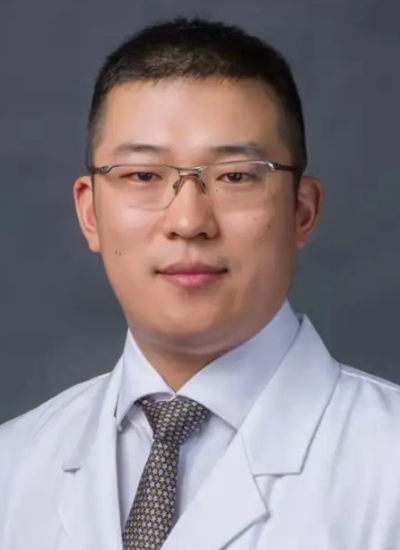 Yu Jiang (姜宇)合作论文数Department of Orthopaedic Surgery, Peking University Third Hospital2
 Alexander Frank Heimann合作论文数1Wei Li合作论文数1Gang Wang合作论文数1Fei Xu合作论文数1Guohong Du合作论文数1Aikeremujiang Muheremu合作论文数1
 Xiaoru Gao合作论文数1




