Figure S1. The overlapped and non-overlapped genes among Tianjin and MSK-IMPACT panels.
Objectives: This study aimed to determine the role and mechanism underlying migration and invasion inhibitory protein (MIIP) modulation in M2 macrophages within the tumor microenvironment and the potential of targeting the MIIP– stimulator of interferon genes (STING) pathway in colorectal cancer (CRC) therapy. Methods: MIIP expression was analyzed for associations with the STING pathway and M2 macrophage infiltration using public datasets and clinical CRC samples. CRC cells were genetically modified using lentiviral vectors to overexpress or silence MIIP and STING. The interactions of genetically modified CRC cells with macrophages were studied in co-culture systems. Techniques, including immunofluorescence staining, RT-qPCR, western blot, ELISA, flow cytometry, and Transwell migration and invasion assays, were used to evaluate the crosstalk between CRC cells and macrophages. An orthotopic mouse CRC model was developed to study the effects of MIIP on M2 macrophage polarization and tumor metastasis through the STING–NFκB2–IL10 axis. The therapeutic significance of a STING antagonist was also assessed in vivo. Results: Analyses of The Cancer Genome Atlas (TCGA) cohort and our CRC cohort revealed low MIIP expression is associated with STING pathway activation, increased M2 macrophage infiltration, and poor clinical outcomes. The results of functional experiments demonstrated that MIIP inhibits IL10 production via the STING–TRAF3–NFκB2 axis in CRC cells, suppressing M2 macrophage polarization in co-culture systems. Conversely, M2 macrophages promoted CRC cell migration and invasion in an IL10-dependent manner. In vitro and in vivo studies confirmed that the MIIP-mediated feedback loop between CRC cells and macrophages depends on the STING–NFκB2–IL10 axis. Furthermore, inhibition of STING expression in a mouse model reduced M2 macrophage polarization and tumor metastasis. Conclusions: This study established MIIP as a crucial regulator of macrophage polarization in the CRC tumor microenvironment, providing new insights into the role in suppressing CRC progression and immune–tumor crosstalk. These findings highlight the potential of targeting the STING pathway as a therapeutic strategy for CRC patients who respond poorly to immune checkpoint inhibitors.
Table S6. The clinicopathologic parameters and molecular variations of RAS-related GISTs in Tianjin cohort and MSK-IMPACT cohort
BackgroundApproximately 20% of patients with stage II colorectal cancer (CRC) experience tumor relapse despite standard surgical treatment. Histopathological analysis holds promise for postsurgical risk stratification and guiding adjuvant chemotherapy (ACT) decisions. The aim of this study was to use deep learning to extract explainable tissue biomarkers from whole-slide images.Methods and findingsIn this retrospective cohort study, we developed and validated SurvFinder, an interpretable deep learning framework designed to autonomously identify tissue-based risk biomarkers from hematoxylin and eosin (H&E)-stained slides. The framework aims to support individualized risk stratification and explore associations with treatment outcomes. The present study included 6,950 H&E slides from 1,604 patients with stage II CRC across four independent cohorts in China. Patients were enrolled from 2012 to 2018 and followed for a minimum of 24 months. The primary outcome of the study was relapse-free survival (RFS). Our analyses identified tertiary lymphoid structures (TLSs) as critical prognostic features in stage II CRC. The multi-view integration of TLS characteristics by SurvFinder consistently demonstrated superior predictive and prognostic accuracy across four multicenter datasets (AUROC with 95% confidence interval [CI]: 0.827 [0.789,0.864], 0.805 [0.749,0.860], 0.805 [0.748,0.861], and 0.712 [0.621,0.804]), surpassing traditional clinical prognostic parameters (hazard ratio [HR]: 8.23, 95% CI: 5.43-12.47; p < 0.001). Using explainable AI (XAI) methods, we ensured model transparency and identified key TLS features-such as their location at the tumor periphery and their maturity state-as significant factors influencing prognosis and the efficacy of adjuvant therapy. The retrospective design without prospective validation and real-world clinical deployment is the main limitation of this study.ConclusionsTogether, these results highlight the potential utility of deep learning-based histopathological analysis for automated risk stratification in stage II CRC. In particular, our findings support the relevance of TLSs as a histological biomarker with potential implications for personalizing ACT decisions.
Inhibitor of nuclear factor kappa-B kinase subunit epsilon (IKBKE), a member of the serine/threonine kinase family, is an important oncogene in glioblastoma. IKBKE is involved in the progression of multiple tumours in glioblastoma (GBM), including tumour invasion, migration, and proliferation. Here, we report an IKBKE–angiomotin-like protein 2 (Amotl2)–yes-associated protein 1 (YAP1) axis in which IKBKE inhibits the protein expression of Amotl2, while Amotl2 regulates the nuclear transport of the YAP1 protein, which has been implicated in brain tumour development and progression. Data analysis of multiple IKBKE mRNA databases of glioma and immunohistochemical analysis of a tissue chip indicated that higher IKBKE expression was associated with higher malignancy and shorter survival in glioma patients. IKBKE downregulation significantly inhibited GBM cell proliferation and restrained tumour growth in a GBM mouse model. Moreover, IKBKE phosphorylates Amotl2, promoting Amotl2 ubiquitination and degradation, leading to YAP1 entry into the nucleus and the activation of downstream genes. In summary, our results are the first to show that IKBKE phosphorylates Amotl2 and that GBM cell proliferation is regulated by the IKBKE↑–Amotl2↓–YAP1↑ axis.
Diagnostic challenges remain in desmoid fibromatosis (DF) due to somewhat frequent β-catenin immunohistochemical negativity, risking misclassification and overtreatment. The present study evaluates the role of CTNNB1 molecular testing in optimizing diagnosis. A single-center, large retrospective analysis of 780 patients with DF was performed. The incidence of DF was higher in females, particularly within the adult demographic. 77.3
Table S4. The clinicopathologic parameters and molecular variations of SDH-deficient GISTs in Tianjin cohort and MSK-IMPACT cohort
Table S3. The clinicopathologic characteristics and comparisons of WT-GISTs from Tianjin and MSK-IMPACT cohorts
Objectives This study aimed to determine the role and mechanism underlying migration and invasion inhibitory protein (MIIP) modulation in M2 macrophages within the tumor microenvironment and the potential of targeting the MIIPu2013 stimulator of interferon genes (STING) pathway in colorectal cancer (CRC) therapy. Methods MIIP expression was analyzed for associations with the STING pathway and M2 macrophage infiltration using public datasets and clinical CRC samples. CRC cells were genetically modified using lentiviral vectors to overexpress or silence MIIP and STING. The interactions of genetically modified CRC cells with macrophages were studied in co-culture systems. Techniques, including immunofluorescence staining, RT-qPCR, western blot, ELISA, flow cytometry, and Transwell migration and invasion assays, were used to evaluate the crosstalk between CRC cells and macrophages. An orthotopic mouse CRC model was developed to study the effects of MIIP on M2 macrophage polarization and tumor metastasis through the STINGu2013NFu03BAB2u2013IL10 axis. The therapeutic significance of a STING antagonist was also assessed in vivo. Results Analyses of The Cancer Genome Atlas (TCGA) cohort and our CRC cohort revealed low MIIP expression is associated with STING pathway activation, increased M2 macrophage infiltration, and poor clinical outcomes. The results of functional experiments demonstrated that MIIP inhibits IL10 production via the STINGu2013TRAF3u2013NFu03BAB2 axis in CRC cells, suppressing M2 macrophage polarization in co-culture systems. Conversely, M2 macrophages promoted CRC cell migration and invasion in an IL10-dependent manner. In vitro and in vivo studies confirmed that the MIIP-mediated feedback loop between CRC cells and macrophages depends on the STINGu2013NFu03BAB2u2013IL10 axis. Furthermore, inhibition of STING expression in a mouse model reduced M2 macrophage polarization and tumor metastasis. Conclusions This study established MIIP as a crucial regulator of macrophage polarization in the CRC tumor microenvironment, providing new insights into the role in suppressing CRC progression and immuneu2013tumor crosstalk. These findings highlight the potential of targeting the STING pathway as a therapeutic strategy for CRC patients who respond poorly to immune checkpoint inhibitors.
Human epidermal growth factor receptor 2 (HER2) is a key biomarker and therapeutic target in several malignancies, including breast, gastric, and other solid tumors. Recent advancements in cancer molecular profiling and the Food and Drug Administration's approval of trastuzumab deruxtecan for HER2u2010positive panu2010tumor indications have highlighted the broader relevance of HER2 alterations across diverse cancers. However, the lack of standardized guidelines for HER2 testing in a panu2010tumor context creates variability in clinical practice, hindering the optimal implementation of HER2u2010targeted therapies beyond traditional indications. To address this gap, a multidisciplinary panel of Chinese experts has developed a consensus providing comprehensive recommendations on diagnostic strategies, testing methodologies, and clinical applications of HER2 overexpression detection. By establishing a unified framework for HER2 overexpression assessment, this consensus aims to enhance the precision of HER2 testing, optimize patient selection for targeted therapies, and improve clinical outcomes across a wide spectrum of HER2 overexpression malignancies.
Genotoxic stress or exogenous DNA damage induces transcription arrest, enabling efficient DNA repair. Transcription activators directly participate in DNA damage repair (DDR), but the trans-regulatory mechanisms linking transcription and DDR remain elusive. Here we reveal that CRTC2 switches from a transcriptional coactivator to a DNA-damage responder. CRTC2 promotes non-homologous end joining (NHEJ) in vitro and in vivo. Mechanistically, PARP1 recruits CRTC2 to DNA breaks, where CRTC2 promotes DNA-PKcs enrichment and DNA-PK holoenzyme assembly, driving NHEJ. DNA-PK phosphorylates CRTC2 at Ser433, dissociating it from transcriptional complexes to suppress target gene transcription and promoting its incorporation into repair complexes, forming a positive feedback loop that enhances NHEJ. CRTC2 loss radiosensitizes liver cancer cells, potentiates irradiation-induced cGAS-STING activation, and promotes antitumor immunity and the abscopal effect. AAV8-mediated targeting of CRTC2 sensitizes tumors to radioimmunotherapy. Thus, CRTC2 couples transcriptional silencing to DNA repair, and its inhibition offers a promising strategy for radioimmunotherapy sensitization.
Table S7. The clinicopathologic parameters of qWT-GIST in Tianjin cohort and MSK-IMPACT cohort
Neurotrophic tyrosine receptor kinase (NTRK) fusions are crucial in tumorigenesis and in guiding targeted therapy with TRK inhibitors. However, their rarity, fusion heterogeneity, and limitations of conventional pan-TRK immunohistochemistry (IHC) impede accurate clinical detection. This multicenter retrospective study analyzed 374 next-generation sequencing/fluorescence in situ hybridization-validated samples (195 NTRK positive and 179 NTRK negative) collected from 12 Chinese centers to investigate fusion heterogeneity and refine the interpretation of pan-TRK IHC. We developed an amplification protocol by combining the traditional pan-TRK IHC (EPR17341) with the OptiView Amplification Kit and established new interpretation criteria. A total of 40 solid tumor types were included, and 23 unique fusion partners were identified. Papillary thyroid cancer was the most common NTRK-positive tumor (49.74%) and harbored all 3 NTRK subtypes. Among NTRK-positive samples, NTRK3 (74.87%) was the most prevalent subtype, followed by NTRK1 (23.59%). ETS variant transcription factor 6 (ETV6) was the most frequent fusion partner identified in 122 of 195 cases. It was uniquely shared across all 3 NTRK subtypes, with its fusion to NTRK1 being reported for the first time. NTRK1 and NTRK3 exhibited marked fusion partner specificity, with no overlap in their associated partners except for ETV6. The optimized pan-TRK IHC protocol significantly improved staining efficiency by enhancing intensity and clarity. Consequently, the newly established criteria (cytoplasmic intensity ≥1 in ≥50% of tumor cells or any nuclear intensity ≥1) exhibited outstanding detection performance, achieving an overall sensitivity of 94.36% and increasing specificity to 79.89% compared with 60.22% under the conventional protocol. Particularly, the detection sensitivity for NTRK3 fusions was significantly enhanced and reached 95.89%. This study contributes to clarifying NTRK fusion distribution in patients and validates a standardized, sensitive pan-TRK IHC strategy for clinical screening.
Background: Accurate oncologic pathology diagnosis remains cognitively demanding, requiring synthesis of heterogeneous evidence within complex guidelines. Although artificial intelligence has shown promise in pathology, most existing systems are image-centric, whereas large language model (LLM)-based approaches for report-level diagnosis remain limited by hallucination, weak guideline grounding, and insufficient entity-level reasoning. Methods: We developed SAGE-Path (Self-reflective Agentic Guideline-grounded Engine for text-based pathology diagnostic assistance), a framework that integrates retrieval-augmented generation with iterative self-reflective reasoning. The system was grounded in a WHO-derived pathology knowledge base comprising 1,811 tumour entities organized into 48,396 retrievable text units. SAGE-Path was evaluated across multiple pathology tasks, including TNM staging, tumour regression grading, rare-tumour diagnosis, metastatic primary-site inference, and complex diagnostic reasoning. Performance was compared with LLM-only baselines and pathologists. Assisted-reader studies were conducted to assess clinical utility. Findings: SAGE-Path achieved 90.3% accuracy in TNM staging and 99.1% accuracy in tumour regression grading, surpassing junior and senior pathologists in these tasks. In rare-tumour diagnosis and metastatic primary-site inference, SAGE-Path improved diagnostic accuracy by approximately 5–6 percentage points compared with LLM-only baselines and reduced diagnostic errors by about 40%. Mechanistically, retrieval and reflection were complementary: together they eliminated correct-to-error transitions observed with reflection alone, producing evidence-grounded reasoning and actionable recommendations. In assisted-reader evaluations, SAGE-Path improved pathologist performance and reduced completion time. Interpretation: By combining authoritative guideline retrieval with structured self-reflective reasoning, SAGE-Path enables reliable and auditable report-level diagnostic reasoning in oncologic pathology. This framework extends computational pathology beyond image-centric analysis and provides a practical approach for supporting complex diagnostic decision-making in routine pathology practice.
Recent investigations reported an association between clear cell ovarian cancer(CCOC) and abnormal immune regulation, but the causal relationship among this association and specific immune cell the features needs further elucidation. The aim of the research conducted had been to investigate the potential causal impact of immune cell traits on CCOC applying a bivariate example Mendelian randomization(MR) journey. We gathered genome-wide association study (GWAS) data on 731 categories of immunological cells and clear cell ovarian cancer from the currently published literature. To find the genetic associations between various immune cell traits and the risk of clear cell ovarian cancer, we employed inverse variance weighting (IVW) as the primary analysis technique and carried out sensitivity analyses to confirm the accuracy of the findings. This study found that six immune features were associated with an increased risk of clear cell ovarian cancer, including various T-cell and B-cell markers. The results of reverse MR analysis show that clear cell ovarian cancer can lower the level of CD3 on CD39 + resting Treg. Sensitivity analysis showed no heterogeneity or level-dependent pleiotropy. This study reveals the possible genetic connected among immunophenotype and the danger of CCOC, providing new genetic ideas for understanding the connection with immune cells and clear cell ovarian cancer, while providing theoretical support for exploring the pathogenesis and immunotherapy of CCOC.
The driver genes of wild-type gastrointestinal stromal tumors (WT-GIST), particularly quadruple WT-GISTs (qWT-GIST), remain unclear. In this study, we collected 119 WT-GISTs from two cohorts and analyzed their clinicopathologic and genomic features, particularly for qWT-GISTs. Next-generation sequencing (NGS) revealed several fusion genes and gene mutations, such as ARID1B, SETD2, and PLCG2, in qWT-GISTs. Further integrated Kyoto Encyclopedia of Genes and Genomes pathway analysis revealed significantly enriched signaling pathways in qWT-GISTs, including the hypoxia-inducible factor-1 (HIF-1). For qWT-GISTs, large tumors or a high mitotic index prompted a shorter recurrence-free survival (RFS) and a high mitotic index or involvement of the HIF-1 pathway prompted a shorter overall survival (OS); however, neither RFS nor OS was prolonged by postoperative adjuvant therapy. In addition, compared with succinate dehydrogenase complex (SDH)-deficient GISTs, qWT-GISTs were less frequently found in the stomach and less frequently presented as high mitotic index; compared with RAS-related GISTs, qWT-GISTs were more frequently found in the stomach. Stratified analyses showed, in patients with low recurrence risk, qWT-GISTs had better RFS than SDH-deficient GISTs. In patients with high recurrence risk or with postoperative adjuvant therapy, qWT-GISTs presented worse OS than SDH-deficient GISTs. In summary, qWT-GISTs exhibited unique clinicopathologic characteristics and outcomes compared with SDH-deficient and RAS-related GISTs, suggesting that they should be managed using different treatment and follow-up strategies, especially stratified management. Considering the rarity and heterogeneity of WT-GISTs, a regulatory detection procedure should be established for WT-GISTs, including NGS for qWT-GISTs, to identify the molecular mechanisms and potential therapeutic targets. IMPLICATIONS:WT-GISTs are heterogenous tumors which should be managed using different treatments and follow-up strategies.
Table S5. The comparison of clinicopathologic characteristics of WT-GISTs between Tianjin and MSK-IMPACT cohorts
This document provides detailed descriptions of patient recruitment procedures for the case-control cohort, methods for genomic feature extraction, model construction and training, statistical analyses, feature importance assessment, and both in silico and experimental evaluations of the limit of detection. It also includes the design of the prospective screening cohort and comprehensive clinical information on the pancreatic cancer–related cases identified therein.
 Wei Zhang (张微)合作论文数School of Medicine, Wake Forest University;Comprehensive Cancer Center of Wake Forest Baptist Medical Center37
 Baocun Sun (孙保存)合作论文数Tianjin Medical University General Hospital36
 He Ren (任贺)合作论文数23Limei Hu合作论文数23
 Jian Wang (王健)合作论文数Tianjin Medical University Cancer Institute & Hospital21
 Kexin Chen (陈可欣)合作论文数Tianjin Medical University Cancer Hospital21
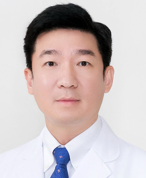 Jihui Hao (郝继辉)合作论文数Tianjin Medical University Cancer Institute & Hospital20
 Song Gao (高嵩)合作论文数Cancer Center, Sun Yat-sen University19
 Xiulan Zhao (赵秀兰)合作论文数天津医科大学总医院19Tiansuo Zhao (赵天锁)合作论文数16Da Yang合作论文数14
 Xishan Hao (郝希山)合作论文数Chinese Academy of Medical Sciences13
 Ping Ji合作论文数12
 Anil K. Sood合作论文数Department of Gynecologic Oncology and Reproductive Medicine, Division of Surgery, The University of Texas MD Anderson Cancer Center;Graduate School of Biomedical Sciences, The University of Texas Health Science Center12Xiuchao Wang合作论文数12Corrine Yingxuan Chua合作论文数11Lynette M Phillips合作论文数11
 Huamin Wang (王华民)合作论文数Department of Pathology, Division of Pathology/Lab Medicine, The University of Texas MD Anderson Cancer Center;MD Anderson UT Health Graduate School of Biomedical Sciences, The University of Texas11
 Ilya Shmulevich合作论文数Institute for Systems Biology11
 Jason B. Fleming合作论文数Department of Surgery, UT Southwestern Medical Center11
 Jilong Yang (杨吉龙)合作论文数Department of Bone and Soft Tissue Oncology, Cancer Institute and Hospital, Tianjin Medical University10David Cogdell合作论文数10Xuebin Zhang合作论文数9Zhang Jingyi (张靖宜)合作论文数9Wenjuan Cai合作论文数9Yanxue Liu (刘岩雪)合作论文数9
 Paul J. Chiao合作论文数Department of Molecular and Cellular Oncology, Division of Basic Science Research, The University of Texas MD Anderson Cancer Center9Liu Yuexin合作论文数8Huang Chongbiao合作论文数8Yuhong Guo合作论文数7Yuexin Liu合作论文数7
 Yang Shengyu合作论文数7
 Jianwei Sun (孙建伟)合作论文数School of Life Sciences, Yunnan University7
 Yumei Feng (冯玉梅)合作论文数Cancer Institute & Hospital, Tianjin Medical University7Matti Nykter合作论文数6Weiyu Bai合作论文数6Xiao-Qing Li合作论文数6Guo Yuhong (郭玉虹)合作论文数6JunLing Shen合作论文数6
 Yichen Yang合作论文数Shanghai Jiao Tong University, Shanghai Jiaotong Univ.6
 Shiwu Zhang (张诗武)合作论文数School of Medicine, Nankai University;Tianjin People's Hospital6
 Fengxia Xue (薛凤霞)合作论文数Internet Hospital Of Tianjin Medical University General Hospital6Yunlong Cui (崔云龙)合作论文数6
 Jonathan C Trent合作论文数Sylvester Comprehensive Cancer Center, University of Miami;Division of Hematology, Department of Medicine, Miller School of Medicine, University of Miami6Chunrong Han合作论文数6
 Muyan Cai (蔡木炎)合作论文数Sun Yat-sen University Cancer Center6Chenglu Lu (卢成陆)合作论文数5Qinggang Hao合作论文数5Lei Sang合作论文数5




