Inflammatory myofibroblastic tumors (IMTs) of the urinary bladder are rare mesenchymal neoplasms with an incompletely defined molecular spectrum. This integrated clinicopathologic and molecular study of 20 bladder IMTs utilized immunohistochemistry (IHC), fluorescence in situ hybridization (FISH), and targeted RNA sequencing to characterize their molecular drivers. Clinically, patients (mean age 45 y) presented with hematuria (85%) and cystoscopic polypoid/nodular masses; despite muscularis propria invovlement (55%) or perivesical soft tissue extension (5%), clinical outcomes were excellent (90% disease-free survival; mean follow-up 32.6 mo), with 2 recurrences managed by repeat resection. Histologically, the tumors exhibited mixed growth patterns: compact spindle cell (75%), myxoid (50%), and hypocellular fibrous (20%), with 45% showing combined features. IHC revealed ALK positivity in 90% (18/20) of cases, predominantly diffuse cytoplasmic staining (17/18), while keratins (AE1/AE3 and/or CAM5.2) were positive in 83.3%. Molecular analysis identified ALK rearrangements in 87.5% (14/16) of FISH-tested cases (signal separation: 13% to 42%) and gene fusions in 88.9% (16/18) of RNA-sequenced cases, with FN1::ALK being the predominant fusion (75%, 12/16). Rare fusions included VCL::ALK, DCTN1::ALK, PPFIBP1::ALK, and a novel TFG::ROS1 (confirmed by RT-PCR and Sanger sequencing). Distinct genotype-phenotype correlations emerged: myxoid morphology strongly associated with FN1::ALK (87.5%, 7/8), while hypocellular fibrous patterns enriched non-FN1 fusions (75%, 3/4). The predominance of FN1::ALK fusions, sharing identical breakpoints (ALK exons 18 to 19) with pseudosarcomatous myofibroblastic proliferations of the urinary bladder, alongside expanded molecular diversity (non-FN1/ROS1 fusions), supports their classification as a biological continuum of ALK-driven bladder mesenchymal neoplasms. These findings broaden the molecular genetic spectrum of bladder IMTs and advocate for histology-guided molecular testing to identify kinase fusions, reinforcing conservative management for these typically indolent tumors.
18F-fluorodeoxyglucose (18F-FDG) is the most widely used radiotracer for positron emission tomography (PET) imaging in clinical oncology, owing to the elevated glycolytic activity of tumor cells. However, transient post-radiotherapy (RT) “metabolic flares” of 18F-FDG uptake are frequently observed in patients and are traditionally attributed to localized inflammatory responses. Whether these flares are linked to immune cell dynamics, particularly tumor-infiltrating T cells, and the mechanisms involved remain poorly understood. Here, we demonstrate that RT markedly upregulates intracellular adhesion molecule-1 (ICAM-1) expression and promotes T cell infiltration in tumors, as observed in both patients and mouse models. Genetic ablation of ICAM-1 significantly attenuates RT-induced metabolic flares in irradiated tumors, primarily due to reduced 18F-FDG uptake by tumor-infiltrating T cells rather than myeloid cells. Mechanistically, ICAM-1 engages with lymphocyte function-associated antigen 1 (LFA-1) to facilitate T cell clustering, thereby promoting their intratumoral accumulation and activating glycolysis and the tricarboxylic acid cycle via the PI3K-AKT-mTOR signaling pathway. These findings identify ICAM-1 as a critical regulator of T cell metabolic reprogramming and tumor infiltration following RT, offering a mechanistic explanation for 18F-FDG PET flares. Clinical monitoring of post-RT tumor ICAM-1 expression may enhance PET interpretation and aid in distinguishing pseudoprogression from true tumor progression.
Metanephric adenoma (MA) is a rare benign renal neoplasm characterized by recurrent BRAF V600E mutations in ∼80% to 90% of cases. The molecular drivers in BRAF V600E‑negative MAs remain poorly understood, although rare kinase fusions have been reported. Here, we present a multi‑institutional series of 7 BRAF V600E‑negative MAs with confirmed kinase fusions. The patients included 5 women and 2 men, with a median age of 43 years (range: 24 to 62 y). All tumors were uncapsulated and exhibited typical MA histology. Notably, leaf‑like structures formed by branching dilated tubules surrounding dense small acini were observed in 4 of 7 cases, and thyroid follicle‑like structures and/or microcystic/reticular patterns were observed in 3 of 7 cases. The majority of cases showed marked paucity of edematous or fibrous stroma. By immunohistochemistry, all cases were diffusely positive for WT1 and CD57, negative for BRAF V600E (clone VE1), and largely negative for CK7 and AMACR. Targeted RNA sequencing identified fusions involving RET (CCDC6::RET in 2 cases, ANKRD26::RET in 1), ALK (STRN::ALK in 2), ROS1 (RDX::ROS1 in 1), and BRAF (CUX1::BRAF in 1). All fusions preserved the kinase domain of the respective genes. Fluorescence in situ hybridization or reverse‑transcriptase PCR confirmed the rearrangements in available cases. In contrast, targeted RNA sequencing of 7 BRAF V600E‑mutant MAs revealed no kinase fusions. All patients underwent partial nephrectomy and remained disease‑free during follow‑up (median: 18 mo, range: 14 to 115 mo). Our findings demonstrate that kinase fusions, including RET, ALK, ROS1, and BRAF, represent alternative drivers in BRAF V600E‑negative MAs, expanding the molecular spectrum of MA. In VE1-negative cases with classic MA histology, targeted RNA sequencing for these fusions may serve as a useful diagnostic adjunct.
Objective:To investigate the clinicopathological and genetic features of acquired cystic kid-ney disease(ACKD)and secondary renal cell carcinoma(RCC)in end-stage renal disease(ESRD)pa-tients undergoing dialysis.Methods:The clinicopathological data of 9 patients with ACKD,of whom,7 had concurrent RCC,of the Department of Pathology,Peking University Third Hospital from 2020 to 2025 were retrospectively analyzed.Immunohistochemistry(IHC)and next-generation sequencing(NGS)were used to detect RCC-related proteins and targeted-drug associated gene variations/microsatel-lite instability(MSI)status.Results:The 9 patients were all male,aged 29-64 years.Causes of ESRD included hypertension,IgA nephropathy,chronic glomerulonephritis,and diabetes.Dialysis dura-tion ranged from 1 to 30 years(median 9.0 years),and 3 patients had kidney transplantation history.Imaging showed reduced kidney size and multiple cysts,including complex cysts.Solid lesions were found in the cyst wall of 5 patients with secondary RCC.The maximum diameter of tumors was 1.3-7.0 cm(median 3.0 cm).Histologically,except for typical morphological changes of ACKD and atypi-cal renal cysts,some cases had papillary adenoma and hemorrhage.Six ACKD-associated RCC(ACKD-RCC)patients and 1 papillary RCC(pRCC)patient were diagnosed.Most ACKD-RCC tumor cells had eosinophilic cytoplasm;5 patients predominantly showed papillary structure,and 1 patient mainly presen-ted sieve-cystic with acinar/solid/micropapillary structures.Oxalate crystals were found in all ACKD-RCC cases,and most patients were accompanied by necrosis and calcification.P504S and CK7 were dif-fusely or focally positive in all the cases,while CAⅨ was negative.The World Health Organization/In-ternational Society of Urological Pathology(WHO/ISUP)nuclear grading of ACKD-RCC and pRCC were 2-3 and 3-4,respectively.Pathological staging of 5 RCC patients was pT1 and of the other 2 patients was pT3a.NGS results identified one PIK3CA point mutation and SETD2 deletion in one ACKD-RCC pa-tient and the pRCC patient,which were accompanied by marked necrosis and pT3a staging.The RCC patients were regularly followed up for 1-32 months postoperatively without additional treatment,and no recurrence or metastasis was observed.Conclusion:ACKD is a common complication in ESRD patients undergoing dialysis.In our cohort,ACKD-RCC was predominantly characterized by papillary architecture histologically with oxalate crystals in all cases.Immunohistochemically,P504S and CK7 were positive.Regardless of secondary occurrent tumor,atypical renal cysts were present in all ACKD cases in this study.NGS detected PIK3CA and SETD2(Tier Ⅱ)variants in 2 RCC patients,respectively,accompa-nied by coagulative necrosis and advanced pathological stage,which may indicate a poor prognosis.This study suggests that imaging screening for RCC should be strengthened in young and middle-aged male ESRD patients with long-term dialysis,and adequate sample examination should be performed to detect potentially precancerous lesions,such as atypical renal cysts.The prognosis of ACKD-RCC requires com-prehensive analysis combining clinicopathological and molecular genetic features.
Synovial sarcoma is a rare malignant mesenchymal neoplasm that typically occurs in soft tissue sites, and the kidney is an uncommon primary location. Previous studies of primary renal synovial sarcoma are limited by small sample sizes, and our understanding remains incomplete. Here, we present the largest multi-institutional case series to date of primary renal synovial sarcoma, with a focus on novel and molecular findings. A total of 70 cases were contributed by multiple institutions. Comprehensive clinical and histopathologic data were collected and analyzed. The mean patient age was 40 years, with a male-to-female ratio of 1.9:1. The most common presenting signs and symptoms were pain and hematuria. The mean tumor size was 11.6 cm (range: 2.3 to 26 cm), with frequent cystic change and necrosis. The mean follow-up was 29 months (range: 2 to 129 mo), and the rates of metastasis, recurrence, and death due to disease were 62.7%, 33.3%, and 50.0%, respectively. Histologically, 56.1% were monophasic synovial sarcoma, 15.1% were biphasic, and 28.8% were poorly differentiated or showed round cell features. Molecularly, SS18::SSX2 fusion was detected in the majority of cases assessed (n=19), followed by SS18::SSX1 fusion (n=6) and a rare SS18::NEDD4 fusion (n=1). Given the morphologic and immunohistochemical overlaps with many other neoplasms, accurate diagnosis with preferably molecular techniques is crucial for appropriate prognostication and treatment of this aggressive tumor.
Metanephric adenoma (MA) is a rare benign renal neoplasm characterized by recurrent BRAF V600E mutations in ∼80% to 90% of cases. The molecular drivers in BRAF V600E‑negative MAs remain poorly understood, although rare kinase fusions have been reported. Here, we present a multi‑institutional series of 7 BRAF V600E‑negative MAs with confirmed kinase fusions. The patients included 5 women and 2 men, with a median age of 43 years (range: 24 to 62 y). All tumors were uncapsulated and exhibited typical MA histology. Notably, leaf‑like structures formed by branching dilated tubules surrounding dense small acini were observed in 4 of 7 cases, and thyroid follicle‑like structures and/or microcystic/reticular patterns were observed in 3 of 7 cases. The majority of cases showed marked paucity of edematous or fibrous stroma. By immunohistochemistry, all cases were diffusely positive for WT1 and CD57, negative for BRAF V600E (clone VE1), and largely negative for CK7 and AMACR. Targeted RNA sequencing identified fusions involving RET ( CCDC6 :: RET in 2 cases, ANKRD26 :: RET in 1), ALK ( STRN :: ALK in 2), ROS1 ( RDX :: ROS1 in 1), and BRAF ( CUX1 :: BRAF in 1). All fusions preserved the kinase domain of the respective genes. Fluorescence in situ hybridization or reverse‑transcriptase PCR confirmed the rearrangements in available cases. In contrast, targeted RNA sequencing of 7 BRAF V600E‑mutant MAs revealed no kinase fusions. All patients underwent partial nephrectomy and remained disease‑free during follow‑up (median: 18 mo, range: 14 to 115 mo). Our findings demonstrate that kinase fusions, including RET , ALK , ROS1 , and BRAF , represent alternative drivers in BRAF V600E‑negative MAs, expanding the molecular spectrum of MA. In VE1-negative cases with classic MA histology, targeted RNA sequencing for these fusions may serve as a useful diagnostic adjunct.
4536 Background: Renal cell carcinoma (RCC) with inferior vena cava tumor thrombus (IVCTT) remains a surgically challenging condition, and evidence supporting neoadjuvant systemic therapy is still evolving. We conducted a phase II study to evaluate the efficacy and safety of apatinib combined with camrelizumab as neoadjuvant treatment in this population. Methods: This single-arm phase II trial enrolled patients with histologically confirmed, resectable RCC with IVCTT (cT3b-T4, N0-1, M0-1; ECOG performance status 0-1). Patients received camrelizumab 200 mg i.v. every 2 weeks for six cycles plus oral apatinib 250 mg once daily, followed by surgical resection when feasible. The primary endpoint was downgrading of Mayo level of tumor thrombus (TT). Secondary endpoints included TT response (RECIST 1.1), response of primary renal tumor, safety (CTCAE v5.0), progression-free survival, and overall survival. Results: Nine patients were included in the intention-to-treat analysis. TT response was observed in 4 patients (44.4%), with disease control achieved in 8 patients (88.9%); one patient (11.1%) experienced progression. Mayo level downgrading occurred in 3 patients (33.3%), all of whom subsequently underwent surgery. Overall, 7 of 9 patients (77.8%) successfully completed neoadjuvant therapy and underwent robotic-assisted radical nephrectomy with thrombectomy. For primary renal tumor, partial response was observed in 1 patient (11.1%), and stable disease in 8 patients (88.9%), resulting in a disease control rate of 100%. Treatment-related adverse events were manageable and consistent with known safety profiles, with grade ≥3 events including hypertension, proteinuria, pneumonitis, and immune-related myasthenia gravis. At a median follow-up of 45 weeks, survival outcomes remain immature. Conclusions: Neoadjuvant apatinib plus camrelizumab demonstrated promising antitumor activity and acceptable safety in patients with RCC and IVCTT, achieving meaningful TT response and Mayo level downgrading, thereby facilitating surgical resection. These findings support further investigation of neoadjuvant combination therapy in this high-risk population. Clinical trial information: ChiCTR2300069990. Patient baseline characteristics. Characteristic N=9 Median age, years, (IQR) 58 (54-66) Sex, n (%) Male 6 (67) Female 3 (33) ECOG PS, n (%) 0 7 (78) 1 2 (22) Clinical T stage, n (%) cT3b 2 (22) cT3c 3 (33) cT4 4 (44) Clinical N stage, n (%) cN0 3 (33) cN1 6 (67) Clinical M stage, n (%) cM0 3 (33) cM1 6 (67) Primary tumor size, cm (IQR) 12.4 (7.3-17.3) Length of the tumor thrombus, cm (IQR) 11.3 (7.2-13.8) Histological subtype on baseline biopsy, n (%) Clear cell RCC 8 (89) TFE3 translocation RCC 1 (11) Mayo level of TT at baseline I 0 II 4 (44) III 2 (22) IV 3 (33) IQR interquartile range, ECOG Eastern Cooperative Oncology Group, PS performance status, RCC renal cell carcinoma.
OBJECTIVE:To characterize the clinicopathological features of long-term survivors among patients with clear cell renal cell carcinoma (ccRCC) and venous tumor thrombus (VTT) who underwent surgical treatment, and to explore potential prognostic factors beyond tumor-related variables, with particular emphasis on host-related inflammatory and nutritional indicators. METHODS:The patients with ccRCC and VTT who underwent radical nephrectomy combined with tumor thrombectomy at Peking University Third Hospital between 2014 and 2019 were retrospectively analyzed. The patients were stratified into a long-term survival group (overall survival ≥60 months) and a non-long-term survival group (overall survival < 60 months). The following parameters were compared between the two groups: clinical characteristics (age, sex, body mass index, Mayo classification), pathological features (T stage, M stage, histological grade, sarcomatoid change, perirenal fat invasion), perioperative parameters (surgical approach, operative time, intraoperative blood loss, complications), and laboratory parameters (platelet count, albumin, neutrophil count, etc.). Optimal cutoff values for continuous variables, such as age, body mass index (BMI), platelet count and serum albumin level, were determined using X-tile software based on the minimum P-value principle. Subsequently, Kaplan-Meier survival analysis with Log-rank test was performed within the long-term survival cohort to evaluate the association of the above-mentioned factors with long-term survival. RESULTS:Long-term survival was achieved in 13.9% of 397 patients with ccRCC and VTT. Significant differences were observed between the long-term survival group (n=55) and the non-long-term survival group (n=342) in baseline platelet count (P=0.040), pathological T stage (P=0.001), histological grade (P=0.006), surgical approach (P < 0.001), as well as receipt of systemic therapy (P=0.028), treatment timing (P=0.003), and treatment regimen (P < 0.001). Regarding T stage, the long-term survival group had a significantly higher proportion of pT3a tumors (63.6% vs. 42.4%) and a significantly lower proportion of pT4 tumors (10.9% vs. 33.0%) compared with the non-long-term survival group. The long-term survival group also had a higher proportion of low-grade tumors (G1/G2: 50.9% vs. 31.9%). Regarding surgical approach, laparoscopic surgery predominated in the long-term survival group (35/55, 63.6%), whereas the non-long-term survival group had a more balanced distribution, including open surgery (102/342, 29.8%), laparoscopic surgery (123/342, 36.0%), and robotic surgery (117/342, 34.2%). Regarding systemic therapy, in the long-term survival group, 26 patients (47.3%) received systemic therapy, all of whom (100.0%) received adjuvant therapy. In the non-long-term survival group, 215 patients (62.9%) received systemic therapy, of whom 160 (74.4%) received adjuvant therapy and 55 (25.6%) received neoadjuvant therapy. Regarding treatment regimen, the long-term survival group was predominantly treated with targeted monotherapy (25/26, 96.2%), whereas the non-long-term survival group had a higher proportion of targeted therapy plus immunotherapy combination (64/215, 29.8%). The median follow-up time for the long-term survival group was 81.7 months. Further subgroup analysis of this group showed that age < 70 years (P=0.038), BMI≥18.5 kg/m2 (P=0.032), platelet count 159×109/L-273×109/L (P=0.003), and albumin level >41.4 g/L (P=0.028) were significantly associated with better long-term survival, whereas Mayo classification of tumor thrombus showed no significant association with long-term survival (P=0.250). CONCLUSION:A subset of patients with ccRCC and VTT can achieve long-term survival. Beyond pathological tumor stage and grade, patient-related inflammatory and nutritional status may be associated with long-term survival. Exploring prognostic factors related to long-term survival may help further optimize risk stratification and treatment strategies for high-risk patients.
Inflammatory myofibroblastic tumors (IMTs) of the urinary bladder are rare mesenchymal neoplasms with an incompletely defined molecular spectrum. This integrated clinicopathologic and molecular study of 20 bladder IMTs utilized immunohistochemistry (IHC), fluorescence in situ hybridization (FISH), and targeted RNA sequencing to characterize their molecular drivers. Clinically, patients (mean age 45 y) presented with hematuria (85%) and cystoscopic polypoid/nodular masses; despite muscularis propria invovlement (55%) or perivesical soft tissue extension (5%), clinical outcomes were excellent (90% disease-free survival; mean follow-up 32.6 mo), with 2 recurrences managed by repeat resection. Histologically, the tumors exhibited mixed growth patterns: compact spindle cell (75%), myxoid (50%), and hypocellular fibrous (20%), with 45% showing combined features. IHC revealed ALK positivity in 90% (18/20) of cases, predominantly diffuse cytoplasmic staining (17/18), while keratins (AE1/AE3 and/or CAM5.2) were positive in 83.3%. Molecular analysis identified ALK rearrangements in 87.5% (14/16) of FISH-tested cases (signal separation: 13% to 42%) and gene fusions in 88.9% (16/18) of RNA-sequenced cases, with FN1::ALK being the predominant fusion (75%, 12/16). Rare fusions included VCL::ALK , DCTN1::ALK , PPFIBP1::ALK , and a novel TFG::ROS1 (confirmed by RT-PCR and Sanger sequencing). Distinct genotype-phenotype correlations emerged: myxoid morphology strongly associated with FN1::ALK (87.5%, 7/8), while hypocellular fibrous patterns enriched non- FN1 fusions (75%, 3/4). The predominance of FN1::ALK fusions, sharing identical breakpoints ( ALK exons 18 to 19) with pseudosarcomatous myofibroblastic proliferations of the urinary bladder, alongside expanded molecular diversity (non- FN1/ROS1 fusions), supports their classification as a biological continuum of ALK -driven bladder mesenchymal neoplasms. These findings broaden the molecular genetic spectrum of bladder IMTs and advocate for histology-guided molecular testing to identify kinase fusions, reinforcing conservative management for these typically indolent tumors.
To retrospectively analyze the clinical and imaging characteristics of classic angiomyolipoma (CAML) with venous tumor thrombus (VTT) and compare them with those of clear cell renal cell carcinoma (ccRCC). Clinical data from six patients with renal CAML complicated by VTT and 18 with ccRCC complicated by VTT, treated at Peking University Third Hospital from April 2018 and June 2022, were retrospectively analyzed. All patients underwent preoperative ultrasound and contrast-enhanced CT. Clinical manifestations, imaging characteristics, surgical findings, and pathological data were collected, and patients were followed up. Enhanced CT showed renal sinus involvement in all CAML cases versus four ccRCC cases (p = 0.002). All primary CAML tumors had fatty components, compared to one ccRCC case (p < 0.001). Enhanced CT also revealed 7 VTTs with fatty components (6 in the CAML group) (p < 0.001). Thrombus lengths in the inferior vena cava (IVC) were 8.05 ± 2.22 cm for CAML and 5.29 ± 2.38 cm for ccRCC, with no significant difference (p = 0.610). The maximum/minimum anteroposterior VTT diameter ratios were 3.98 and 1.09, respectively (p < 0.001); coronal diameter ratios were 4.00 and 1.12, respectively (p < 0.001). Ultrasound revealed that, except for one Mayo Level 0 case, the involved IVC in the CAML group had continuous, intact walls with blood flow signals in the residual lumen, while in the ccRCC group, most VTTs had unclear boundaries and only one case showed blood flow signals in the residual lumen (p = 0.001). Intraoperative blood loss was significantly lower in CAML cases (p = 0.017). No CAML patient had VTT invading the venous wall, unlike 8 ccRCC patients (p = 0.016). All patients were followed for 21–74 months (median: 34.5 months, mean: 36.6 months). All were alive with normal renal function, and no tumor recurrence or metastasis was observed. Renal CAML with VTT is characterized by three imaging features: the presence of fatty components, a unique geometric growth pattern, and the absence of venous wall invasion, potentially serving as valuable indicators for differentiating CAML from ccRCC lesions.
Primary Ewing sarcoma (ES) of the kidney is rare. We describe the clinicopathologic features of primary renal ES with emphasis on gene fusion partners. A multi-institutional study was conducted to obtain clinicopathologic data on primary ES of the kidney. All tumors with available tissue underwent NGS to determine fusion partners. Twenty-four patients (8 male, 16 female) were identified. Mean age was 33.2 (±12.3). Mean tumor size was 10.5 cm (±4.2). Clinical presentation was available in 21 patients: flank/abdominal pain (13, 61.9%), hematuria (4, 19%), mass (2, 9.5%), hypertension (1, 4.8%), and incidental (1, 4.8%). For 23 nephrectomies, 2 (8.7%) were ypT0 (post-neoadjuvant therapy), 3 (13%) pT1, 15 (65.2%) pT2, 1 (4.4%) pT3, and 2 (8.7%) pT4. Four (16.7%) had metastatic disease at presentation. Of 18 patients with available follow-up, 9 (50%) were alive with disease, 7 (38.9%) alive with no disease, and 2 (11.1%) died of disease (mean follow-up 34 mo). Metastatic disease was documented in 9/18 patients, including lung (3), adrenal (2), bone (2), retroperitoneum (2), liver (2), lymph node (1), and ureter (1). FISH was performed in 14 tumors and real-time quantitative PCR in 1, confirming EWSR1 rearrangements. NGS was performed in 17 tumors, showing EWSR1::FLI1 in 16 (94.1%) and EWSR1::ETV4 in 1. Primary renal ES is a rare neoplasm occurring in a wide age range. Most tumors invaded adjacent tissues. Although they share similar histologic and molecular features with their counterpart in the bone/soft tissue, we document the first case of a rare EWSR1::ETV4 fusion in the kidney.
Primary Ewing sarcoma (ES) of the kidney is rare. We describe the clinicopathologic features of primary renal ES with emphasis on gene fusion partners. A multi-institutional study was conducted to obtain clinicopathologic data on primary ES of the kidney. All tumors with available tissue underwent NGS to determine fusion partners. Twenty-four patients (8 male, 16 female) were identified. Mean age was 33.2 (±12.3). Mean tumor size was 10.5 cm (±4.2). Clinical presentation was available in 21 patients: flank/abdominal pain (13, 61.9%), hematuria (4, 19%), mass (2, 9.5%), hypertension (1, 4.8%), and incidental (1, 4.8%). For 23 nephrectomies, 2 (8.7%) were ypT0 (post-neoadjuvant therapy), 3 (13%) pT1, 15 (65.2%) pT2, 1 (4.4%) pT3, and 2 (8.7%) pT4. Four (16.7%) had metastatic disease at presentation. Of 18 patients with available follow-up, 9 (50%) were alive with disease, 7 (38.9%) alive with no disease, and 2 (11.1%) died of disease (mean follow-up 34 mo). Metastatic disease was documented in 9/18 patients, including lung (3), adrenal (2), bone (2), retroperitoneum (2), liver (2), lymph node (1), and ureter (1). FISH was performed in 14 tumors and real-time quantitative PCR in 1, confirming EWSR1 rearrangements. NGS was performed in 17 tumors, showing EWSR1::FLI1 in 16 (94.1%) and EWSR1::ETV4 in 1. Primary renal ES is a rare neoplasm occurring in a wide age range. Most tumors invaded adjacent tissues. Although they share similar histologic and molecular features with their counterpart in the bone/soft tissue, we document the first case of a rare EWSR1::ETV4 fusion in the kidney.
We present the case of a 59-year-old Chinese man diagnosed with stage III clear cell renal cell carcinoma who developed 2 suspicious lung lesions 5 years after follow-up. Pathological evaluation revealed 2 distinct types of cancer: lung adenocarcinoma in situ and clear cell renal carcinoma with lung metastasis. Lung tissue samples were sequenced using a panel of 1267 cancer-related genes. The analysis revealed completely different molecular profiles between the 2 lung lesions and similar clonal mutations in the superior lingular lobe and kidney. This indicates multiple metachronous primary tumors.
Tandem repeats (TRs) are genomic regions that tandemly change in repeat number, which are often multiallelic. Their characteristics and contributions to gene expression and quantitative traits in rice are largely unknown. Here, we survey rice TR variations based on 231 genome assemblies and the rice pan-genome graph. We identify 227,391 multiallelic TR loci, including 54,416 TR variations that are absent from the Nipponbare reference genome. Only 1/3 TR variations show strong linkage with nearby bi-allelic variants (SNPs, Indels and PAVs). Using 193 panicle and 202 leaf transcriptomic data, we reveal 485 and 511 TRs act as QTLs independently of other bi-allelic variations to nearby gene expression, respectively. Using plant height and grain width as examples, we identify and validate TRs contributions to rice agronomic trait variations. These findings would enhance our understanding of the functions of multiallelic variants and facilitate rice molecular breeding. Tandem repeats (TRs) have unique ability to drive a range of phenotype variations. Here, the authors survey rice TR variations based on 231 genome assemblies and the rice pan-genome graph, identify TR variations associated with expressed genes, and reveal expression TRs contributed to rice agronomic trait variations.
Background: Warthin-like Mucoepidermoid carcinoma (MEC) is a new and rare morphological variant of MEC, with only a few case reports in the literature. The clinicopathological, molecular features and bio-behaviors of Warthin-like MEC has not been studied extensively. We reappraisal all Warthin-like MEC patients diagnosed and treated at our hospital. Methods: Patient characteristics including clinicopathological features, genetic aberrations, treatment, and prognostic information were assessed and evaluated. Results: Twenty-nine Warthin-like MEC patients were identified, 19 patients were female (65.5 %), and 10 were male (34.5 %). The patients’ age varied widely from 8 to 68 years (mean 42.3 years). Genetic aberrations of MAML2 rearrangement were detected in all Warthin-like MEC patients, which suggesting this genetic event is the unique feature of Warthin-like MEC. Twenty-five patients (86.2 %) were assessed as having a low-stage disease (I/II), and four (13.8 %) as having high-clinical stage disease (III/IV). More than half of the patients (16/29) underwent only partial sialoadenectomy; 2 patients underwent extended sialoadenectomy, and 11 patients underwent extended sialoadenectomy with cervical lymph node dissection. After a median follow-up time of 73 months (5–128 months), Twenty-eight patients were alive without recurrence at the end of the follow-up period, one patient died 1 year after surgery due to lung metastasis. Conclusion: Our data suggested that most Warthin-like MEC exhibited mild clinicopathological course and less aggressive bio-behavior, and an aggressive bio-behavior seemed to be very rare. In addition, in the salivary gland, MAML2 rearrangement seems to be a unique molecular feature of salivary Warthin-like MEC.
ObjectiveGATA binding protein 3 (GATA3) and forkhead box A1 (FOXA1) have been individually implicated in the progression of upper tract urothelial carcinoma (UTUC). This study aims to evaluate the prognostic value of GATA3/FOXA1 co-expression in UTUC patients.MethodsWe collected 108 UTUC pathological tissue samples with complete follow-up data and 24 normal control urothelial tissues. We created a 132-site microarray and performed immunohistochemistry (IHC) to measure GATA3 and FOXA1 expression levels. Kaplan-Meier survival and Cox regression analyses were conducted to assess UTUC prognosis.ResultsGATA3 expression was positively correlated with FOXA1 (P=0.031). Absence of GATA3/FOXA1 co-expression (GATA3-/FOXA1-) was associated with tumor extensive necrosis (P=0.001) after Bonferroni correction for multiple comparisons. GATA3-/FOXA1- was associated with shorter Disease-Free Survival (DFS) (P=0.001) and Cancer-Specific Survival (CSS) (P<0.001) than other combination groups. Multivariate analyses identified extensive necrosis as an independent prognostic factor for CSS (P=0.030).ConclusionsOur study revealed a positive correlation between GATA3 and FOXA1 expression in UTUC. GATA3-/FOXA1- is linked to tumor extensive necrosis and poor prognosis in UTUC and may serve as a potential biomarker for UTUC patients.
 Ming Zhou合作论文数Department of Anatomic Pathology, Cleveland Clinic17Cristina Magi-Galluzzi合作论文数15
 Qiu Rao (饶秋)合作论文数Medical School of Nanjing University10
 Weigang Fang (方伟岗)合作论文数School of Basic Medical Sciences, Peking University;Peking University Clinical Research Institute10
 Hongquan Zhang (张宏权)合作论文数School of Basic Medical Sciences, Peking University9
 Reza Alaghehbandan合作论文数Department of Pathology and Laboratory Medicine, Faculty of Medicine, The University of British Columbia8
 Lulin Ma (马潞林)合作论文数Peking University Third Hospital8
 Sean Williamson合作论文数Department of Pathology, Cleveland Clinic8QiuYuan Xia (夏秋媛)合作论文数7
 Jun Zhan (战军)合作论文数School of Basic Medical Sciences, Peking University Health Science Center7
 Maria Tretiakova合作论文数Department of Pathology, University of Chicago7
 Jie Zheng (郑杰)合作论文数School of Basic Medical Sciences, Peking University Health Science Center7Guo-Qing Ru合作论文数6
 Longwen Chen合作论文数6
 Przybycin Christopher合作论文数6
 Shudong Zhang (张树栋)合作论文数Department of Urology, Peking University Third Hospital6Jesse K. Mckenney合作论文数6Ondrej Hes合作论文数6Eva Comperat合作论文数5WanJie Heng (衡万杰)合作论文数5
 Xiaojun Tian (田晓军)合作论文数Department of Urology, Peking University Third Hospital5
 Sara M Falzarano合作论文数5
 Anthony Gill合作论文数Department of Anatomical Pathology, Royal North Shore Hospital;Faculty of Medicine and Health, University of Sydney5
 Kiril Trpkov合作论文数Department of Pathology and Laboratory Medicine, Cuming School of Medicine, University of Calgary;Southern Alberta Institute of Urology;Tom Baker Cancer Center5
 Liqun Zhou (周利群)合作论文数Peking University First Hospital5Dengfeng Cao合作论文数School of Medicine, Washington University5TANG Shi-ying (唐世英)合作论文数4XiKang Wu (吴西抗)合作论文数4José I López合作论文数4
 Binbin Li (李彬彬)合作论文数Environmental Research Center, Duke Kunshan University;Nicholas School of the Environment, Duke University4Xiao-Tong Wang合作论文数4Delia Perez-Montiel合作论文数4XiangLei He (何向蕾)合作论文数4XiaoDong Teng (滕晓东)合作论文数4Carver Paula合作论文数4
 Liang Cheng合作论文数Department of Surgery, Warren Alpert Medical School of Brown University;Department of Pathology and Laboratory Medicine, Warren Alpert Medical School of Brown University4Rajal B. Shah合作论文数4
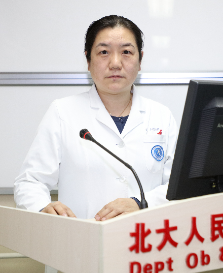 Yue Wang (王悦)合作论文数Peking University People's Hospital4
 Hailan Feng (冯海兰)合作论文数Peking University School and Hospital of Stomatology4
 Steven C. Smith合作论文数Pacific Northwest National Laboratory4
 Xiaodong Teng合作论文数4Fumiyoshi Kojima合作论文数3Mingxin Xu合作论文数3Zhentao Lei合作论文数3Yanting Zhou合作论文数3Xuelian Pei合作论文数3Yan Li (李燕)合作论文数3Xiaona Yin合作论文数3




