Ten new palladium(II) complexes [PdCl(L1−10)]Cl have been synthesized by the reaction of palladium(II) chloride and ten 4′-(substituted-phenyl)-2,2′:6′,2′′-terpyridine ligands bearing hydrogen(L1), p-hydroxyl(L2), m-hydroxyl (L3), o-hydroxyl (L4), methyl (L5), phenyl (L6), fluoro (L7), chloro (L8), bromo (L9), or iodo (L10). Their structures were confirmed by FT-IR, 1H NMR, elemental analysis and/or single crystal X-ray diffraction analysis. Their in vitro anticancer activities were investigated based on five cell lines, including four cancer cell lines (A549, Eca-109, Bel-7402, MCF-7) and one normal cell line (HL-7702). The results show that these complexes possess a strong killing effect on the cancer cells but a weak proliferative inhibition on the normal cells, implying their high inhibitory selectivity for the proliferation of the cancer cell lines. Flow cytometry characterization reveals that these complexes affect cell proliferation mainly in the G0/G1 phase and induce the late apoptotic of the cells. The quantity of palladium(II) ion in extracted DNA was determined by ICP-MS, which proved that these complexes target genomic DNA. And the strong affinity of the complexes with CT-DNA were confirmed by UV–Vis spectrum and circular dichroism (CD). The possible binding modes of the complexes with DNA were further explored by molecular docking. As the concentration of complexes 1–10 gradually increases, the fluorescence intensity of bovine serum albumin (BSA) decreases by a static quenching mechanism.
An entry from the Cambridge Structural Database, the world’s repository for small molecule crystal structures. The entry contains experimental data from a crystal diffraction study. The deposited dataset for this entry is freely available from the CCDC and typically includes 3D coordinates, cell parameters, space group, experimental conditions and quality measures.
An entry from the Cambridge Structural Database, the world’s repository for small molecule crystal structures. The entry contains experimental data from a crystal diffraction study. The deposited dataset for this entry is freely available from the CCDC and typically includes 3D coordinates, cell parameters, space group, experimental conditions and quality measures.
Six new zinc(II) complexes were prepared by the reaction of ZnBr2 or ZnI2 with 4′-(substituted-phenyl)-2,2′:6′,2′′-terpyridine compounds, bearing p-methylsulfonyl (L1), p-methoxy (L2) and p-methyl (L3), which were characterized by elemental analysis, FT-IR, NMR and single crystal X-ray diffraction. The antiproliferative properties against Eca-109, A549 and Bel-7402 cell lines and the cytotoxicity test on RAW-264.7 of these compounds were monitored using a CCK-8 assay, and the studies indicate that the complexes show higher antiproliferative activities than cisplatin. The interactions of these complexes with CT-DNA and proteins (BSA) were studied by UV-Vis, circular dichroism (CD) and fluorescent spectroscopy, respectively. The results indicate that the interaction of these zinc(II) complexes with CT-DNA is achieved through intercalative binding, and their strong binding affinity to BSA is fulfilled through a static quenching mechanism. The simulation of the complexes with the CT-DNA fragment and BSA was studied by using molecular docking software. It further validates that the complexes interact with DNA through intercalative binding mode and that they have a strong interaction with BSA.
本研究采用超高速离心法从人胚胎干细胞培养液上清分离提纯出外泌体,应用透射电镜、免疫印记和RT-PCR方法检测其生物学特性.结果 表明,在透射电镜下观察到人胚胎干细胞外泌体呈茶盏状,大小不均匀,直径30~100 nm,有完整的膜结构,内含低密度物质;人胚胎干细胞外泌体表达外泌体标志抗原CD63、TSG101和多能性转录因子OCT4、SOX2、NANOG、KLF4;干细胞外泌体呈剂量性依赖方式促进小鼠胚胎成纤维细胞增殖.本研究为进一步研究人胚胎干细胞外泌体的生物学特征和功能提供有价值的生物学信息.
为了研究蛋白质磷酸化修饰状况在体细胞被诱导为多能性干细胞过程中发挥的作用,试验采用TiO2富集磷酸化肽段技术和高通量质谱技术,对第三代牛胎儿成纤维细胞的磷酸化修饰蛋白质组进行定性检测和生物信息学分析.结果表明:在牛胎儿成纤维细胞样品中共鉴定出363种磷酸化蛋白,共502个磷酸化位点.这些磷酸化蛋白参与多种细胞信号传导通路,在代谢进程和细胞定位等生物学进程中发挥重要作用.说明对牛胎儿成纤维细胞磷酸化修饰蛋白质组的定性研究可为进一步探讨牛体细胞重编程为诱导性多能干细胞的机理和提高重编程效率奠定理论基础.
本文简述了研究生助教工作的开展,研究生助教在动物生物化学实验中的积极作用及面临的困难,积极探索研究生助教在提高本科实验教学质量中的作用。
Hemagglutinin(HA) is an important surface antigen protein of avian influenza virus(AIV),and plays an important role in diagnosis,prevention and treatment of AIV.The purpose of this study is to develop an easier and more efficient way to express HA recombinant protein.The open reading frame(ORF) of H5N1 AIV HA gene was analyzed using bioinformatics software.Its codon bias to Escherichia coli,rare codon's contributions in the sequence and the antigenicity of HA protein were used to construct a recombinant expression plasmids of HA epitopes,pET-32a(+)-HA.When these recombinant plasmids were induced overnight with 1 mmol/L IPTG solution,they could be highly expressed in E.coli Rosetta-gami B(DE3),and the desired recombinant proteins of 48.1 kD could be got.After purified by 6×His-tagged protein purification kit,the recombinant proteins were mixed with equal amount of Freund's Adjuvant Complete/Incomplete to make the antigen.Then every seven days this antigen was hypodermically injected into two-months-old SPF chickens with a dose of 200 μg per chicken,totally three times.Serums were collected from chickens after last injection.Western-Blot results showed that the recombinant protein could hybridize with both the serum from the highly-immunized chicken and the serum from H5N1 AIV positive chicken.Specific bands were present on the nitrocellulose membrane.This indicates that the recombinant protein expressed by HA antigen gene segment has great immunogenicity and antigenicity and keeps the antigen activity of HA protein.This study suggests that the recombinant protein has practical value in the diagnosis,prevention and treatment of H5N1 AIV.
Inhibin α-subunit gene could feedback suppress synthesis and secretion of FSH, regulate animal reproduc tion trait at its species level. To investigate feasibility of increasing buffalo reproduction trait by silencing inhibin gene, buffalo inhibin-α gene RNAi vectors were constructed and screened in this study. According to CI)S sequence of buffalo INH-α gene cloned in our lab, 5 pair of siRNA oligonucleotides were designed, synthsized, annealed and di redtly cloned into pSilencer4.1-CMV neo expression vector. The constructed vectors were transfected to in vitro culture buffalo granulose cells, relative transcription level of INH-α gene mRNA were determined by Real time PCR 48 h late. Then the high efficiency knock down vector was chosen to detect the variation of inhibition ratios at 24,48,72 and 96 h. The results showed that:5 buffalo inhibin a-subunit RNAi vectors were constructed including pSileneer4.1 145 ,pSilencer4.1-308 ,pSilencer4.1-769, pSilencer4.1-1013 and pSilencer4. 1-1053, which were conformed correctly by restriction and sequencing analysis. The relative transcription levels of inhibin-α mRNA in buffalo granulose cells transfected with pSilencer4.1 308,pSilencer4.1-769,pSilencer4.1-1013 and pSilencer4.1 1053 decreased. The inhibi- tion ratios were 58.8% ,44.9% ,67.4% and 43.9% ,respectively. Then pSilencer4.1-1013 was transfected and qRT PCR were performed at 2448,72 and 96 h, inhibition ratios were 27.1% ,61.0% ,57.0% and 41.2% ,respectively. In conclusion, effective RNAi vectors for buffalo inhibin-α gene have been successfully constructed, which is of great important in producing INH-α silencing transgenic buffalo.
Total RNA was extracted in germinal vesicle(GV)oocytes and 8-cell embrgos of pig and catte by using QIAGEN RNeasy Micro lxit,RNAPrep Micro Kit and Huashun rapid.RNA extracting reagent box(trace).Through comparasion and analysis of the putity and denxity of total RNA,the better way of extracting was found.The results indicated that trace amount of total RNA with high quality can be extracted from small number of oocytes and embryos with QIAGEN RNeasy Micro Kit.
In this study,mRNA isolated from embryos was subjected to reverse transcription polymerase chain reaction to investigate the expression of eNOS,iNOS mRNA in normal pathenogenetic activation porcine embryos.The results showed that eNOS was expressed only in two-cell and four-cell stage embryos,while iNOS was expressed around early embryo development stages.These studies determined that inducible NOS(iNOS) and endothelial NOS(eNOS) were differentially distributed in cells along the development,higher level of eNOS was detected at four-cell stage embryo but iNOS at morula/blastocyst stage.The study provides the first evidence for the presence of mRNA of NOS isoforms during porcine embryogenesis.Furthermore,the expression of NOS during the embryo development suggest that NO may be crucial importance for reproductive events that occur in this period.
The objective of this study was to investigate the effect of addition of Ghrelin to in vitro culture medium on preimplantation development of buffalo in vitro fertilized and parthenogenetic embryos.The final concentration of Ghrelin,0,0.5,5,50,500 μg/L,respectively,were added in the culture medium during the early embryo development stage.The results showed that,no prominent different was found between the Ghrelin treated group and the control in parthenogenetic embryos or in vitro fertilized embryos.In in vitro fertilized group,the blastocyst rate was remarkably(22.17% vs 6.67%,P0.05) increased when 500 μg/L of Ghrelin was added in vitro culture medium compared with the control,while there was no significant difference among the other Ghrelin treated group.In parthenogenetic embryos,the blastocyst rate was significantly(32% vs 14.47%,P0.05)enhanced when 50 μg/L of Ghrelin was added to culture medium compared with the control.The results suggest that Ghrelin can enhance blastocyst formation of buffalo in vitro fertilized and parthenogenetic embryos while has no effect on the early cleavage.
Effects of porcine sperm cytosolic factor(different extraction methods,different albumen concentrations and different micro-injection doses) on parthenogenetic activation(PA) of porcine oocytes were investigated in 3 experiments.The results showed that there was a significant activation effect of oocytes after injection of cytosolic sperm factor extracted either by repeated frozen-thawed or ultrasonic fragmentation method.The optimal micro-injection dose for PA was 2-4 pL per oocyte.Porcine oocytes could be effectively activated by micro-injection of 2-4 pL porcine sperm extract,in which albumen concentration was above 1.35 mg/mL.
The Inhibin alpha subunit gene was amplified by RT-PCR from total RNA of swamp buffalo ovary and cloned into pMD 18-T vector.The recombinant vector pMD 18-T-INHA was identified by PCR,restriction endonuclease analysis and sequenced.The INHA CDS was amplified and cloned into pET-30a(+) expression vector.The recombinant pET-30a-INHA expression plasmid was confirmed to be constructed successfully by restriction endonuclease analysis and nucleotide sequenced.Then the recombinant plasmid was transformed into BL21(DE3) and induced expression by IPTG.The result show the buffalo INHA CDS contain 1 083 bp,encoded 360 aa,and exhibited a high homology with bovine(96%),human(80%),pig(87%).This result indicated the INHA gene was relatively conservative in the progress of evolution.The recombinant protein INHA was expressed successfully when induced by 1 mmol/L IPTG at 37 ℃ for 4 hours.Most of the expression products were inclusion body with the molecular weigh of about 40 000 identified by SDS-PAGE.
A pair of primers was designed and synthesized according to the gene sequence of cow Ghrelin.RT-PCR method was used to amplify Ghrelin gene from the whole RNA of abomasum tissue in swamp buffalo.Sequence analysis revealed that the gene was 351 bp in length,encoding 116 amino acids and exhibited a high homology with those from cow(97%),sheep(95%),pig(81%),mouse(75%) and human(73%).The result indicated that Ghrelin gene is highly conservative in evolution.This study lays a foundation for further analysis of structure,expression and regulation of Ghrelin gene in swamp buffalo.
In order to enrich the donor variety of the somatic cell nuclear transfer,methods for isolation and culture of porcine testis leydig cell were investigated in this study.Porcine testis leydig cells isolated by enzymatic digestion and steel net filtration were round and large.It began to attach clumps after 4~6 h of culture,proliferated to form monolayers at 48 h of culture,and confluented on 3~5 d of culture.These results will provide a basis for isolation and in vitro culture of porcine testis eydig cells.
The objective of this study was to develop a culture system which could support buffalo spermatogonia differentiation into spermatids in vitro. Testes from 3- to 5-month-old buffaloes were decapsulated and seminiferous tubules were enzymatically dissociated to recover spermatogonia and sertoli cells. The cells were cultured in modified Dulbecco modified Eagle medium supplemented with different concentrations of foetal bovine serum, retinol, testosterone for 2 months at 37 degrees C. Spermatogonia and sertoli cells were identified with an antibody against c-kit or GATA4, respectively. The viability of spermatogonia in the media supplemented with different concentrations of serum was all significantly higher (p < 0.05) compared with that in the medium without serum. A-paired or A-aligned spermatogonia and spermatogonial colonies (AP-positive) were observed after 7-10 days of culture and spermatid-like cells with a flagellum (6-8 microm) appeared after 30 days of culture. For cultured conditions, retinol could not significantly promote the formation of spermatid-like cells (p > 0.05), whereas supplementation of testosterone could significantly promote (p < 0.05) the formation of spermatid-like cells after 41 days of culture. The expression of the spermatid-specific marker gene (PRM2) was identified after 30 days of culture by RT-PCR. Yet, the transition protein 1 (TP1, a haploid makers) was not detected. Meanwhile, spermatids developed in vitro were also confirmed by Raman spectroscopy. These results suggest that buffalo spermatogonia could differentiate into spermatids in vitro based on the analysis of their morphology, PRM2 expression and Raman spectroscopy. Yet, the normality of the spermatid-like cells was not supported by TP1 expression.
The objective of this study was to examine the telomerase activity in swamp buffalo oocytes and early embryos derived from in vitro fertilization(IVF).The telomerase activity of immature and matured oocytes,IVF embryos at the 2-4,8-16 cell,morula and blastocyst stages was assayed using a telomere repeat amplification protocol(TRAP).The telomerase activity was higher in the immature oocytes than that in the matured oocytes(P 0.05).After in vitro fertilization,the telomerase activity decreased as the zygotes cleaved to the 2-4 cell and 8-16 cell stages,increased again when the embryos developed to the morula stage(P 0.05),and reached the highest level at the blastocyst stage.When the telomerase activity was calculated on the individual cell,telomerase activity declined gradually as the immature oocytes matured to Metaphase II and developed to the blastocyst stage after in vitro fertilization.These results indicate that the changes of telomerase activity in the buffalo oocytes and early embryos are related to their maturation,embryonic developmental block and totipotency decline during the embryogenesis.
The transfection efficiency of electroporation and lipofectin mediated methods was compared. The efficiency of electroporation was line up with the electrical field intensity in which 20%~50% of cells died when the field intensity was set at 40 to 50 V/mm. Employment of 8 μL lipofectin and 2 μg DNA resulted in more cells transfected. Quality of DNA and state of cell were associated with transfection efficiency of lipofectin. At the same culture condition, lipofectin reagent method can obtain more transgenic colonies.
To set up a suitable culture system for preparing porcine somatic cells,dissociation,culture,purification and subculturing of porcine cumulus cells and fetal fibroblasts were carried out.The formation of porcine cumulus cell monolayers was in 5-6d.The porcine fetal fibroblasts were prepared by the tissue explant culture and enzymatic digestive culture methods,and their monolayers formation was needed 10 to 11d and 8 to 9d respectively.The growth characters of the above 2 cell lines were stable.The methods could provide rich material and resources of the donor cells for somatic cell nuclear transfer in vitro.
 Deshun Shi (石德顺)合作论文数Guangxi Key Laboratory of Animal Breeding & Disease Control and Prevention, College of Animal Science and Technology, Guangxi University;State Key Laboratory for Conservation and Utilization of Subtropical Agro-Bioresources14
 Wei Nong (农微)合作论文数6Li-Xia Pan合作论文数School of Medicine, Tsinghua University, Beijing 100084, China4Rongping Liu合作论文数4Jiahe Li合作论文数4ShiJian Tan (谭世俭)合作论文数4YaQiong Huang (黄雅琼)合作论文数4
 Kuiqing Cui (崔奎青)合作论文数College of Animal Science and Technology, Guangxi University4Xinjie Jia合作论文数3
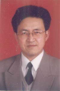 Zhen Ma (马震)合作论文数College of Chemistry and Chemical Engineering, Guangxi University3
 Hongming Liu (刘宏鸣)合作论文数College of Chemistry and Chemical Engineering, Guangxi University3
 Hailan Chen (陈海兰)合作论文数College of Animal Science and Technology, Guangxi University3
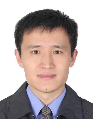 Qingyou Liu (刘庆友)合作论文数College of Animal Science and Technology, Guangxi University3
 Xiaoli Wang (王晓丽)合作论文数College of Animal Science and Technology, Guangxi University3
 Ben Huang (黄奔)合作论文数College of Animal Science and Technology, Guangxi University3Xinmin Zhang (张新民)合作论文数3Hesheng Jiang合作论文数2SuFang Yang (杨素芳)合作论文数2Gangbin Tang (汤刚彬)合作论文数2Zhiyuan Wang合作论文数1Siran Zhu合作论文数1Xie Li-ji合作论文数1WU Qing-ping (吴青萍)合作论文数1Huahai Chen合作论文数1Yafen Guo合作论文数1Hao Yan合作论文数1Xing Liang合作论文数1Jinzhang Jiang合作论文数1Haifeng Yan合作论文数1XIE Qin (谢琴)合作论文数1Wan Xie (谢婉)合作论文数1GuoBao Lan (兰国宝)合作论文数1BingKun Xie (谢炳坤)合作论文数1GuiHua Bian (卞桂华)合作论文数1JianRong Jiang (蒋建荣)合作论文数1ZhiCai Li (李志才)合作论文数1JianGuang Mo (莫建光)合作论文数1AnGen Lu (卢安根)合作论文数1YaoShan Pang (庞耀珊)合作论文数1
 Jiabo Liu (刘加波)合作论文数1Dan Wang (王丹)合作论文数1
 Zhixun Xie (谢芝勋)合作论文数Guangxi Veterinary Research Institute1Yi Mo合作论文数1Tong Li合作论文数1Zhiqiang Wang合作论文数1
 Xu Zhou (周虚)合作论文数College of Animal Sciences, Jilin University1
 Chunjin Li (李纯锦)合作论文数College of Animal Sciences, Jilin University1
 Mengmei Li (李梦梅)合作论文数College of Animal Science and Technology, Guangxi University1
 Qinyang Jiang (蒋钦杨)合作论文数College of Animal Science and Technology, Guangxi University1




