Mitochondrial dysfunction is a key factor in exacerbating pressure overload-induced cardiac hypertrophy and is linked to increased morbidity and mortality. ECSIT, a crucial adaptor for inflammation and mitochondrial function, has been reported to express multiple transcripts in various species and tissues, leading to distinct protein isoforms with diverse subcellular localizations and functions. However, whether an unknown ECSIT isoform exists in cardiac cells and its potential role in regulating mitochondrial function and pathological cardiac hypertrophy has remained unclear. This study identified a 42-kDa ECSIT isoform encoded by the transcript variant Ecsit-X4, which is highly expressed in the mitochondria of adult cardiomyocytes but down-regulated in hypertrophic human heart samples and TAC-treated mouse hearts. AAV9-mediated Ecsit-X4 gene therapy, administered either before or after TAC surgery, significantly attenuated cardiac hypertrophy. Cardiomyocyte-specific Ecsit deficiency worsened TAC-induced cardiac hypertrophy, while Ecsit-X4 compensation independently rescued hypertrophic phenotypes in EcsitcKO mice. Mechanistically, ECSIT-X4 localized to the mitochondria and interacted with STAT3, leading to increased STAT3 levels and enhanced serine 727 phosphorylation in cardiomyocyte mitochondria, thereby promoting strong mitochondrial bioenergetics. This study identified a novel transcript variant of ECSIT localized in the mitochondria of adult cardiomyocytes and highlights its potential as a therapeutic target for heart failure.
Ferroptosis is a type of non-apoptotic regulated cell death characterized by iron accumulation and lipid peroxidation. Cisplatin is an effective chemotherapy drug with several serious side effects including acute kidney injury (AKI). Asprosin is a peptide contributing to metabolism regulation and metabolic disorders. This study aimed to determine the role and mechanism of asprosin in AKI. Cisplatin was used to induce cell damage in mouse renal tubular epithelial (TCMK-1) cells and AKI in C57BL/6 mice. Cisplatin caused asprosin upregulation in cisplatin-treated TCMK-1 cells and mice. In TCMK-1 cells, asprosin overexpression led to iron overload and lipid peroxidation, while asprosin knockdown attenuated cisplatin-induced iron overload, lipid peroxidation and ferroptosis. Exogenous asprosin promoted cell damage and ferroptosis, which were attenuated by ferroptosis inhibitors. Asprosin-induced iron overload, lipid peroxidation, cell damage and SMAD1/5/8 phosphorylation were prevented by bone morphogenetic protein (BMP) type I receptor inhibitor. Integrin antagonist prevented asprosin-induced SMAD1/5/8 phosphorylation, and asprosin can specifically bind to integrin (33. Inhibition of integrin (33 reduced the asprosin-induced increases in Fe2+ and MDA levels. Asprosin knockdown relieved cisplatin-induced hepcidin upregulation, while hepcidin knockdown attenuated asprosin-induced iron overload, lipid peroxidation and ferroptosis. In cisplatin-induced AKI mice, specific knockdown of asprosin in the kidney not only attenuated renal dysfunction and damage, but also alleviated iron overload, lipid peroxidation and ferroptosis. These results indicated that excessively increased asprosin promotes TCMK-1 cells ferroptosis and damage via integrin (33/BMP/hepcidin-mediated iron overload and lipid peroxidation. Silencing of asprosin attenuates renal injury and dysfunction in cisplatin-induced AKI by inhibiting ferroptosis.
Vascular adventitial fibroblasts (VAFs) contribute to vascular remodelling in hypertension. However, the mechanisms by which VAFs regulate vascular smooth muscle cells (VSMCs) in vascular remodelling are not well known. Here we report the crucial roles of extracellular nanoparticles exomeres (EMs) derived from VAFs in promoting VSMCs proliferation, migration and vascular remodelling in normotensive Wistar-Kyoto rats (WKY) and spontaneously hypertensive rats (SHR). VSMCs' proliferation and migration were enhanced by EMs of SHR via the uptake of EMs in VSMCs, but not by EMs of WKY. Proteomics analysis showed that increased osteopontin (OPN) content may be responsible for the roles of EMs of SHR, which was confirmed by the fact that EMs of SHR pretreated with OPN knockdown lost their roles in promoting VSMCs proliferation and migration. OPN successively promoted the phosphorylation of FAK, PI3K and AKT via acting on integrin αVβ3. Inhibition of integrin αVβ3, FAK, PI3K or AKT almost abolished the effects of EMs of SHR on VSMCs proliferation and migration. Knockdown of OPN in the carotid artery attenuated local vascular remodelling in SHR. Repetitive intravenous injection of EMs of SHR increased blood pressure and promoted vascular remodelling in WKY and SHR. We conclude that EMs from VAFs of SHR promote VSMCs proliferation, migration and vascular remodelling via transferring OPN and subsequently activating integrin αVβ3/FAK/PI3K/AKT signalling pathway.
Long-term hyperglycemia can lead to diabetic cardiomyopathy (DCM), a main lethal complication of diabetes. However, the mechanisms underlying DCM development have not been fully elucidated. Heat shock protein A12A (HSPA12A) is the atypic member of the Heat shock 70kDa protein family. In the present study, we found that the expression of HSPA12A was upregulated in the hearts of mice with streptozotocin-induced diabetes, while ablation of HSPA12A improved cardiac systolic and diastolic dysfunction and increased cumulative survival of diabetic mice. An increased expression of HSPA12A was also found in H9c2 cardiac cells following treatment with high glucose (HG), while overexpression of HSPA12A-enhanced the HG-induced cardiac cell death, as reflected by higher levels of propidium iodide cells, lactate dehydrogenase leakage, and caspase 3 cleavage. Moreover, the HG-induced increase of oxidative stress, as indicated by dihydroethidium staining, was exaggerated by HSPA12A overexpression. Further studies demonstrated that the HG-induced increases of protein kinase B and forkhead box transcription factors 1 phosphorylation were diminished by HSPA12A overexpression, while pharmacologically inhibition of protein kinase B further enhanced the HG-induced lactate dehydrogenase leakage in HSPA12A overexpressed cardiac cells. Together, the results suggest that hyperglycemia upregulated HSPA12A expression in cardiac cells, by which induced cell death to promote DCM development. Targeting HSPA12A may serve as a potential approach for DCM management.
BACKGROUND: The infiltration of macrophages into the lungs is a common characteristic of perivascular inflammation, contributing to vascular remodeling in pulmonary hypertension (PH). Peli1 (pellino E3 ubiquitin-protein ligase 1) plays a critical role in regulating the production of proinflammatory cytokines and the polarization of macrophages in various diseases. However, the role of Peli1 in PH remains to be investigated. METHODS: The expression and biological function of Peli1 were investigated in both human and experimental models of PH. Peli1-deficient mice and bone marrow transplant mice were utilized to explore the roles of Peli1 in macrophages in vivo. Proteomic analysis and molecular biology techniques were used to uncover the underlying mechanisms. RESULTS: The upregulation of Peli1 in the lungs and alveolar macrophages was observed in hypoxia-treated mice. Peli1 knockout mice and myeloid Peli1-deficient mice significantly ameliorated hypoxia-induced right ventricular systolic pressure, right ventricular hypertrophy, and pulmonary vascular remodeling. Mechanistically, Peli1 facilitated the ubiquitination and subsequent proteasomal degradation of Foxp1 (forkhead box p1), thereby alleviating its suppression of IL ( interleukin ) -6 transcription and contributing to macrophage activation. Furthermore, myeloid Foxp1 deficiency partially eliminates the protective effect of myeloid Peli1 deficiency in hypoxia-induced PH mice. CONCLUSIONS: Our findings demonstrate that the Peli1-Foxp1-IL-6 pathway plays a crucial role in macrophage activation and recruitment during the development of PH, underscoring the potential of Peli1 as a therapeutic target for PH.
BACKGROUND:Sympathetic hyperactivity contributes to the pathogenesis of hypertension. However, it is unclear whether the excessive sympathetic activity is an independent and crucial factor for vascular remodeling in hypertension. This study focused on the effect of local sympathetic denervation with superior cervical ganglionectomy (SCGx) on vascular remodeling. METHODS:Surgical bilateral SCGx was performed in 9-week-old male Wistar-Kyoto rats (WKY) and spontaneously hypertensive rats (SHR). Control rats received sham-operation without SCGx. All measurements were made 4 weeks after the surgery. RESULTS:The effectiveness of SCGx was confirmed by the eye features of Horner syndrome, greatly reduced tyrosine hydroxylase (TH) contents in the superior cervical ganglion (SCG)-innervated arteries in the head. Although SCGx had no significant effects on blood pressure and heart rate in WKY and SHR, it attenuated vascular remodeling of facial artery and superficial temporal artery in SHR, two representative SCG-innervated extracranial arteries, without significant effects on non-SCG-innervated thoracic aorta and mesenteric artery. SCGx-treated SHR had more auricular blood flow and retina microvasculature than sham-operated SHR. However, SCGx had only a mild effect in attenuating the vascular remodeling of basilar artery and middle cerebral artery, two representative SCG-innervated intracranial arteries, in SHR. SCGx-treated SHR exhibited upregulation of α-smooth muscle actin, downregulation of proliferating cell nuclear antigen, and attenuation of oxidative stress and inflammation in facial artery and superficial temporal artery. CONCLUSIONS:Sympathetic denervation by SCGx in SHR attenuated local vascular remodeling, suggesting that sympathetic overactivity is a crucial pathogenic factor of vascular remodeling in SHR.
Chemerin is an adipokine that contributes to metabolism regulation. Nucleus tractus solitarius (NTS) is the first relay station in the brain for accepting various visceral afferent activities for regulating cardiovascular activity. However, the roles of chemerin in the NTS in regulating sympathetic activity and blood pressure are almost unknown. This study aimed to determine the role and potential mechanism of chemerin in the NTS in modulating sympathetic outflow and blood pressure. Bilateral NTS microinjections were performed in anaesthetized adult male Sprague-Dawley rats. Renal sympathetic nerve activity (RSNA), mean arterial pressure (MAP) and heart rate (HR) were continuously recorded. Chemerin and its receptor chemokine-like receptor 1 (CMKLR1) were highly expressed in caudal NTS (cNTS). Microinjection of chemerin-9 to the cNTS increased RSNA, MAP and HR, which were prevented by CMKLR1 antagonist α-NETA, superoxide scavenger tempol or N-acetyl cysteine, nicotinamide adenine dinucleotide phosphate (NADPH) oxidase inhibitors diphenyleneiodonium or apocynin. Chemerin-9 increased superoxide production and NADPH oxidase activity in the cNTS. The increased superoxide production induced by chemerin-9 was inhibited by α-NETA. The effects of cNTS microinjection of chemerin-9 on the RSNA, MAP and HR were attenuated by the pretreatment with paraventricular nucleus (PVN) microinjection of NMDA receptor antagonist MK-801 rather than AMPA/kainate receptor antagonist CNQX. These results indicate that chemerin-9 in the NTS increases sympathetic outflow, blood pressure and HR via CMKLR1-mediated NADPH oxidase activation and subsequent superoxide production in anaesthetized normotensive rats. Glutamatergic inputs in the PVN are needed for the chemerin-9-induced responses.
Objective: and design Mild vascular inflammation promotes the pathogenesis of hypertension. Asprosin, a newly discovered adipokine, is closely associated with metabolic diseases. We hypothesized that asprosin might led to vascular inflammation in hypertension via NLRP3 inflammasome formation. This study shows the importance of asprosin in the vascular inflammation of hypertension. Methods: Primary vascular smooth muscle cells (VSMCs) were obtained from the aorta of animals, including spontaneously hypertensive rats (SHR), Wistar-Kyoto rats (WKY), NLRP3-/- and wildtype mice. Studies were performed in VSMCs in vitro, as well as WKY and SHR in vivo. Results: Asprosin expressions were up-regulated in VSMCs and media of arteries in SHR. Asprosin overexpression promoted NLRP3 inflammasome activation via Toll-like receptor 4 (TLR4), accompanied with activation of NF kappa B signaling pathway in VSMCs. Exogenous asprosin protein showed similar roles in promoting NLRP3 inflammasome activation. Knockdown of asprosin restrained NLRP3 inflammasome and p65-NF kappa B activation in VSMCs of SHR. NLRP3 inhibitor MCC950 or NF kappa B inhibitor BAY11-7082 attenuated asprosin-caused VSMC proliferation and migration. Asprosin-induced interleukin-1 beta production, proliferation and migration were attenuated in NLRP3-/- VSMCs. Local asprosin knockdown in common carotid artery of SHR attenuated inflammation and vascular remodeling. Conclusions: Asprosin promoted NLRP3 inflammasome activation in VSMCs by TLR4-NF kappa B pathway, and thereby stimulates VSMCs proliferation, migration, and vascular remodeling of SHR.
Proliferation of renal tubular epithelial cells (TEC) is essential for restoring tubular integrity and thereby to support renal functional recovery from kidney ischemia/reperfusion (KI/R) injury. Activation of transcriptional factor c-Myc promotes TEC proliferation following KI/R; however, the mechanism regarding c-Myc activation in TEC is incompletely known. Heat shock protein A12A (HSPA12A) is an atypic member of HSP70 family. In this study, we found that KI/R decreased HSPA12A expression in mouse kidneys and TEC, while ablation of HSPA12A in mice impaired TEC proliferation and renal functional recovery following KI/R. Gain-of-functional studies demonstrated that HSPA12A promoted TEC proliferation upon hypoxia/reoxygenation (H/R) through directly interacting with c-Myc and enhancing its nuclear localization to upregulate expression of its target genes related to TEC proliferation. Notably, c-Myc was lactylated in TEC after H/R, and this lactylation was enhanced by HSPA12A overexpression. Importantly, inhibition of c-Myc lactylation attenuated the HSPA12A-induced increases of c-Myc nuclear localization, proliferation-related gene expression, and TEC proliferation. Further experiments revealed that HSPA12A promoted c-Myc lactylation via increasing the glycolysis-derived lactate generation in a Hif1α-dependent manner. The results unraveled a role of HSPA12A in promoting TEC proliferation and facilitating renal recovery following KI/R, and this role of HSPA12A was achieved through increasing lactylation-mediated c-Myc activation. Therefore, targeting HSPA12A in TEC might be a viable strategy to promote renal functional recovery from KI/R injury in patients. HSPA12A facilitated renal functional recovery from kidney ischemia/reperfusion (KI/R) injury. This protective effect of HSPA12A was mediated through directly interacting with c-Myc as well as upregulating the Hif1α-mediated lactate generation, thereby increasing c-Myc lactylation and nuclear localization to drive expression of genes related to cell proliferation, and ultimately promoting TEC proliferation.
Myocardial ischemia/reperfusion (MI/R) injury is a major cause of adverse outcomes of revascularization following myocardial infarction. Anaerobic glycolysis during myocardial ischemia is well studied, but the role of aerobic glycolysis during the early phase of reperfusion is incompletely understood. Lactylation of Histone H3 (H3) is an epigenetic indicator of the glycolytic switch. Heat shock protein A12A (HSPA12A) is an atypic member of the HSP70 family. In the present study, we report that, during reperfusion following myocardial ischemia, HSPA12A was downregulated and aerobic glycolytic flux was decreased in cardiomyocytes. Notably, HSPA12A KO in mice exacerbated MI/R-induced aerobic glycolysis decrease, cardiomyocyte death, and cardiac dysfunction. Gain- and loss-of-function studies demonstrated that HSPA12A was required to support cardiomyocyte survival upon hypoxia/reoxygenation (H/R) challenge and that its protective effects were mediated by maintaining aerobic glycolytic homeostasis for H3 lactylation. Further analyses revealed that HSPA12A increased Smurf1-mediated Hif1α protein stability, thus increasing glycolytic gene expression to maintain appropriate aerobic glycolytic activity to sustain H3 lactylation during reperfusion and, ultimately, improving cardiomyocyte survival to attenuate MI/R injury.
Glucagon-like peptide-1 (GLP-1) and its analogs are widely used for diabetes treatment. The paraventricular nucleus (PVN) is crucial for regulating cardiovascular activity. This study aims to determine the roles of GLP-1 and its receptors (GLP-1R) in the PVN in regulating sympathetic outflow and blood pressure. Experiments were carried out in male normotensive rats and spontaneously hypertensive rats (SHR). Renal sympathetic nerve activity (RSNA) and mean arterial pressure (MAP) were recorded. GLP-1 and GLP-1R expressions were present in the PVN. PVN microinjection of GLP-1R agonist recombinant human GLP-1 (rhGLP-1) or EX-4 increased RSNA and MAP, which were prevented by GLP-1R antagonist exendin 9-39 (EX9-39) or GLP-1R antagonist 1, superoxide scavenger tempol, antioxidant N-acetylcysteine, NADPH oxidase (NOX) inhibitor apocynin, adenylyl cyclase (AC) inhibitor SQ22536 or protein kinase A (PKA) inhibitor H89. PVN microinjection of rhGLP-1 increased superoxide production, NADPH oxidase activity, cAMP level, AC, and PKA activity, which were prevented by SQ22536 or H89. GLP-1 and GLP-1R were upregulated in the PVN of SHR. PVN microinjection of GLP-1 agonist increased RSNA and MAP in both WKY and SHR, but GLP-1 antagonists caused greater effects in reducing RSNA and MAP in SHR than in WKY. The increased superoxide production and NADPH oxidase activity in the PVN of SHR were augmented by GLP-1R agonists but attenuated by GLP-1R antagonists. These results indicate that activation of GLP-1R in the PVN increased sympathetic outflow and blood pressure via cAMP-PKA-mediated NADPH oxidase activation and subsequent superoxide production. GLP-1 and GLP-1R upregulation in the PVN partially contributes to sympathetic overactivity and hypertension.
Introduction: Cardiac fibrosis is the main driver for adverse remodeling and progressive functional decline in nearly all types of heart disease including myocardial infarction (MI). The activation of cardiac fibroblasts (CF) into myofibroblasts is responsible for cardiac fibrosis. Unfortunately, no ideal approach for controlling CF activation currently exists. Objectives: This study investigated the role of Heat shock protein A12A (HSPA12A), an atypical member of the HSP70 family, in CF activation and MI-induced cardiac fibrosis. Methods: Primary CF and Hspa12a knockout mice were used in the experiments. CF activation was indicated by the upregulation of myofibroblast characters including alpha-Smooth muscle actin (αSMA), Collagen, and Fibronectin. Cardiac fibrosis was illustrated by Masson’s trichrome and picrosirius staining. Cardiac function was examined using echocardiography. Glycolytic activity was indicated by levels of extracellular lactate and the related protein expression. Protein stability was examined following cycloheximide and MG132 treatment. Protein-protein interaction was examined by immunoprecipitation-immunoblotting analysis. Results: HSPA12A displayed a high expression level in quiescent CF but showed a decreased expression in activated CF, while ablation of HSPA12A in mice promoted CF activation and cardiac fibrosis following MI. HSPA12A overexpression inhibited the activation of primary CF through inhibiting glycolysis, while HSPA12A knockdown showed the opposite effects. Moreover, HSPA12A upregulated the protein expression of transcription factor p53, by which mediated the HSPA12A-induced inhibition of glycolysis and CF activation. Mechanistically, this action of HSPA12A was achieved by acting as a scaffolding protein to bind p53 and ubiquitin specific protease 10 (USP10), thereby promoting the USP10-mediated p53 protein stability and the p53-medicated glycolysis inhibition. Conclusion: The present study provided clear evidence that HSPA12A is a novel endogenous inhibitor of CF activation and cardiac fibrosis. Targeting HSPA12A in CF could represent a promising strategy for the management of cardiac fibrosis in patients.
Enhanced proliferation and migration of vascular smooth muscle cells (VSMCs) contributes to vascular remodeling in hypertension. Adventitial fibroblasts (AFs)-derived extracellular vesicles (EVs) modulate vascular remodeling in spontaneously hypertensive rat (SHR). This study shows the important roles of EVs-mediated miR-21-3p transfer in VSMC proliferation and migration and underlying mechanisms in SHR. AFs and VSMCs were obtained from aorta of Wistar-Kyoto rat (WKY) and SHR. EVs were separated from AFs culture with ultracentrifugation method. MiR-21-3p content in the EVs of SHR was increased compared with those of WKY. MiR-21-3p mimic promoted VSMC proliferation and migration of WKY and SHR, while miR-21-3p inhibitor attenuated proliferation and migration only in the VSMCs of SHR. EVs of SHR stimulated VSMC proliferation and migration, which were attenuated by miR-21-3p inhibitor. Sorbin and SH3 domain containing 2 (SORBS2) mRNA and protein levels were reduced in the VSMCs of SHR. MiR-21-3p mimic inhibited, while miR-21-3p inhibitor promoted SORBS2 expressions in the VSMCs of both WKY and SHR. EVs of SHR reduced SORBS2 expression, which was prevented by miR-21-3p inhibitor. EVs of WKY had no significant effect on SORBS2 expressions. SORBS2 overexpression attenuated the roles of miR-21-3p mimic and EVs of SHR in promoting VSMC proliferation and migration of SHR. Overexpression of miR-21-3p in vivo promotes vascular remodeling and hypertension. These results indicate that miR-21-3p in the EVs of SHR promotes VSMC proliferation and migration via negatively regulating SORBS2 expression.
The polarization of macrophages to the M1 or M2 phenotype has a pivotal role in inflammatory response following myocardial ischemia/reperfusion injury. Peli1, an E3 ubiquitin ligase, is closely associated with inflammation and autoimmunity as an important regulatory protein in the Toll-like receptor signaling pathway. We aimed to explore the function of Peli1 in macrophage polarization under myocardial ischemia/reperfusion injury and elucidate the possible mechanisms. We show here that Peli1 is upregulated in peripheral blood mononuclear cells from patients with myocardial ischemia/reperfusion, which is correlated with myocardial injury and cardiac dysfunction. We also found that the proportion of M1 macrophages was reduced and myocardial infarct size was decreased, paralleling improvement of cardiac function in mice with Peli1 deletion in hematopoietic cells or macrophages. Macrophage Peli1 deletion lessened M1 polarization and reduced the migratory ability in vitro. Mechanistically, Peli1 contributed to M1 polarization by promoting K63-linked ubiquitination and nuclear translocation of IRF5. Moreover, Peli1 deficiency in macrophages reduced the apoptosis of cardiomyocytes in vivo and in vitro. Together, our study demonstrates that Peli1 deficiency in macrophages suppresses macrophage M1 polarization and alleviates myocardial ischemia/reperfusion injury by inhibiting the nuclear translocation of IRF5, which may serve as a potential intervention target for myocardial ischemia/reperfusion injury.
目的 探讨问题导向学习(PBL)在医学本科生病理生理学教学中的应用效果.方法 2022 年9 月至 2023 年 6 月选择南京医科大学五年制二年级由同一名病理生理教师授课的临床医学平行班 104 人为研究对象,一个班采用PBL+传统教学模式(试验组,n=54),另一个班采用传统讲座教学模式(对照组,n=50).通过期末测评及教学满意度问卷调查对比两组学生的教学效果.结果 试验组学生病理生理学课程期末成绩高于对照组,教学满意度优于对照组(P<0.05).结论 PBL与病理生理学课程体系融合起到了一种动态的协同作用,加深学生对疾病过程的理解,增强了学生对专业知识应用的能力,提高了学习效率.
Mood instability, a subjective emotional state defined as rapid mood oscillations of up and down, is a symptom that occurs in several psychiatric disorders, particularly major depressive disorder and bipolar disorder. Heat shock protein A12A (HSPA12A) shows decreased expression in the brains of schizophrenia patients. However, the causal effects of HSPA12A in any psychiatric disorders are completely unknown. To investigate whether HSPA12A affects mood stability, Hspa12a- knockout mice ( Hspa12a − / − ) and wild-type (WT) littermates were subjected to tests of open field, forced swimming, elevated plus maze, and sucrose preference. Cerebral lactate levels were measured in cerebrospinal fluid (CSF). Adult hippocampal neurogenesis (AHN) was assessed by BrdU labeling. We found that acute mood stress increased hippocampal HSPA12A expression and CSF lactate levels in mice. However, Hspa12a − / − mice exhibited behaviors of mood instability (anhedonia, lower locomotor activity, antidepression, and anxiety), which were accompanied by impaired AHN, decreased CSF lactate levels, and downregulated hippocampal glycolytic enzyme expression. By contrast, HSPA12A overexpression increased lactate production and glycolytic enzyme expression of primary hippocampal neurons. Intriguingly, lactate administration alleviated the mood instability and AHN impairment in Hspa12a − / − mice. Further analyses revealed that HSPA12A was necessary for sustaining cerebral lactate homeostasis, which could be mediated by inhibiting GSK3β in hippocampal neurons, to maintain AHN and mood stabilization. Taken together, HSPA12A is defined as a novel regulator of mood stability and exerts therapeutic potential for mood disorder. Our findings establish a framework for determining mood disorder and AHN relevance of cerebral lactate homeostasis.
Rationale: Liver ischemia-reperfusion (LI/R) injury is characterized by two interconnected phases: local ischemia that causes hepatic cell damage to release damage-associated molecular pattern (DAMPs), and DAMPs that recruit immune cells to elicit inflammatory cascade for further injury of hepatocytes. High-mobility group box 1 (HMGB1) is a representative DAMP. Studies in macrophages demonstrated that HMGB1 is secreted after lactylation during sepsis. However, whether lactylation mediates HMGB1 secretion from hepatocytes after LI/R is known. Heat shock protein A12A (HSPA12A) is an atypical member of HSP70 family. Methods: Gene expression was examined by microarray analysis and immunoblotting. The hepatic injury was analyzed using released ALT and AST activities assays. Hepatic macrophage chemotaxis was evaluated by Transwell chemotaxis assays. Inflammatory mediators were evaluated by immunoblotting. HMGB1 secretion was examined in exosomes or serum. HMGB1 lactylation was determined using immunoprecipitation and immunoblotting. Results: Here, we report that LI/R decreased HSPA12A expression in hepatocytes, while hepatocyte-specific HSPA12A overexpression attenuated LI/R-induced hepatic dysfunction and mortality of mice. We also noticed that hepatocyte HSPA12A overexpression suppressed macrophage chemotaxis to LI/R-exposed livers in vivo and to hypoxia/reoxygenation (H/R)-exposed hepatocytes in vitro. The LI/R-increased serum HMGB1 levels of mice and the H/R-increased HMGB1 lactylation and secretion levels of hepatocytes were also inhibited by hepatocyte HSPA12A overexpression. By contrast, HSPA12A knockout in hepatocytes promoted not only H/R-induced HMGB1 lactylation and secretion of hepatocytes but also the effects of H/R-hepatocytes on macrophage chemotaxis and inflammatory activation, while all these deleterious effects of HSPA12A knockout were reversed following hepatocyte HMGB1 knockdown. Further molecular analyses showed that HSPA12A overexpression reduced glycolysis-generated lactate, thus decreasing HMGB1 lactylation and secretion from hepatocytes, thereby inhibiting not only macrophage chemotaxis but also the subsequent inflammatory cascade, which ultimately protecting against LI/R injury. Conclusion: Taken together, these findings suggest that hepatocyte HSPA12A is a novel regulator that protects livers from LI/R injury by suppressing glycolysis-mediated HMGB1 lactylation and secretion from hepatocytes to inhibit macrophage chemotaxis and inflammatory activation. Therefore, targeting hepatocyte HSPA12A may have therapeutic potential in the management of LI/R injury in patients.
Cardiac fibrosis is an essential pathological process in pressure overload (PO)-induced heart failure. Recently, myocyte-fibroblast communication is proven to be critical in heart failure, in which, pathological growth of cardiomyocytes (CMs) may promote fibrosis via miRNAs-containing exosomes (Exos). Peli1 regulates the activation of NF-κB and AP-1, which has been demonstrated to engage in miRNA transcription in cardiomyocytes. Therefore, we hypothesized that Peli1 in CMs regulates the activation of cardiac fibroblasts (CFs) through an exosomal miRNA-mediated paracrine mechanism, thereby promoting cardiac fibrosis. We found that CM-conditional deletion of Peli1 improved PO-induced cardiac fibrosis. Moreover, Exos from mechanical stretch (MS)-induced WT CMs (WT MS-Exos) promote activation of CFs, Peli1-/- MS-Exos reversed it. Furthermore, miRNA microarray and qPCR analysis showed that miR-494-3p was increased in WT MS-Exos while being down regulated in Peli1-/- MS-Exos. Mechanistically, Peli1 promoted miR-494-3p expression via NF-κB/AP-1 in CMs, and then miR-494-3p induced CFs activation by inhibiting PTEN and amplifying the phosphorylation of AKT, SMAD2/3, and ERK. Collectively, our study suggests that CMs Peli1 contributes to myocardial fibrosis via CMs-derived miR-494-3p-enriched exosomes under PO, and provides a potential exosomal miRNA-based therapy for cardiac fibrosis.
 Qi Chen (陈琪)合作论文数School of Basic Medical Sciences, Nanjing Medical University65
 Guoqing Zhu (朱国庆)合作论文数School of Basic Medical Sciences, Nanjing Medical University51
 Chuanfu Li (李传福)合作论文数45
 Yuming Kang (康玉明)合作论文数School of Basic Medical Sciences, Health Science Center, Xi'an Jiaotong University31Li Liu (刘莉)合作论文数School of Life Sciences, Nanjing Normal University28Jiantao Li (李建涛)合作论文数23
 Zhengnian Ding (丁正年)合作论文数Jiangsu Province Hospital;Nanjing Medical University21Tuanzhu Ha合作论文数21Xiaojin Zhang (张晓金)合作论文数18Lingli Que合作论文数18Linli Que (阙琳利)合作论文数18
 Xiaoqing Xiong (熊晓青)合作论文数School of Basic Medical Sciences, Nanjing Medical University16
 Juejin Wang (王觉进)合作论文数School of Basic Medical Sciences, Nanjing Medical University15
 Qiuyue Kong合作论文数14XiaoHong Qi (戚晓红)合作论文数13Fen Zheng合作论文数11CuiZhen Wu (吴翠贞)合作论文数11AiDong Chen (陈艾东)合作论文数11Jing Li (李菁)合作论文数11
 Yebo Zhou (周业波)合作论文数南京医科大学11
 Xingya Gao (高兴亚)合作论文数School of Basic Medical Sciences, Nanjing Medical University10Chao Ye合作论文数10Bing Zhou合作论文数10Li Jing合作论文数10
 Ying Han (韩莹)合作论文数School of Basic Medical Sciences, Nanjing Medical University9
 Xingsheng Ren合作论文数9
 Hai-Jian Sun合作论文数Yong Loo Lin School of Medicine, National University of Singapore9Hao Li (李皓)合作论文数南京医科大学9Feng Zhang (张峰)合作论文数9Dan Chen合作论文数8Hao Cheng合作论文数8Ying Tong合作论文数8
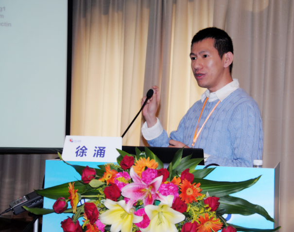 Yong Xu (徐涌)合作论文数南京医科大学基础医学院8Yu-Long Hu合作论文数8Xiaolu Wang合作论文数7Xiaofei Cao合作论文数7Nan Wu合作论文数7Zhou Hong合作论文数7Jim Lee Kelley合作论文数7Xuan Xu合作论文数7Jing-Xiao Wang合作论文数6Tongyan Liu合作论文数6Ming-Xia Zhao合作论文数6Yun Qiu合作论文数6Lin Guo合作论文数6Li JIANG (蒋莉)合作论文数6Surong Jiang (蒋素荣)合作论文数College of Mathematics, Nanjing University of Aeronautics and Astronautics6Meiling Wu合作论文数6Ying-Hao Zang合作论文数5




