Prosthodontics is rapidly entering the digital era, with computer-aided design and manufacturing (CAD/CAM) soon becoming the mainstream method for fixed restoration design. Recent advances in image recognition, data analytics, and decision systems are accelerating the use of artificial intelligence (AI), shifting workflows from simple automation to adaptive, learning-based design. Nevertheless, clear clinical guidance remains limited. This expert consensus sets out core principles, technology categories, and priority use cases for AI in fixed restorations, and proposes Standardized Operating Procedures that span from preoperative planning, digital impression processing, tooth preparation evaluation, to functional design and personalized esthetic design. It also defines quality control checkpoints and ethical safeguards that highlight the central role of the clinician in reviewing and validating AI outputs. Recommendations are provided for data governance, including security, privacy, and auditability. Finally, the document outlines near-term development needs such as interoperable data standards, transparent model reporting, and clinically oriented validation metrics. The goal is to support standardized, safe, and effective clinical adoption of AI in fixed dental restorations.
Conventional periodontitis therapy is limited by the lack of an all-in-one material with the desired functions of toxin neutralization, pathogen eradication, and osteopromotion. Exploiting the innate capacity of macrophages to recognize and neutralize pathological stimuli, this study develops novel engineered macrophage-based materials: phytate/Zn2 +-loaded gelated macrophages (PZ-GMs). These materials are fabricated at room temperature by polymerizing polycationic hydrogels and loading phytate/Zn2 + into live macrophages, innovatively integrating the natural immune neutralization function of intact macrophage membranes with the bioactivity of intracellular hydrogels. PZ-GMs are found to eradicate >99.99% of pathogens, remove >90% of bacterial toxins, and eliminate >80% of related inflammatory factors, while simultaneously promoting bone repair through in situ mineralization and osteoinduction. They can be further incorporated into a hydrogel designed for acid-triggered release, ensuring targeted delivery to inflamed sites. In a murine periodontitis model, PZ-GMs facilitate periodontal regeneration via a dual mechanism: transforming the pathological microenvironment induced by bacteria and toxins and fostering the remodeling of an osteogenic microenvironment. Furthermore, oral microbiome homeostasis is restored. Collectively, PZ-GMs represent a promising all-in-one neutralization-eradication-osteopromotion material for highly efficient periodontitis therapy.
BACKGROUND:Previous studies have indicated that older adults with the symptom of dry mouth have an increased risk of death. B vitamins are strongly linked to oral and systemic health. However, the relationship among dry mouth, vitamin B2 and mortality remains unclear. OBJECTIVES:This study aimed to investigate the potential relationship among dry mouth, vitamin B2 and mortality. METHODS:A total of 6183 participants from the US National Health and Nutrition Examination Survey (NHANES) 2011-2014 with information on dry mouth were included at baseline. 4324 normal individuals and 684 individuals with dry mouth were followed up until 31 December 2019. The Cox proportional hazard model was used to estimate the hazard ratios (HRs) and 95% confidence intervals (CIs) for the associations among dry mouth, vitamin B2 and all-cause mortality. RESULTS:Compared to the population without dry mouth, the risk of all-cause mortality was significantly higher in the population with dry mouth. The multivariable-adjusted HR and 95% CI was 1.41 (1.05, 1.90). The intake of vitamin B2 can significantly reduce the risk of death in participants with dry mouth, but not in participants without dry mouth. The HR and 95% CI of all-cause mortality in the highest vitamin B2 intake group was 0.37 (0.19, 0.73) compared to the lowest intake group. CONCLUSIONS:Individuals with dry mouth suffered a significantly higher risk of all-cause mortality compared to those without dry mouth. Higher intake of vitamin B2 was apparently associated with a lower risk of all-cause mortality in the population with dry mouth.
Digital technologies have become an integral part of complete denture restoration. With advancement in computer-aided design and computer-aided manufacturing (CAD/CAM), tools such as intraoral scanning, facial scanning, 3D printing, and numerical control machining are reshaping the workflow of complete denture restoration. Unlike conventional methods that rely heavily on clinical experience and manual techniques, digital technologies offer greater precision, predictability, and efficacy. They also streamline the process by reducing the number of patient visits and improving overall comfort. Despite these improvements, the clinical application of digital complete denture restoration still faces challenges that require further standardization. The major issues include appropriate case selection, establishing consistent digital workflows, and evaluating long-term outcomes. To address these challenges and provide clinical guidance for practitioners, this expert consensus outlines the principles, advantages, and limitations of digital complete denture technology. The aim of this review was to offer practical recommendations on indications, clinical procedures and precautions, evaluation metrics, and outcome assessment to support digital restoration of complete denture in clinical practice.
Aim or purpose: Periodontitis involves immune dysregulation driving neutrophil extracellular traps (NETs) and osteoclast activation, damaging periodontal tissue. Herein, we investigated a treatment using the hydrogel microneedle patch with multilevel structures, including a pH/ROS-responsive base for drug controlled-release, a degradable tip for drug sustained-release and an antibacterial substrate. Dehydroepiandrosterone (DHEA) and mangiferin are loaded into microneedles to stimulate endothelial cell-derived developmental endothelial locus-1 (DEL-1) release, thereby suppressing neutrophil recruitment, inhibiting NETs formation and reversing osteoblast-osteoclast imbalance, ultimately reducing periodontal tissue damage. Materials and methods: In vitro experiments were conducted using bone marrow mesenchymal stem cell (BMSCs), human umbilical vein endothelial cells (HUVECs) and neutrophils isolated from mouse. NETs formation was visualized via confocal microscopy. Quantitative real-time polymerase chain reaction, western blot assay and transcriptome sequencing were used for the assessment of associated gene expression. In vivo immunomodulation properties were validated through rat model with periodontitis (Ethic committee. 2025000725). Micro-CT and immunohistochemical staining were utilized for the measurement of the periodontitis damage. Results: The dual-layer microneedle patch exhibited adequate mechanical strength, pH/ROS-responsive properties, and excellent biocompatibility, fulfilling clinical application requirements. Under DHEA/mangiferin stimulation, HUVECs significantly upregulated DEL-1 secretion, which markedly suppressed neutrophils adhesion, NETs formation and enhanced inflammatory osteogenic regeneration. In vivo results demonstrated that drug-loaded microneedle patch effectively reduced periodontal tissue destruction and enhanced alveolar bone repair. Conclusions: The DHEA/mangiferin-loaded microneedle patch demonstrated superior periodontal immunomodulatory efficacy and tissue-protective functions, offering a novel therapeutic strategy for clinical periodontitis management.
The development of scaffolds for repairing critical-sized bone defects heavily relies on establishing a neuro-vascularized network for proper penetration of nerves and blood vessels. Despite significant advancements in using artificial bone-like scaffolds infused with various agents, challenges remain. Natural bone tissue consists of a porous bone matrix surrounded by a neuro-vascularized periosteum, with unique piezoelectric properties essential for bone growth. Drawing inspiration from this assembly, we developed a periosteum-bone-mimicking bilayer scaffold with piezoelectric properties for regeneration of critical-sized bone defects. The periosteum-like layer of this scaffold features a double network hydrogel composed of chelated alginate, gelatin methacrylate, and sintered whitlockite nanoparticles, emulating the viscoelastic and piezoelectric properties of the natural periosteum. The bone-like layer is composed of a porous structure of chitosan and bioactive hydroxyapatite created through a biomineralization process. Unlike conventional bone-like scaffolds, this bioinspired bilayer scaffold significantly enhances osteogenesis, angiogenesis, and neurogenesis combined with low-intensity pulsed ultrasound-assisted piezoelectric stimulation. Such a scheme enhances neuro-vascularized bone regeneration in vivo. The results suggest that the bilayer scaffold could serve as an effective self-powered electrical stimulator to expedite bone regeneration under dynamic physical stimulation.
Introduction and aims As an experimental teaching method, emerging learning methods including problem-based learning (PBL), case-based learning, team-based learning and flipped classroom (FC) have been widely applied in dental education. This study aims to evaluate the effect of these teaching methods on dental education performance compared to traditional lecture-based learning (LBL). Methods The search was carried out in April 2024 in PubMed, EMBASE, Web of Science, and Cochrane Library. All randomized controlled trials were included and the methodological quality assessment was based on the guidelines described in the Cochrane Handbook for Systematic Reviews, followed by a meta-analysis using Stata 14.0 software. Using standard mean deviation (SMD) and 95% confidence interval (95% CI) to determine the effectiveness of emerging teaching methods and LBL in all dental disciplines. Meta-regression was used to analyse sources of heterogeneity. Sensitivity analysis was performed to determine the stability, and Begg's analysis was used to determine whether there is publication bias. Results A total of 29 randomized controlled trials including 3502 students were included. The results indicate that emerging educational methods have a significantly positive effect on achieving higher scores (SMD = 0.48, 95% CI = 0.34-0.62, P < .001), whether it was theoretical scores (SMD = 0.52, 95% CI = 0.32-0.72, P < .001) or skill scores (SMD = 0.45, 95% CI = 0.15-0.76, P < .001). Compared to LBL, PBL (SMD = 0.33, 95% CI = 0.01-0.65, P = .045) and FC (SMD = 0.50, 95% CI = 0.31-0.69, P < .001) can both significantly improve students’ academic performance. Conclusion Compared to LBL, emerging educational methods (PBL, case-based learning, and FC) have significantly improved the learning effectiveness of dental students. These emerging educational methods can be advocated and popularized as routine teaching methods. Clinical Relevance This study is the first meta-analysis of the effects of emerging teaching methods in dental education which shows great impact of emerging teaching methods on students’ development.
OBJECTIVES:To evaluate the effects of repeated cut-off and rescan procedures on the trueness of three intraoral scanners (IOS). METHODS:A tooth model (#16) with a standard class II cavity was prepared, and the complete-arch was scanned using a laboratory scanner (D2000, 3Shape A/S) to obtain a reference scan. Then the typodont was scanned with three IOSs (3Shape TRIOS 3, CEREC Omnicam, and Medit i500) under two rescanning strategies (full-cut and partial-cut), with varying numbers of repeated cut-off and rescanning procedures (0, 1, 3, 5, 7, or 10). The trueness discrepancy between the reference and experimental digital scan was estimated using root mean square (RMS) calculations. Three regions of interest were selected to represent the rescanning, identification, and non-rescan area. And the discrepancies were analyzed using a linear mixed model (α=0.05). RESULTS:Cut-off and rescanning procedures significantly decreased the trueness of digital scans in all test conditions compared to the reference. However, no progressive increase in discrepancy was observed under any rescan conditions. Significant influences on trueness were found based on the IOS used, with the 3Shape system exhibiting lower RMS values. The partial-cut strategy showed lower RMS values compared to the full-cut strategy, albeit without statistical significance. CONCLUSIONS:While repeated cut-off and rescanning procedures led to a decline in the quality of digital impressions, they did not result in discrepancy accumulation with repeated rescanning. CLINICAL SIGNIFICANCE:To ensure high scanning accuracy in dental practice, it is advisable to minimize the rescanning area when correcting imperfections in digital scans. Additionally, selecting an appropriate scanner can help mitigate the negative effects of the rescanning technique.
The wear behavior of dental restorative materials is highly related to the biolubricating medium in the oral environment. Bacteria, along with their metabolic products, are essential substances in the oral cavity and have not been studied as a potential factor affecting lubrication performance during mastication. In this study, the effects of the Streptococcus mutans bacterial cells and their metabolites were investigated on the wear behavior of resin composites, polymer-infiltrated ceramic networks and zirconium-lithium silicate glass-ceramics. A reciprocating friction test and quantitative analysis of the wear morphology were utilized to determine the coefficient of friction (COF) and wear resistance of the test materials. The results showed that the bacterial metabolite medium significantly reduces the COF and wear rate of the three restorative materials and provide better protection against superficial abrasion. When tested under lactic acid medium, a key acid production in bacterial metabolites, similar wear reduction results were observed in the three materials, which confirmed that lactic acid should be accountable for the excellent lubricating property of bacterial metabolites. Furthermore, the resin composite with lower wettability exhibited a more significant wear reduction than the other two materials when lubricating with a bacterial metabolite medium. These findings provide novel insights into the biological basis of lubrication mechanisms in the oral cavity under high-loading and low-velocity conditions.
A rare case of extensive multiple idiopathic cervical root resorption with potential genetic predisposition was presented. A heathy 19-year-old Chinese male with no contributory medical or family/social history complained of pain during mastication that lasted for several months. Oral examination identified 7 missing teeth and external cervical root resorption involving 9 teeth. Comparison of orthopantomograms taken in May 2021 and February 2022 identified that cervical root resorption occurred in 22 teeth. Resorption commenced at the cementoenamel junction and progressed rapidly over the 9-month period. Laboratory test results were within normal limits. Trio-basedwhole-exome sequencing showed amissensemutation c.5630 C > T in the filamin A (FLNA) gene at chromosome X of the subject. This is suggestive of the possibility of sex-linked recessive inheritance. This is the first study to report FLNA mutation in human subjects with cervical root resorption involving multiple teeth.
This study systematically compared the mechanical performances and polymerization shrinkage of two novel dual-cured resin composites (DCRC) with one conventional packable light-cured resin composite (LCRC) for their application as core build-up material by micro-hardness test, flexural strength test, push-out test, and digital image correlation analysis. The LCRC had a significantly higher micro-hardness (p<0.05) whereas the bond strength demonstrated no difference. The mean values of three materials ranged from 35.16 and 64.82 for the Vickers hardness and from 4.66 MPa to 11.53 MPa for the bond strength. The flexure strength of the three materials was not statistically different from each other. LCRC demonstrated 1.88% of volumetric shrinkage while the two DCRC showed 5.06% and 4.91%, respectively. In general, the DCRC demonstrated a comparable flexural strength and bond strength as the LCRC, however, the significant polymerization shrinkage of DCRC should be emphasized.
Objectives. To obtain an accurate picture of the temporal development of bond strength between resin composites and tooth structures during cure for assessing debonding at the tooth-composite interface. Methods. An assembly of uncured composite sandwiched between a glass block and a dentin slab with a layer of pre-cured adhesive was used in this study. A conventional composite was compared against a bulk-fill composite. The rate of bond formation was determined by measuring the tensile bond strength of specimens of different thicknesses at different time points during cure. The changing light irradiance exiting the composite as it cured was also recorded. Mode of fracture was analyzed by examining the fracture surfaces. Results. Photo-bleaching occurred in both resin composites. The development of the dentin-composite bond strength was initially dictated by the developing cohesive strength of the resin composite, and its final value was capped by the strength of the preformed dentin-adhesive bond. The higher interfacial irradiance in the bulk-fill composite did not lead to faster development of the overall bond strength. This was caused by its slower rate of cohesive strength development as reflected in the longer time for its interfacial irradiance to plateau and the greater proportion of cohesive failure seen in the initial stage of polymerization. The law of reciprocity did not hold for the development of dentin bond strength. Significance. The results from this study, when compared with the development of shrinkage stress, can be used as a basis for ensuring the integrity of the dentin-composite interface during cure. (C) 2020 The Academy of Dental Materials. Published by Elsevier Inc. All rights reserved.
Objectives. Polymerization shrinkage of resin composite restorations can cause debonding at the tooth-restoration interface. Theory based on the mechanics of materials predicts that debonding at the pulpal floor would half the shrinkage displacement at the occlusal surface. The aim of this study is to test this theory and to examine the possibility of detecting subsurface resin composite restoration debonding by measuring the superficial shrinkage displacements. Methods. A commercial dental resin composite with linear shrinkage strain of 0.8% was used to restore 2 groups of 5 model Class-II cavities (8-mm long, 4-mm wide and 4-mm deep) in aluminum blocks (8-mm thick, 10-mm wide and 14-mm tall). Group I had the restorations bonded to all cavity surfaces, while Group II had the restorations not bonded to the cavity floor to simulate debonding. One of the proximal surfaces of each specimen was sprayed with fine carbon powder to allow surface displacement measurement by Digital Image Correlation. Images of the speckled surface were taken before and after cure for displacement calculation. The experiment was simulated using finite element analysis (FEA) for comparison. Results. Group I showed a maximum occlusal displacement of 34.7 +/- 6.7m and a center of contraction (COC) near the pulpal floor. Group II had a COC coinciding with the geometric center and showed a maximum occlusal displacement of 17.4 +/- 3.8 m. The difference between the two groups was statistically significant (p-value=0.0007). Similar results were obtained by FEA. The theoretical shrinkage displacement was 44.6 and 22.3 m for Group I and II, respectively. The lower experimental displacements were probably caused by slumping of the resin composite before cure and deformation of the adhesive layer. Significance. The results confirmed that the occlusal shrinkage displacement of a resin composite restoration was reduced significantly by pulpal floor debonding. Recent in vitro studies seem to indicate that this reduction in shrinkage displacement could be detected by using the most accurate intraoral scanners currently available. Thus, subject to clinical validation, the occlusal displacement of a resin composite restoration may be used to assess its interfacial integrity. (C) 2017 The Academy of Dental Materials. Published by Elsevier Ltd. All rights reserved.
Commercially available dental adhesives fail to chemically unite the demineralized collagen matrix with resinous materials within the resin-dentin interface. Sub-micron separations between the collagen fibrils and polymerized resin provide the backdrop for bond deterioration. Here, novel isocyanate-terminated urethane methacrylate precursors (UMP) were synthesized with the capacity to bond chemically to dentin collagen via covalent and hydrogen bonds. Collagen grafted with UMP also copolymerized with other methacrylate resin monomers, thereby producing a monoblock of chemically-linked biocomposite. The viscosity, degree of conversion and biocompatibility of UMP are comparable with commercially available resin monomers. An experimental adhesive containing 40% UMP demonstrated co-polymerization capability, good infiltration capacity and achieved higher immediate bond strength to dentin than the control commercially available adhesive. Improvement of dentin bonding by incorporation of UMP into dentin adhesives justifies future evaluation of the potential of these UMP-based adhesives in extending the longevity of resin-dentin bonds. Statement of Significance Composite-adhesive restorations have become an indispensable treatment modality in contemporary restorative dentistry. While the inability of these adhesives to bond chemically with collagen undermines the bond quality. This study describes a novel isocyanate-terminated urethane multi methacrylate precursors (UMP) which can bridge dentinal matrix collagen with adhesive resin by covalent and hydrogen bonds. Furthermore, an experimental UMP-based adhesive shows better co-polymerization capability, good infiltration capacity and higher immediate bond strength than the putatively effective adhesive Single Bond 2. The new chemical bonding mechanism based on UMP would theoretically produce more stable bonding interface that are more resistant to degradation. (C) 2018 Acta Materialia Inc. Published by Elsevier Ltd.
To fabricate a sustained-release delivery system of bone morphogenetic protein (BMP-2) on titanium surface, explore the effect of BMP-2 concentration on the loading/release behavior of BMP-2 and evaluate the cell compatibility of the system in vitro, pure titanium specimens were immersed into supersaturated calcium phosphate solutions (SCP) containing 4 different concentrations of BMP-2: 0, 50, 100, 200 and 400 ng/mL. Biomimetic calcium phosphate coating was formed on titanium surface and BMP-2 was incorporated into the coating through co-deposition. The release profile of BMP-2 suggested that BMP-2 were delivered sustainably up to 20 days. CCK-8 and ALP assay showed that 200 group and 400 ng/mL BMP-2 group have significant effect on promoting MC3T3-E1 cell proliferation and differentiation. The BMP-2 incorporated into the hybrid coating released in a sustained manner and significantly promoted the proliferation and differentiation of MC3T3-E1 on the titanium surface.
Objective: To explore the effect of calcium concentration in the electrolyte solution on the physicochemical properties and biocompatibility of coatings formed by micro-arc oxidation (MAO) on titanium surfaces.Methods: The surfaces of pure titanium plates were modified by MAO in an electrolytic solution containing calcium acetate (CA; C4H6CaO4) at concentrations of 0.05, 0.1, 0.2, or 0.3 M and beta-glycerophosphate disodium salt pentahydrate (beta-GP; C3H7Na2O6P center dot 5H(2)O) at a fixed concentration of 0.02 M. Surface topography, elemental characteristics, phase composition, and roughness were investigated by scanning electron microscopy, energy-dispersive X-ray analysis, X-ray diffraction, and a surface roughness tester, respectively. To assess the cytocompatibility and osteoinductivity of the surfaces, MC3T3-E1 preosteoblasts were cultured on the surfaces in vitro, and cell morphology, adhesion, proliferation, and differentiation were observed.Results: The porous MAO coating was composed primarily of TiO2 rutile and anatase. The amount of TiO2 rutile, the Ca/P ratio, and the surface roughness of the MAO coating increased with increasing CA concentration in the electrolyte solution. Ca-3(PO4)(2), CaCO3, and CaTiO3 were formed on MAO-treated surfaces prepared with CA concentrations of 0.2 and 0.3 M. Cell proliferation and differentiation increased with increasing CA concentration, with MC3T3-E1 cells exhibiting favorable morphologies for bone-implant integration.Conclusions: MAO coating improves the surface characteristics and cytocompatibility of titanium for osseointegration. Higher CA concentration in the MAO electrolyte solution has a positive effect on the surface properties, chemical composition, and cell response. (C) 2014 Elsevier B.V. All rights reserved.
OBJECTIVE:To explore the effect of changes of processing time on the surface properties of titanium coating formed by micro-arc oxidation (MAO).METHODS:Forty-four disc-shaped pure titanium specimens with 10 mm diameter and 1 mm thickness were equally divided into 4 groups and processed by MAO technique in electrolytes containing 0.2 mol/L calcium acetate (CA) and 0.02 mol/L β-glycerol phosphate disodium salt pentahydrate (β-GP). The processing time were set at 1 min, 5 min, 10 min and 15 min respectively. The topograph of the MAO film surface and the film-substrate interface was observed by a scanning electron microscopy (SEM), and the composition was analyzed by an energy dispersive spectroscope (EDS) incorporated in the SEM. The phase and the microstructure of the film were analyzed by X-ray diffraction (XRD). The roughness of the film was measured using a roughness tester. The surface static contact angle was detected by a contact angle measurement instrument and the surface energy was calculated accordingly.RESULTS:With the increase of processing time from 1 min to 15 min, the pore size increased from (1.30 ± 0.07) µm to (1.55 ± 0.09) µm, and film thickness increased from (10.2 ± 1.1) µm to (20.9 ± 2.9) µm. The content of the Ca in the film increased accordingly, and Ca/P increased from 1.99 to 2.45, and the surface energy increased from 24.62 mJ/m(2) to 39.49 mJ/m(2). Meanwhile, the XRD pattern indicated that rutile increased but anatase and titanium decreased gradually. At the time of 15 min, part of the MAO film peeled off.CONCLUSIONS:Processing time has impact on the thickness, surface topography, crystal component and surface energy of titanium MAO coating. MAO film treated for 5 - 10 min demonstrated favorable surface properties.
Objective. To investigate the effect of indenter size on the failure mode of amalgam and its interaction with thickness.Methods. Dental silver amalgam(Lojic+, SDI) discs, 10 mm diameter, thickness h = 0.4-8.0 mm, were stored in air at 37 degrees C for 7 days before testing in 'Hertzian contact', resting freely on a 30% glass fibre-reinforced nylon 6,6 substrate (E: 10 GPa) at 23 degrees C in air at a crosshead speed of 0.2 mm/min. The load at the first crack, detected acoustically, was recorded. Two series of tests were conducted. In the first, a 3 or 20 mm diameter hard steel ball was used to apply the load to discs with h = 0.4-8.0 mm. The second was performed on discs with h = 2.0 and 5.0 mm with indenter diameters D = 2-40 mm.Results. The failure loads for these two groups were substantially different, with most values for D = 3 mm being lower than those for D = 20 mm. The transitions of the main failure mode with increasing thickness were similar, i.e., from RC-dominant for h small to CC-dominant for h large, except that this occurred over a greater range for the larger indenter. For h = 2 mm, failure load was constant for D = 2-40 mm; all specimens consistently failed from bottom RC. For h = 5 mm, failure load increased with D with a quadratic relationship up to about 10 mm (r(2) = 0.85), after that, values were near constant; the failure mode gradually changed with D from PD- or CC-prevailing for D small (<= 5 mm) to RC-prevailing for D very large (30 and 40 mm).Significance. The failure mode of amalgam is dependant on the indenter size; indenter size and coating thickness have reciprocal effects. (C) 2010 Academy of Dental Materials. Published by Elsevier Ltd. All rights reserved.
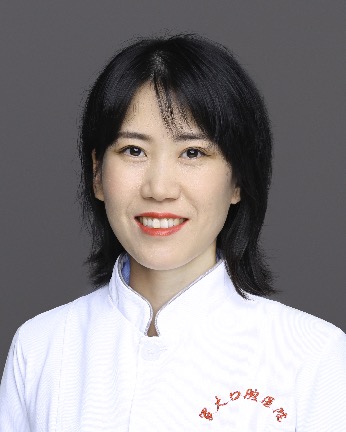 Li-Na Niu (牛丽娜)合作论文数The Third Affiliated Hospital of Air Force Medical University6
 Jiawen Guo (郭嘉文)合作论文数4
 Franklin Tay合作论文数Department of Endodontics, Dental College of Georgia, Augusta University4
 Alex Fok合作论文数Department of Restorative Sciences, Division of Biomaterials, School of Dentistry, University of Minnesota;Minnesota Dental Research Center for Biomaterials and Biomechanics, University of Minnesota4Jiajun Yang合作论文数3Jihua Chen (陈吉华)合作论文数Air Force Medical University3
 Chengyun Ning (宁成云)合作论文数School of Materials Science and Engineering, South China University of Technology3Shuailin Jia (贾帅林)合作论文数2
 Bo Yang合作论文数2Ming Fang合作论文数2Sui-dan WU (吴穗丹)合作论文数2
 Sai Ma合作论文数2Fu Wang (王富)合作论文数2Kai Jiao (焦凯)合作论文数中国人民解放军第四军医大学2
 Aregawi Wondwosen A合作论文数Department of Biosystems, University of Leuven2
 Ling Zhang (张凌)合作论文数2
 Joao Batista Novaes Jr合作论文数1Ruihan Ge合作论文数1Zhaoxia Bing合作论文数1Xuefei Pang合作论文数1Rongchen Xu合作论文数1
 Elissa Talma合作论文数Struct Engn Dept, Univ Fed Minas Gerais1
 Wenkai Jiang合作论文数Novogene Bioinformat Inst1Wen Niu合作论文数1Xiaojing Zhu合作论文数1Fan Yu合作论文数1Gang Chang合作论文数1Wen Qin合作论文数1Dongmei Li合作论文数1Wei Zhou合作论文数1Yun Xie (谢芸)合作论文数1Xinchun Zhang合作论文数1Xueyang Zhao (赵雪阳)合作论文数1Si-qin YANG (杨斯琴)合作论文数1Jing Li合作论文数1DianCai Zhao (赵殿才)合作论文数1
 Zhe Wu (吴哲)合作论文数School & Hospital of Stomatology, Guangzhou Medical University1Xiang Sun (孙翔)合作论文数1Fang Li (李芳)合作论文数1
 Lin Niu (牛林)合作论文数Health Science Center, Xi'an Jiaotong University;Hospital of Stomatology, Xi'an Jiaotong University1
 Tsoi, James Kit Hon (徐傑漢)合作论文数Faculty of Dentistry, The University of Hong Kong1
 Jianfeng Ma (麻健丰)合作论文数School & Hospital of Stomatology, Wenzhou Medical University;Alberta Institute, Wenzhou Medical University1Liao Hongbing合作论文数1
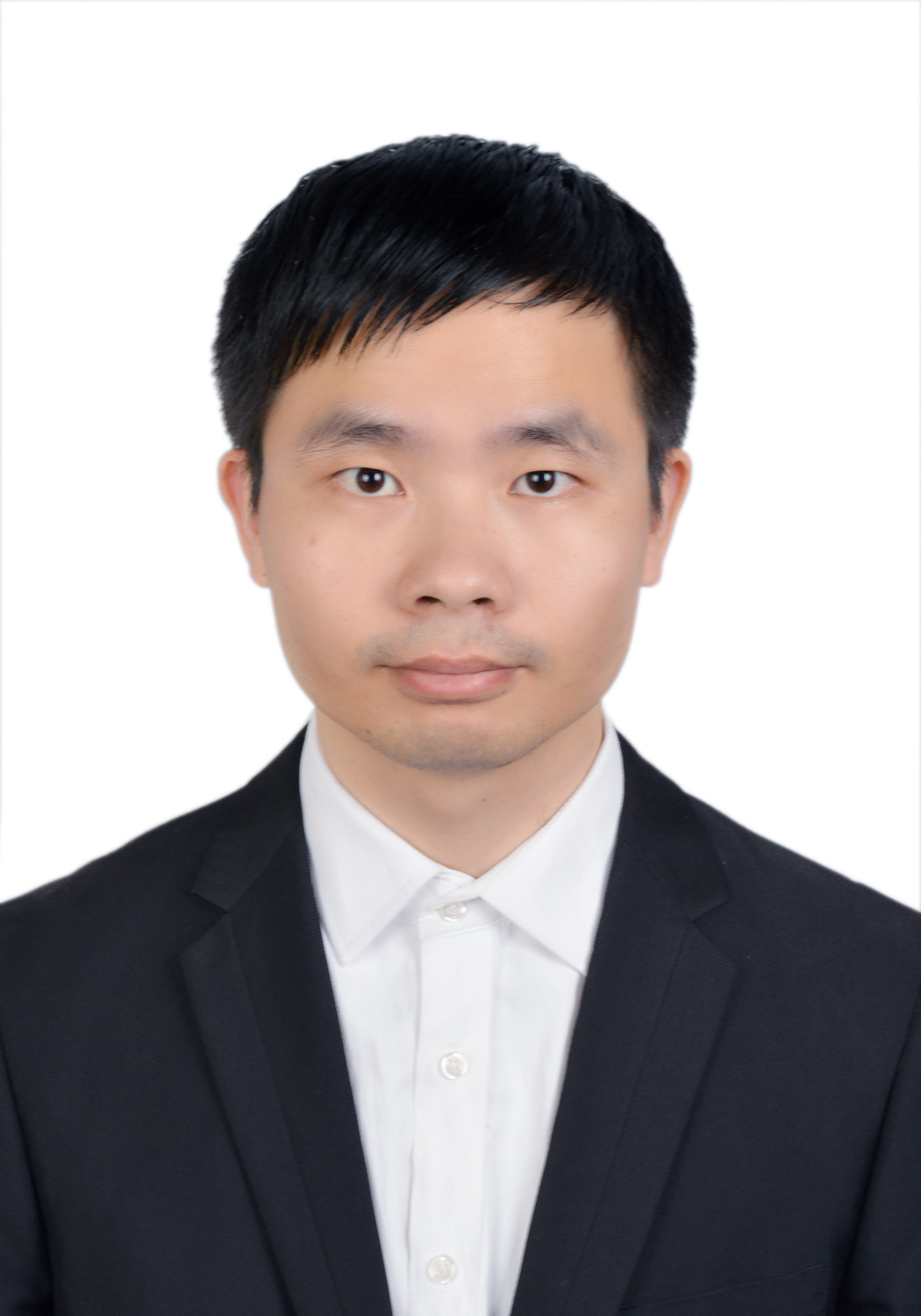 Yongkang Bai (白永康)合作论文数School of Chemical Engineering and Technology, Xi'an Jiaotong University1
 Xiaojian Wang (王小健)合作论文数College of Chemistry and Materials Science, Jinan University1
 Huade Zheng (郑华德)合作论文数School of Materials Science and Engineering, South China University of Technology1Ling Li (李龄)合作论文数1




