AIM:Carpal tunnel syndrome (CTS) is a compressive neuropathy commonly encountered in hand surgery, and decisions regarding whether operative decompression should be conducted rely on accuracy and clinical interpretability of imaging findings. Ultrasound is commonly used to directly visualize the median nerve in this condition, but remains operator-dependent and is limited by inconsistent diagnostic thresholds. This study aimed to develop a surgically oriented ultrasound-based artificial intelligence (AI) model that provides strong discriminative power, reliable probability calibration for preoperative counseling, and anatomy-aware explainability aligned with the nerve targeted in carpal tunnel release. METHODS:In this retrospective study, adults with suspected CTS, who had ultrasound image of adequate quality and met criteria for the clinical standard, were included; cases with prior carpal tunnel surgery or non-diagnostic images were excluded. We analyzed 2900 wrist ultrasound examinations, reserving an a priori 20% test set (n = 580 images) at the patient level. DenseNet-121 was utilized as the reference baseline. The proposed model used ConvNeXt-T augmented with Convolutional Block Attention Modules (CBAM), optimized with a supervised contrastive warm-up before standard fine-tuning; probabilities were post-hoc calibrated by temperature scaling. Images underwent de-identification, normalization, and ultrasound-appropriate augmentation. Primary outcome was discrimination (receiver operating characteristic (ROC), area under the curve (AUC), average precision (AP)) with bootstrap bands; secondary outcomes included accuracy, precision, recall, F1 score (harmonic mean of precision and recall), confusion matrices, probability distributions, class-conditional score separation, and Grad-CAM++ agreement with expert-defined regions of interest. Test set labels used a fixed 0.5 threshold. RESULTS:The proposed model outperformed the baseline in terms of discrimination (AUC 0.904 vs 0.821; AP 0.907 vs 0.831). Aggregate metrics also favored the proposed approach (accuracy 0.91 vs 0.83; precision 0.89 vs 0.81; recall 0.88 vs 0.82; F1 score 0.88 vs 0.81). Confusion matrices showed concurrent reductions in false positives (58→33, -43%) and false negatives (52→35, -33%): baseline true-negatives (TN)/false-positives (FP)/false-negatives (FN)/true-positives (TP) = 232/58/52/238; proposed TN/FP/FN/TP = 257/33/35/255. Predicted-probability histograms and class-conditional densities indicated more confident, better-separated outputs with calibration. Grad-CAM++ overlays were more compact and nerve-concordant relative to expert contours, supporting anatomy-aligned interpretability for surgical planning. CONCLUSIONS:A calibrated, explainable ConvNeXt-CBAM ultrasound classifier delivers reliable probabilities and anatomically faithful saliency that are directly actionable for surgical triage, timing of decompression, and preoperative counseling in CTS. These findings support ultrasound-based AI as a practical adjunct to clinical assessment and nerve conduction studies in hand surgery, warranting prospective, multicenter validation and workflow integration.
BackgroundPostoperative vascular compromise remains a clinically important threat after digital replantation or revascularization, but its timing, clinical presentation, risk factors, and post-compromise salvage profile are often less systematically reported than final survival.MethodsWe performed a single-center retrospective cohort study using digit-level records. The primary outcome was postoperative vascular compromise at the digit level. Compromise type, the operation-start–referenced interval to first documentation of compromise, recorded management components, and salvage success, defined as complete survival or partial survival with digit preservation and without secondary amputation, were summarized. Logistic regression with patient-cluster robust standard errors was used for the vascular-compromise model; salvage models were exploratory complete-case analyses.ResultsThis single-center retrospective study enrolled 671 digits from 484 patients. Postoperative vascular compromise occurred in 128 of 671 digits (19.1%). Venous congestion was the most common presentation (71.9%), followed by arterial insufficiency (21.1%) and mixed compromise (7.0%). The median operation-start–referenced interval to first documentation of vascular compromise was 15.88 h (IQR, 8.03–34.52), and 112 of 128 events (87.5%) were first documented within 48 h after the recorded operation-start timestamp. In the parsimonious multivariable vascular-compromise model, complete amputation (OR, 2.853; 95% CI, 1.780–4.575), vein grafting (OR, 2.963; 95% CI, 1.615–5.437), and longer operative duration (OR per hour, 1.079; 95% CI, 1.010–1.152) were associated with higher odds of compromise, whereas a greater number of arterial anastomoses was associated with lower odds (OR, 0.526; 95% CI, 0.331–0.835). Among compromise digits, salvage success was achieved in 81 of 128 digits (63.3%). Any recorded thrombolysis was associated with lower odds of salvage success in an exploratory complete-case model (N = 84; OR, 0.243; 95% CI, 0.089–0.662), a finding likely affected by confounding by indication.ConclusionsIn this single-center retrospective cohort, approximately one in five replanted or revascularized digits developed postoperative vascular compromise, most commonly venous congestion. Complete amputation, vein grafting, longer operative duration, and fewer arterial anastomoses were associated with compromise and may help identify digits requiring intensified early surveillance. Post-compromise treatment-outcome findings should be interpreted as exploratory associations rather than causal effects.
Repair after peripheral nerve injury (PNI) faces major obstacles due to microenvironmental imbalance and neuronal loss. Ferroptosis, an iron-dependent cell death driven by lipid peroxidation, has emerged as a key pathological event in PNI, linking oxidative stress, mitochondrial dysfunction, and inflammation to regenerative failure. Targeting ferroptosis protects vital cells-such as Schwann cells and neurons-and ameliorates the regenerative niche, offering a promising therapeutic strategy. This review elucidates the mechanisms of ferroptosis in PNI, detailing its roles in Schwann cells, dorsal root ganglion neurons, and macrophages via core pathways including Nrf2/HO-1/GPX4 and ACSL4. We further evaluate current intervention strategies and their therapeutic efficacy. This synthesis provides novel insights into PNI pathology and guides the development of innovative treatments.
Background Kienböck’s disease, characterized by lunate avascular necrosis, requires effective interventions to relieve pain and restore function. This study evaluated arthroscopic lunate decompression with K-wire fixation for Lichtman stage II–IIIa patients. Methods A retrospective cohort of 32 patients (18 stage II, 14 stage IIIa) treated at Ningbo No. 6 Hospital (2018–2022) was analyzed, with a 12-month follow-up. The sample size was determined via a power calculation (α = 0.05, power = 0.80) on the basis of VAS score reduction. The primary outcomes were visual analog scale (VAS) pain scores and Mayo wrist scores; the secondary outcomes included grip strength, wrist range of motion (ROM), and the lunate height index. Paired t tests, independent samples t tests, and Cohen’s d effect sizes were used. Results The VAS score decreased to 2.91 ± 0.82 at 3 months and 2.35 ± 0.70 at 6 months, from 7.33 ± 0.95 to 1.73 ± 0.44 at 12 months (mean difference: 5.60, 95% CI: 5.12–6.08, P < 0.001, Cohen's d = 5.89), exceeding the minimal clinically important difference (MCID, 1.5 points). The Mayo wrist score improved from 44.58 ± 8.56 to 86.01 ± 7.85 (mean difference: 41.43, 95% CI: 37.92–44.94, P < 0.001; Cohen's d = 4.85). The grip strength reached 86.17 ± 7.25% of that of the contralateral side. Stage II patients had lower VAS scores (1.57 ± 0.38) than did stage IIIa patients (1.94 ± 0.43, P = 0.017). The complications included transient nerve numbness, pin-site infections (15.6% each), and wrist stiffness (12.5%), all of which were resolved conservatively. Conclusion Arthroscopic lunate decompression with K-wire fixation significantly reduces pain and improves function in patients with stage II–IIIa KD, with superior pain relief in patients with stage II disease. This minimally invasive approach shows promise but requires multicenter randomized controlled trials for long-term validation.
Peripheral nerve injuries beyond 5 cm lack effective treatments. Functional nerve guidance conduits (NGCs) have emerged as transformative tools orchestrating regeneration by leveraging Schwann cell (SC)-centric mechanisms. This review comprehensively analyzes how NGC designs modulate SC behavior through three synergistic axes: physical cues, biochemical signaling, and bioelectric regulation. By enhancing microenvironmental regulation, next-generation NGCs aim to surpass autograft efficacy, offering scalable solutions for functional nerve recovery and improved patient outcomes.
Objective:To compare the clinical effecacy of artificial dermal regeneration matrix (as "artificial dermis" hereafter) and flap transfer in the treatment of soft tissue defects around interphalangeal joint.Methods:Through postoperative follow-up at outpatient clinic, a retrospective study was conducted on the clinical data of 60 patients who had soft tissue defects around the interphalangeal joints and received treatment in the Department of Hand Surgery, the Sixth Hospital of Ningbo from January 2018 to January 2022. According to applied surgical procedures, patients were divided into a flap transfer group (group A, n=30) and an artificial dermis group (group B, n=30). Group A included 19 males and 11 females aged 44.83 years ± 11.56 years including 5 patients with simple soft tissue defects, and 6 with soft tissue defects and fractures, 10 with soft tissue defects with tendon and (or) ligament injuries, 3 with soft tissue defects and vessel and (or) nerve injuries, and 6 with soft tissue defects and over 2 other types of compound injuries. The defect areas ranged from 2.5 cm×1.2 cm to 5.0 cm×1.6 cm. After emergency debridement and treatment for bone and tendon injuries, the patients in group A received transfers of free flap or transfers with adjacent digit flaps or island flaps. Postoperative functional exercise started from 1-6 weeks after surgery. Patients in group B included 17 males and 13 females, aged 44.70 years ± 11.20 years and there were 6 patients with simple soft tissue defects, 6 with soft tissue defects and fractures, 9 with soft tissue defects and tendon and (or) ligament injuries, 5 with soft tissue defects and vessel and (or) nerve injuries, and 4 with soft tissue defects and over 2 other types of compound injuries. The defect area ranged from 3.1 cm ×1.3 cm to 4.5 cm × 1.8 cm. Debridement and treatment of the bone and tendon injury in group B were the same as what in group A, but the patients in group B received artificial dermis coverage other than transfer of flap. After an artificial dermis had completely vascularised, a split-thickness skin graft was performed over the neo-dermis in the second phase surgery. Postoperative functional exercise started from 1-2 weeks after artificial dermis grafting surgery. The interval time of flap transfer or split-thickness skin grafting, survival rate of flap transfer or split-thickness skin grafting, Vancouver Scar Scale(VSS), TPD and total active movement(TAM) were compared between the 2 groups. The count data were analysed by Chi-square test. All measured data were analysed by independent sample t test or Mann-Whitney U test. P<0.05 indicates a statistically significant difference. Results:The interval time of flap transfer in group A and artificial dermis grafting in group B were 2.93 days ± 2.48 days and 19.87 days ± 3.35 days, respectively. There was a statistically significant difference between the 2 groups( P<0.05). All patients were entered in postoperative outpatient follow-up for 11-14 months(12.00 months ± 0.93 months). The appearance of flaps of some patients in group A was bloated with slightly limited digit movement. In group B, the digit movement was normal, without obvious scar hyperplasia in both of the recipient and donor sites. The survival rate of flap transfer or artificial dermis graft, TPD, VSS score and TAM score for group A and group B were 96.00% ± 9.32% and 98.17% ± 3.07%, respectively, 8.67 mm ± 2.01 mm and 9.50 mm ± 1.81 mm, 3.40 ± 1.07 and 3.17 ± 0.91 and 18.30 ± 1.97 and 18.93 ± 1.64, respectively. There were no significant differences between the 2 groups( P>0.05). Conclusion:In comparison with the transfer of flap, an artificial dermis is also effective and satisfactory in the treatment of soft tissue defects around the interphalangeal joint. Further large scale and multi-centre investigations are required.
OBJECTIVE:To explore clinical effects of the stageⅠrepair of full-thickness skin defect at dorsal skin of middle phalanx fingers using artificial dermis combing with digital artery perforator fascial flaps.METHODS:From January 2019 to May 2020, 21 patients(27 middle phalanx fingers)with full-thickness skin defect were repaired at stageⅠusing artificial dermis combing with digital artery perforator fascial flaps. All patients were emergency cases, and were accompanied by the exposure of bone tendon and the defects of periosteum and tendon membrane. Among patients, including 11 males and 10 females aged from 18 to 66 years old with an average age of (39.00±8.01) years old;9 index fingers, 10 middle fingers and 8 ring fingers;range of skin defect area ranged from (2.5 to 3.5) cm×(1.5 to 3.0) cm;range of exposed bone tendon area was (1.5 to 2.0) cm×(1.0 to 2.0) cm. The time from admission to hospital ranged from 1 to 6 h, operation time started from 3 to 8 h after injury.RESULTS:All patients were followed up from 6 to12 months with an average of (9.66±1.05) months. The wounds in 26 cases were completely healed at 4 to 6 weeks after operation. One finger has changed into wound infection with incompletely epithelialized dermis, and achieved wound healing at 8 weeks after dressing change. All fingers were plump with less scars. The healed wound surface was similar to the color and texture of the surrounding skin. These fingers have excellent wearability and flexibility. According to the upper limb function trial evaluation standard of Hand Surgery Society of Chinese Medical Association, the total score ranged from 72 to 100. 26 fingers got excellent result and 1 good.CONCLUSION:StageⅠrepair of full-thickness skin defect at dorsal skin of middle phalanx fingers using artificial dermis combing with digital artery perforator fascial flaps is easy to operate with less trauma. It has made satisfactory recovery of appearance and function of fingers. It could provide an effective surgical method for clinical treatment of full-thickness skin loss of fingers with tendon and bone exposure.
Objective:To retrospectively analyze hand skin and soft tissue infections (SSTI) caused by nontuberculosis mycobacterium (NTM) in Ningbo and analyze the clinical manifestations, strain identification results and treatment outcomes of NTM infection.Methods:Fifty-two cases of hand SSTI patients with NTM culture positive in Ningbo, Zhejiang Province from 2013 to 2020 were analyzed retrospectively. Their age, occupation, pathogenic factors, infection site, course of disease, culture results and strain characteristics were analyzed.Results:There were 52 patients (57 specimens) in this group. Seven kinds of mycobacteria were found after the identification of culture positive strains, including 42 cases of Mycobacterium marinum (73.68%), 2 cases of Mycobacterium chelonei, 1 case of Mycobacterium Kansasii, 2 cases of Mycobacterium fortuitum, 2 cases of Mycobacterium gilvum, 2 cases of Mycobacterium chubuense, 1 case of Mycobacterium nematode and 5 cases of unknown typing. The pathological reports of mycobacterial infection often suggest chronic inflammatory cell infiltration with granulomatous inflammation. All the patients started empirical medication at the same time of mycobacterium culture, mainly macrolides plus levofloxacin. Eight mild patients received conservative treatment with oral antimycobacterial drugs, with an average of 3.5 months. Due to the poor effect of conservative treatment, the remaining 44 cases were hospitalized for surgical treatment. After operation, they were treated with oral antimycobacterial drugs, and recovered after an average of 2.7 months. Among them, 5 patients with recurrent infection underwent secondary surgery or more, and only 1 case underwent finger amputation. According to the trial standard of upper limb function evaluation issued by Hand Surgery Society of Chinese Medical Association, the hand function was evaluated as excellent in 36 cases, good in 10 cases, fair in 4 cases and poor in 2 cases.Conclusion:Mycobacterium marinum is still the most common pathogenic strain of NTM infection in Ningbo, Zhejiang Province. The pathology is mainly granulomatous inflammation. For cases with poor conservative treatment effect, thorough lesion clearance surgery and antimycobacterial drug treatment can achieve good clinical efficacy.
肩关节脱位合并臂丛神经损伤可定义为因直接或间接暴力引起的肩关节脱位导致包括但不限于臂丛神经根干部及束支部麻痹或损伤,是肩关节脱位少见但严重的并发症之一,对上肢功能造成严重影响。但是由肩关节脱位合并臂丛神经的损伤容易被忽视,笔者就肩关节脱位合并臂丛神经损伤的流行病学、病因及损伤机制、诊断、治疗等的进展方面进行阐述,为临床诊治提供参考。
Objective To investigate the application and the clinical efficacy of wrist arthroscopy in the treatment of carpal intraosseous ganglion cysts (IGCs). Methods A retrospective case study method was adopted to analyze the clinical data of 28 patients with carpal IGCs admitted to the Sixth Hospital of Ningbo from April 2012 to January 2019. A hypodensity in the bone was shown by X-ray before the operation, with hypodensity and cystic change in the bone being confirmed by computed tomography and magnetic resonance imaging. Arthroscopic open window of the wrist, cystectomy, and autologous iliac bone graft implantation were conducted. Regular postoperative X-ray combined with CT follow-ups were conducted to observe the healing after bone implantation. Patients were followed up regularly and assessed by the Modified Mayo Wrist Score in four aspects of the postoperative pain, wrist mobility, grip, and function to provide an objective overall assessment of the therapeutic outcome. Results All 28 patients were followed up for 8–16 months, with an average follow-up duration of 10 months. After the operation, pain disappeared completely for 25 patients, and 3 cases showed significant improvement. All cases were pathologically confirmed as ganglion cysts and had first-stage bony healing after bone grafting with an average healing time of 10.8 weeks. The grip returned to normal for all patients, and wrist flexion and extension were the same as the healthy wrist for 25 patients, with a Modified Mayo Wrist Score of excellent in 19 cases and good in 9 cases. No recurrence was observed. Conclusion In patients with symptomatic carpal IGCs, the application of arthroscopic open window, cystectomy, and autologous bone graft implantation could achieve satisfactory clinical therapeutic effects.
目的 根据手功能评定结果制定个性化的手术方案,探讨手功能评定对手部复杂损伤的前瞻性指导意义.方法 选择39例手外伤患者,在进行手术前,设计不同的手术方案,通过前瞻性手功能评定方法,评估每种手术方案后患肢所获得的手失能百分比结果,根据手失能百分比结果选择适当的手术方案,确定优先保留指或优势指,个性化地设计利用损伤严重的废弃指或劣势指移位修复保留指或优势指,使手功能最大化.结果 39例患者中,选择吻合血管的游离复合组织移植及手指再植28例,选择带蒂皮瓣移位修复及手指移位重建11例.皮瓣及手指全部成活,随访6~12个月,术后上肢部分功能评定:优5例、良21例、可8例、差5例.结论 治疗手部多个指体复杂损伤时,根据手功能评定方法,前瞻性评估手部功能,可更有效地制定合理的手术方案,充分利用废弃指或劣势指的组织移位重建指体、修复组织缺损,最大程度保留手部功能,减少多余供区,避免多次手术,减少患者痛苦,缩短治疗时间.
Objective Scaphoid fracture was the most common carpal fracture and the most challenging. The purpose of this study was to investigate and compare the clinical effects of closed reduction and percutaneous cannulated screw internal fixation under fluoroscopy and arthroscopy‐assisted percutaneous cannulated screw internal fixation in the treatment of Herbert B2‐type of acute scaphoid fractures. Methods A retrospective controlled study was conducted on 29 patients with Herbert B2‐type acute scaphoid fracture with a displacement of >1 mm admitted to our hospital from January 2017 to June 2021. Patients were divided into two groups, 11 patients were treated with closed reduction percutaneous cannulated screw internal fixation under fluoroscopy and 18 patients were treated with percutaneous cannulated screw internal fixation assisted by arthroscopy. The operative time, intraoperative fluoroscopy times, fracture healing time, complications, and postoperative wrist function score of the two groups were compared. Results All patients were followed up for 6–18 months (mean follow‐up duration: 10.38 ± 2.69 months). The respective operation times in the arthroscopy group and fluoroscopy group was 51.50 ± 6.69 min and 56.73 ± 11.48 min, respectively (p > 0.05). The number of fluoroscopies performed in the arthroscopy group was (6.83 ± 1.30), which was less than that in the fluoroscopy group (10.91 ± 2.62) (p < 0.05). All fractures in the arthroscopy group healed after the operation, and the fracture healing time was 11.44 ± 1.25W. Ten patients in the fluoroscopy group healed. The fracture healing time was 13.60 ± 2.32 W. The fracture healing time in arthroscopy group was less than that in the fluoroscopy group (p < 0.05). One patient in the fluoroscopy group had nonunion and healed after bone grafting and internal fixation. At the postoperative 6‐month follow‐up, the modified Mayo wrist function score was used to evaluate the clinical results. The wrist function score of patients in the arthroscopy group was 90 (85, 95), which was >80 (80, 90) in the fluoroscopy group (z = 2.74, p < 0.05). Conclusion For Herbert B2‐type acute scaphoid fracture with fracture displacement > 1 mm, the arthroscopy‐assisted percutaneous cannulated screw internal fixation has less fluoroscopy times, short fracture healing time, and good recovery effect of wrist function compared to the fluoroscopy.
Objective:To investigate the surgical indications and treatment of acute open distal radioulnar joint (DRUJ) instability caused by distal radius fracture combined with fracture at the base of styloid process or recess of ulna.Methods:From January 2014 to June 2019, 13 patients with acute DRUJ instability caused by distal radius fracture combined with fracture at the base of styloid process or recess of ulna were treated by one-stage operation. After the reduction and volar plate fixation of radius fracture, the DRUJ impact test was performed comparing with the contralateral side during the operation. In case of instability, the stable structure of bone and soft tissue can be repaired by fixation of ulna styloid process fracture or anchor repair of triangular fibrocartilage complex, so as to achieve the purpose of early rehabilitation training.Results:After operation, 13 patients achieved healing of the distal radius and ulnar styloid process fractures. All the patients were follow-up for 6 to 36 months. The comprehensive evaluation of the DRUJ impact test, grip strength, wrist active range of motion, pain score, DASH score showed that the clinical efficacy was good.Conclusion:The one stage fixation of radius fracture and repair of ulnar styloid process fracture involving triangular fibrocartilage complex injury is helpful to restore the stability of bone and soft tissue structure of DRUJ, facilitate early rehabilitation exercise, improve the clinical efficacy of acute DRUJ instability and reduce the incidence of complications.
Background Few studies have investigated families in which multiple individuals over three or more generations are affected by radial polydactyly and syndactyly. This report describes an extremely rare family in which nine individuals across six generations were affected by complex radial polydactyly. Methods We investigated a six-generation pedigree with radial polydactyly including triplication, triphalangism, hypoplasia, and symphalangism. There was a total of 34 individuals (including their spouses) in the family and 11 individuals had polydactyly. The average age of the patients ranged from 7 months to 96 years. The characteristic feature of the malformation in these patients was described. Two patients underwent surgical resection for radial supernumerary thumbs. The Bilhout-Cloquet technique and On-top-plasty technique were used to reconstruct the nail and the joints. Results The patients in this family presented with thumb duplication and triphalangism in both hands, including a variety of deformities, such as triplication, triphalangism, hypoplasia, and symphalangism. Syndactyly and ulnar polydactyly were also frequently observed. Two patients who underwent surgical treatment showed good hand and thumb function at the 8- and 2-year post-operative follow-up, respectively. Conclusions The present study reported various mixed phenotypes including triplication, triphalangism, hypoplasia, and symphalangism within the same family which may represent a rare type of polydactyly. Surgical resection of extra digits to achieve mobility of the thumb is the main treatment option for radial polydactyly. Given the ulnar thumb is better developed, the radial thumb is typically resected in patients with radial polydactyly. These reconstructive principles are fit for this Chinese family as well.
Objective:To explore the clinical efficacy of Kapandji technique assisted by wrist arthroscopy for the treatment of Die-punch collapse fracture of distal radius.Methods:From January 2012 to December 2018, 31 patients with Die-punch collapse fracture of distal radius were treated. All the fractures were confirmed by CT examination. During the operation, Kirschner wire was used to pry for the reduction of the collapsed fracture block of distal radius under wrist arthroscopy to achieve the smooth articular cartilage surface. The internal ligaments of wrist joint, especially scapholunate ligament, lunatotriquetral ligament and triangular fibrocartilage complex, were further examined and repaired. All the fractures were fixed with Kirschner wire and external fixator.Results:During the operation, there were 5 cases of scapholunate ligament injury, 2 cases of lunatotriquetral ligament injury and 10 cases of triangular fibrocartilage complex injury. All the patients achieved primary healing and were follow-up for 6 to 24 months with an average of 11.3 months. The fracture healing time was 6 to 8 weeks with an average of 7 weeks. The palmar inclination angle with 6° to 17° and the ulnar deviation angle was 15° to 23° by measuring the postoperative X-ray films. According to Gartland-Werley evaluation criteria, 22 cases were excellent, 7 cases were good and 2 cases were fair. The excellent and good rate was 93.5%.Conclusion:The recovery of Die-punch collapse fracture of distal radius by Kapandji technique assisted with arthroscopy can accurately reduce the collapsed joint surface, reduce the complications of traumatic osteoarthritis, and also clarify the damage degree of the structure of the ligament and triangular fibrocartilage complex of the wrist joint, which is convenient for early treatment, so as to prevent chronic wrist pain or instability.
Objective To explore the use of wrist arthroscopy for treatment of aseptic necrosis of lunate bone and its curative effect. Methods From March 2012 to January 2016, 12 patients with aseptic necrosis of lunate bone underwent necrotic lunate bone extirpation assisted by wrist arthroscopy, and the scaphocapitate joint treated with fusion‐stabilization were selected. Among these patients, 10 patients were male and two patients were female. The age of these patients ranged from 25 to 42 years old, with an average age of 32 years old. These lesions were located in the right hand in eight patients, and in the left hand in four patients. Lichtman stage: stage IIIA in two patients, and stage IIIB in 10 patients. All patients were diagnosed by anterior lateral radiographs and magnetic resonance imaging (MRI). From the second day after the surgery, patients underwent active motion for the metacarpophalangeal joint and interphalangeal joints. When the X‐ray film indicated that the fracture had healed, patients began to resume their daily activities and work. After surgery, the plain films of all planes, including the axial plane of the scaphoid, were taken every month, until fracture healing. All patients were followed up for a mean duration of 8 months, and the Mayo wrist score was used to assess the range of motion of the wrist joint and grip force during the follow‐ups for objective function evaluation. Results Fracture healing was obtained in all patients within 9–12 weeks after the operation, and average healing time was 10 weeks. The duration of follow‐up ranged between 6–14 months, and the mean duration was 8 months. According to the modified Mayo wrist score, the objective function of the wrist joint was evaluated as follows: flexion and extension range of the wrist joint was 40°–110°, with an average of 105°, which was 80% of that of the unaffected side. Radioulnar deviation was 45°‐80°, with an average of 55°, which was 82% of that of the unaffected side. Grip force was 25–48 kg, with an average of 40 kg, which was 84% of that of the unaffected side. The modified Mayo wrist score was good in eight patients, acceptable in three patients, and poor in one patient. Conclusions Wrist arthroscopy is an effective and feasible tool for the treatment of aseptic necrosis of the lunate bone, and the right kind of surgical procedure should be selected for different stages of the disease, and wrist arthroscopy is applied timely, in order to achieve the desired therapeutic effect.
Objective:To compare the clinical efficacy between wrist arthroscopy-assisted ulnar head Wafer resection and ulnar shortening osteotomy in the treatment of ulnar impaction syndrome.Methods:From March 2012 to February 2017, 45 patients with ulnar impaction syndrome were treated at Department of Hand Surgery, No. 6 Hospital of Ningbo. They were 26 males and 19 females, aged from 28 to 48 years (average, 38 years). The right side was affected in 31 cases and the left side in 14. They were divided into 2 groups according to different surgical methods: 22 patients were subjected to arthroscopy-assisted ulnar head Wafer resection (the resection group) and 23 to open ulnar shortening osteotomy and internal fixation (the osteotomy group). Preoperative X-rays were taken to evaluate the height of positive ulnar variances and MRI was used to initially assess the damages to triangular fibrocartilage disc complex (TFCC), the semilunare and the tri-quetrum. Arthroscopy was conducted to evaluate intra-articular conditions, remove hyperplastic synovial membrane and repair the injured TFCC. In the resection group, the patients underwent arthroscopic ulnar head Wafer resection while in the osteotomy group, the patients underwent open ulnar shortening osteotomy and plate fixation. The platelet-rich plasma (PRP) was injected into the wrist joint in all cases after operation. Regular follow-ups and X-rays were performed to observe healing of the ulna. The wrist function was evaluated by the modified Mayo scoring system.Results:There was no significant difference in the general data between the resection group and the osteotomy group, showing comparability beween groups( P>0.05). Twenty patients in the resection group were followed up for an average time of 13.7 months. Their modified Mayo scores were 80.3±6.2; 7 of them were rated as excellent, 11 as good and 2 as fair, yielding a good and excellent rate of 90.0%(18/20). Twenty-two patients in the osteotomy group were followed up for an average time of 14.3 months. Three of them reported slight pain at 6-month follow-up. Their modified Mayo scores were 85.1±5.9; 6 of them were rated as excellent, 13 as good and 3 as fair, yielding a good and excellent rate of 86.4%(19/22). There was a significant difference in the modified Mayo scores between the 2 groups ( P<0.05). Conclusion:In treatment of ulnar impaction syndrome with the height of positive ulnar variances ≤3 mm, wrist arthroscopy-assisted ulnar head Wafer resection can obtain better clinical results than ulnar shortening osteotomy.
The triangular fibrocartilage complex (TFCC) is an important stable structure of the distal radioulnar joint. The injury of TFCC is a common cause of ulnar wrist pain. TFCC injury can lead to wrist weakness, pain, limited activity and so on, affecting daily life and work. With the deepening of the researches on anatomy and biomechanics of TFCC, the injury classification of TFCC is also changing, and the repair methods are improving, but there is still no "gold standard" . The authors review the anatomy, clinical manifestations, diagnostic methods, classification and treatment of TFCC injuries, hoping to provide a reference for clinical treatment of TFCC injuries.
手指指腹中小面积皮肤缺损在临床上较为常见,常伴有骨或肌腱等深层组织损伤或外露,行游离植皮难以成活,通常需行皮瓣修复.但无论植皮还是皮瓣,都必然会导致额外的供区损伤.近年来皮肤组织工程及材料学的发展,使得应用人工真皮材料修复手指部位中小面积皮肤缺损创面逐渐增多[1~4].本次研究将本院应用人工真皮(皮耐克)修复手指部位中小面积皮肤缺损的效果报道如下.
Polydactyly and syndactyly are digital abnormalities in limb-associated birth defects usually caused by genetic disorders. In this study, a five-generation Chinese pedigree was found with triphalangeal thumb polysyndactyly syndrome (TPTPS), showing an autosomal dominant pattern of inheritance. We utilized linkage analysis and whole genome sequencing (WGS) for the genetic diagnosis of this pedigree. Linkage analysis was performed using a genome-wide single nucleotide polymorphism (SNP) chip and three genomic regions were identified in chromosomes 2, 6, and 7 with significant linkage signals. WGS discovered a copy number variation (CNV) mutation caused by a large duplication region at the tail of chromosome 7 located in exons 1-5 of the LMBR1 gene, including the zone of polarizing activity regulatory sequence (ZRS), with a length of approximately 180 kb. A real-time polymerase chain reaction (PCR) assay confirmed the duplication. The findings of our study supported the notion that large duplications including the ZRS caused TPTPS. Our study showed that linkage analysis in combination with WGS could successfully identify the disease locus and causative mutation in TPTPS, which could help elucidate the molecular mechanisms and genotype-phenotype correlations in polydactyly.
 Hong Chen (陈宏)合作论文数Ningbo University School of Medicine37
 Xin Wang (王欣)合作论文数18XueYuan Li (李学渊)合作论文数17Peng Wei (魏鹏)合作论文数17WeiWen Zhang (章伟文)合作论文数15DanYa Zhou (周丹亚)合作论文数14RuiBin Hu (胡瑞斌)合作论文数12Xinkun He (何信坤)合作论文数11HuiZong Yuan (袁辉宗)合作论文数10Jian Ruan (阮健)合作论文数10JiHai Xu (徐吉海)合作论文数9KeYue Yang (杨科跃)合作论文数5JiaDong Pan (潘佳栋)合作论文数5JianWu Qi (戚建武)合作论文数5Tao Sun (孙涛)合作论文数5
 Li Miaozhong (李苗钟)合作论文数4Feng Zhu (竺枫)合作论文数4Jihai Xu合作论文数3YiTong Chai (柴益铜)合作论文数3KeJie Wang (王科杰)合作论文数3YaoPeng Huang (黄耀鹏)合作论文数3LiMing Zhou (周黎明)合作论文数3Yi Li (李一)合作论文数3Jian Zhang (张健)合作论文数3XiaoFeng Wang (王晓峰)合作论文数3Xiaokun Chen合作论文数2LI Li (李丽)合作论文数2Jian-bo Xue合作论文数2Ai-hua PU (蒲爱华)合作论文数2JianLong Zhou (周坚龙)合作论文数2JinRong Ni (倪进荣)合作论文数2ShuMing Ye (叶曙明)合作论文数2LiBing Cai (蔡利兵)合作论文数2WeiGang Yin (尹维刚)合作论文数2
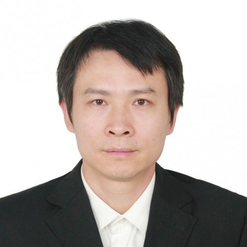 Jin Mei (梅劲)合作论文数Ningbo University School of Medicine2Yabin Pan (潘亚斌)合作论文数2Xueyuan Li (李学渊)合作论文数2Juehua Jing (荆珏华)合作论文数2JIANG Lüetao (蒋略韬)合作论文数1Hongwei Chen (陈红卫)合作论文数1Songou Zhang合作论文数1Mu Hu合作论文数1Ning Jiang合作论文数1




