目的 评估早期和晚期减压固定对胸腰段骨折合并脊髓损伤预后的改善情况.方法 对 2010年 1 月至 2018 年 12 月,T11~L2 发生创伤性脊髓损伤的 320 例患者进行回顾性分析,男 227 例,女 93 例,平均年龄(32.55±11.43)岁.对伤后 24 h 内接受早期手术(组 1,n = 145)、24~72 h 内接受手术(组 2,n = 46)和伤后 72 h 以上接受手术(组 3,n = 34)的患者进行分组分析.比较术后 1 年时 Frankel 分级的变化.结果 组 1 有 81 例、组 2 有 46 例、组 3 有 34 例患者的 Frankel 分级改善≥1 级(P<0.001).组 1 有 33 例、组 2 有 13 例、组 3 有 11 例患者的 Frankel 分级改善≥2 级(P = 0.070).Logistic 回归分析显示,早期手术和初始脊髓损伤程度与神经功能预后显著相关(P<0.001).结论 手术减压越早预后越好,72 h 内为有效手术减压的窗口期,在此期间,手术减压可能对不完全脊髓损伤具有保护作用.
[目的]探讨阿仑膦酸钠(alendronate,AL)和特立帕肽(teriparatide,TPTD)对双足鼠脊柱侧弯进展的作用.[方法]将45只C57BL/6J雌性4周龄小鼠行双前肢和尾离断,建立双足鼠脊柱侧弯模型,随机分为3组,每组15只.对照组、AL组和TPTD组,分别给予生理盐水、AL和TPTD,连续12周.行影像和骨密度(bone mineral density,BMD)检测.[结果]随时间推移,三组动物Cobb角均显著增加(P<0.05).术后8周和12周,脊柱侧弯Cobb角由大至小依次为:对照组>AL组>TPTD组,总体差异有统计学意义(P<0.05);两两比较,对照组的Cobb角显著大于AL组和TPTD组(P<0.05),而AL组与TPTD组间差异无统计学意义(P>0.05).随时间推移,三组动物BMD均显著增加(P<0.05).术后4、8和12周BMD值由低至高均为:对照组<AL组<TPTD组,总体差异有统计学意义(P<0.05).两两比较方面,对照组的BMD与AL组和TPTD组的差异均有统计学意义(P<0.05),而AL组与TPTD组间的BMD差异均无统计学(P>0.05).[结论]在双足鼠脊柱侧弯模型早期给予AL和TPTD可有效减小鼠脊柱侧弯Cobb角,同时增加生长期小鼠脊柱BMD值.
Objective: To evaluate the efficiency of chitosan/collagen scaffold combined with umbilical cord mesenchymal stem cells (UCMSCs) transfected with insulin- like growth factor- 1 (IGF-1) genes in the treatment of osteochondral defects. Methods: 30 New Zealand rabbits were used to create osteochondral defect animal models. They were randomly distributed to two groups. In the experimental group (n = 15), chitosan/collagen scaffold combined with UCSCs transfected with IGF-1 was grafted into the defect by mosaicplasty technique. In the control group, the defect was filled by chitosan/collagen scaffold containing UCSCs. Three months after the surgery, histological scores, biomechanical measures and the level of proteoglycan expression were carried out to evaluate the efficiency of two treatment methods. Results: Animals in the experimental group showed significantly better histological recovery than control group measured with overall evaluation, cartilage and subchondral evaluation scores (P < 0.01). Level of proteoglycan was significantly higher in the experimental group than the control group (P < 0.01). In the biomechanical analysis, maximum bending load was significantly higher in the experimental group than control group (P < 0.01). Conclusion: Chitosan/collagen scaffold combined with umbilical cord mesenchymal stem cells transfected with IGF-1 is an efficient measure in the treatment of osteochondral defect.
Purpose: The goal of this study was to evaluate the efficiency and safety of debridement, bone grafting, and internal fixation via different approaches in patients with lumbosacral tuberculosis. Method: This is a retrospective study of patients with lumbosacral tuberculosis and treated by posterior, anterior or combined approaches in our center from January 2001 to January 2016. Intraoperative time and blood loss, alleviation of pain and restoration of functional abilities, and change in lordosis angle were recorded and analyzed. Results: A total of 72 patients were included in the final analysis. The results indicate that posterior approach is superior to anterior and combined approaches concerning intraoperative blood loss and time of surgery (P<0.01). The lumbosacral lordosis angle was significantly different only at the last follow up (P=0.04). Although pain and functional scores were significantly improved with the surgery, no significant differences were found among the three groups (P>0.05). Conclusion: Although anterior, posterior, or combined approaches are all safe and reliable in the treatment of lumbosacral tuberculosis, the posterior approach could be a safer and less invasive method compared to the other approaches.
BACKGROUND: Kyphosis in ankylosing spondylitis is a kind of spinal sagittal imbalance; due to center of gravity displaced and complicated biomechanical properties of the spine, the spinal biomechanics after kyphosis correction is little reported. OBJECTIVE: To establish a three-dimensional finite element model of the spine after osteotomy for kyphosis. METHODS: A three-dimensional finite element model of kyphosis in ankylosing spondylitis was established, simulating three kinds of osteotomy orthopedic programs (osteotomy angle in 20°, 30° and 40°), and the orthopedic effect and biomechanics were analyzed.RESULTS AND CONCLUSION: (1) The three-dimensional finite element model of finite element model of kyphosis in ankylosing spondylitis was established successfully, and simulated three kinds of osteotomy orthopedic programs at the angles of 20°, 30°, and 40°. (2) The best osteotomy angle was 30°, the stress distribution was less, and the stress on the T12, L1, L2, L4, L5, S1and rod was 7.346 1, 11.952, 72.783, 81.368, 28.144, 41.114, and 109.69 MPa, respectively. (3) Under 30°osteotomy angle, the postoperative Cobb angle is 1.4°, which not only obtains better orthopedic effect, but also reduces the incidence of complications caused by stress concentration.
目的 探讨以PDCA质量循环管理模式联合PBL教学方法在骨外科住院医师规范化培训中的教学效果.方法 选择新疆医科大学第六附属医院2016-2017级骨外科住院医师规范化培训医师,随机分为PDCA循环联合PBL教学联合组和传统教学对照组,各40人,比较教学结束时两组规培生的理论知识、临床操作与思维有无差异.结果 理论方知识笔试方面,联合组(87.56±3.12)分,对照组(82.27±4.25)分,成绩分别有统计学差异(P<0.05),在临床操作及思维分析方面,联合组表现优良率为67.2%,而对照组仅为52.5%,且差异有统计学意义(P<0.05).结论 相对于传统教学方法,PDCA循环结合PBL教学法对提高骨外科住培生的理论知识掌握、临床操作及诊疗技能有较好的效果,有一定的推广和应用价值.
}~4j ;# # j +# F ¡¢£¤¡¥ ¦§; ̈©¢a «¬ ® ̄°±2 3 ́3v#μ¶· ̧F 16 CTo ̧¡¥ !"¦§; ̈©¢a »E L NO\ ¢1⁄4 cg m, «¬ " F 1⁄2¡¥3⁄4HI "¦§; ̈©¢a «¿¢1⁄4Y ÀÁ¡¥ L Âcg ÂÃÄÅ{  ¢aÆÇÈÉ; ̈©¬£¤ Ansys workbench 15.0 ¢aÊˬÌN%Í4*Ω 948 874 1 564 477JN% g '( ¢aÊˬÌN% Í4*Ω 931 969 1 548 812JN%ÆÏÐÑÑÒ8ÓÔ ÂÕÖvc 4× 6g  ØÙÚB¬Û® 40.946 67.26 493.64 304.05 75.359 146.31 MPa n ÜÚB® 391.01 MPaÆÝVÞßà 2 ÄÅ{ Y +Âá¡}~4â ã ×6g ¿äåæ®àç èR{NO`R{é êÕÖvë«êì#í î×6g F
BACKGROUND: The full three-dimensional finite element model can not only establish a realistic three-dimensionalmodel, but also can simulate the osteotomy on the model. Analysis of biomechanics guides the clinical operation.OBJECTIVE: To establish a three-dimensional finite element model of ankylosing spondylitis kyphosis and provide an effective digital platform for further studies.METHODS: A 30-year-old male patient with ankylosing spondylitis kyphosis participated voluntarily in the current study. CTimages obtained from CT transverse scanning from C1 to the sacrococcyx were imported into Mimics 17.0 software toestablish a three-dimensional geometric model of the posterior spine. The geometric model was then imported into Studio Geomagic 2013 software. For the subsequent optimization of image processing, the posterior spine convex geometry wasestablished on the three-dimensional geometric model. We used Unigraphics NX 8.5 to establish the spinal kyphosis surfacemodel, then added modeling of calcification of the ligaments, partial resection of useless sacral bone, and finally, imported themodel into ANSYS 15.0 finite element analysis software, then added the ligaments and set the parameters of the material,generating a complete three-dimensional finite element model of ankylosing spondylitis.RESULTS AND CONCLUSION: A three-dimensional finite element model of complete ankylosing spondylitis wassuccessfully established. Using the ten-node approach, we generated 398370 tetrahedral elements, and 668538 nodes.This will provide a reliable digital platform for the next step of biomechanical analysis.
BACKGROUND: Patients with severe ankylosing kyphosis are often treated with orthopedic osteotomy, but with high surgical risk, treatment schemes are individualized, and the biological morphology and mechanical properties of the deformed spine are complex. Meanwhile, the finite element model is more adaptable for biomechanical analysis of the spinal deformity. However, its application in the treatment of ankylosing kyphosis in China was little reported.OBJECTIVE: To establish an ankylosing kyphosis finite element model so as to investigate the relationship between different osteotomy methods and the deformation and stress distribution of the spine.METHODS: A three-dimensional finite element model of ankylosing kyphosis was established, and then imported into ANASYS software, to simulate vertebral column decancellation and vertebral column resection at T12 and L1 levels,respectively. The vertebrae and screw were loaded with the same load, and deformation and stress nephograms were obtained.RESULTS AND CONCLUSION: (1) The deformation of the osteotomy at T12 level was significantly larger than that at L1 level. (2) The stress distribution of the decancellation osteotomy concentrated on the pedicle screw and titanium bar; the stress concentration of vertebral column resection was the intervertebral cage. (3) Individualized surgical treatment of ankylosing kyphosis may influence the selection of the osteotomy segment and osteotomy operation. The osteotomy segment may affect the stability of the spine, and stress distribution of internal fixation is likely related to the method of internal fixation. (4) This conclusion may provide theoretical reference for the biomechanical study of analysing ankylosing kyphosis.
Objective To investigate the effect and prognosis of anterior posterior surgery combined with internal fixation on treatment of patients with severe idiopathic thoracolumbar scoliosis.Methods A total of 60 patients with severe idiopathic thoracolumbar scoliosis were randomly divided into two groups.The anterior and posterior group was treated with anterior and posterior internal fixation,and posterior group was treated with posterior combined with internal fixation.Effect,preoperative and postoperative indicators and condition during follow-up were observed.Results There were significant differences in operation time,blood loss,length of stay between two groups (P<0.05).There was no significant difference of fusion segments between two groups (P>0.05).There were significant differences in the main bending angle Cobb,kyphosis and trunk shift between two groups (P<0.05).There were no significant differences in the final follow-up Cobb angle loss,the degree of kyphosis loss,the correction rate of Cobb angle and the correction rate of kyphosis between two groups (P>0.05).Conclusion Anterior combined with posterior internal fixation is similar to posterior internal fixation in treatment of patients with severe idiopathic thoracolumbar scoliosis,but the operation time and hospital stay are shorter by posterior internal fixation,and the amount of bleeding is greater and the risk is higher.
Objective To establish a three-dimensional (3D) finite element (FE) model of ankylosing spondylitis (AS) kyphosis that is a digital platform for further studies. Methods A 30-year-old man with AS kyphosis underwent computed tomography transverse scanning from T1 to the sacrococcyx. The images were imported into Mimics® 17.0 software to establish a 3D model of the posterior spine, which was then imported into Studio Geomagic 2013 software. Posterior spine convex geometry was established on the 3D geometric model for subsequent optimization of image processing. Unigraphics NX 8.5 produced the spinal kyphosis surface model. Modeled calcification of ligaments and partial resection of useless sacral bone were added. The model was imported into ANSYS 15.0 FE analysis software. Ligaments were added. Parameters were set to generate a 3D FE model of AS. Results and Conclusion A 3D FE model of AS was successfully established, providing a reliable digital platform for subsequent biomechanical analysis.
In order to elucidate the value of positron emission tomography (PET)/computed tomography (CT) in the clinical diagnosis and treatment of osseous and soft tissue malignancies, two authors independently searched the PubMed, Medline, Elsevier, Embase and Cochrane Library databases for literature published between January 2003 and February 2016, using the key words 'PET/CT', 'positron emission tomography/computed tomography', 'osseous sarcoma', 'bone tumor', 'soft tissue sarcoma' and 'neoadjuvant', to identify prospective and retrospective studies on the applicability of PET/CT on the clinical diagnosis of bone and soft tissue lesions, and evaluation of their response to neoadjuvant therapies. Data were independently extracted by the two authors and any disagreements were resolved by a third author when necessary. Extracted data were analyzed by Meta-Disc 1.6 software. As a result, 16 trials with a total of 883 patients and 2,214 lesions were included in the present study. The overall diagnostic accuracy of PET/CT exhibited a sensitivity and specificity of 0.90 (0.86-0.92) and 0.89 (0.85-0.92), respectively, and the effect of neoadjuvant therapy was assessed with a sensitivity and specificity of 0.79 (0.30-0.93) and 0.79 (0.69-0.89), respectively. Thus, it may be concluded from the present study that PET/CT is a reliable imaging method to be applied in the diagnosis and treatment of osseous and soft tissue malignancies.
To evaluate the efficacy and safety of Halo-pelvic ring traction in the treatment of severe kyphotic deformity secondary to spinal tuberculosis.Eighty patients with severe kyphotic deformity due to spinal tuberculosis were included in the study. Forty of those patients (experimental group) received Halo-pelvic ring traction before surgery and the rest (control group) received surgical treatment directly. Two groups were compared by means of the duration of surgery, intraoperative blood loss, correction of Cobb angle, change in patient height, and American Spinal Injury Association (ASIA) impairment scale.Halo-pelvic traction group achieved significantly (P<. 05) better results than direct surgical treatment group by means of the time of surgery (244+/-58minutes vs 276+/-47minutes, P=.036), intraoperative blood loss (950+/-236mL 1150+/-305mL, P=.018), correction of Cobb angle (68.3+/-12.6 vs 55.6+/-13.8, P=.001), change in patient height (9.4+/-4.0cm vs 6.8+/-3.8cm, P=.024). The mean improvement of ASIA scale was more in the experimental group than in the control group (0.23+/-0.07 vs 0.15+/-0.06); however, the difference is not statistically significant (P=.09).Halo-pelvic ring traction before osteotomy can be applied in patients with severe spinal kyphotic deformity due to spinal tuberculosis to increase efficacy and safety of surgical treatment.
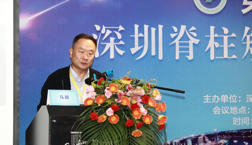 Yuan Ma (马原)合作论文数新疆医科大学第六附属医院脊柱外科9
 JiangTao Sui (眭江涛)合作论文数6Aikeremujiang Muheremu合作论文数4Yong Ma合作论文数4Xu Zhu (朱旭)合作论文数4Li Li (李栎)合作论文数4Kaisaier Aihemaiti合作论文数2Hui Li (李辉)合作论文数2HuiZhong Tian (田慧中)合作论文数2Jiang Xie (谢江)合作论文数2Jie Dai (代杰)合作论文数2Maimaitiaili Niyazi合作论文数1Junjie Cheng合作论文数1Shuo Shan合作论文数1Aierxiding Abulaiti合作论文数1Hui LI (李辉)合作论文数1Yanji Duan合作论文数1Jun-jie CHENG (程俊杰)合作论文数1Yong Ou (欧勇)合作论文数1Aierken Amudong (艾尔肯.阿木冬)合作论文数新疆医科大学第一附属医院1Zhenwei Liu (刘振伟)合作论文数1




