Antibody-based therapeutics encompass diverse modalities for targeting tumor cells. Among these, antibody-drug conjugates (ADCs) and extracellular targeted protein degradation (eTPD) specifically depend on efficient lysosomal trafficking for activity. A major limitation of ADCs is their reliance on antigens with efficient internalization, while eTPD approaches, although capable of trafficking diverse targets to lysosomes, lack cytotoxic potency. To address this, we developed degrader-drug conjugates (DDCs), leveraging the endocytic and recycling activities of eTPD to enhance lysosomal delivery. We utilized fast internalizers, the low-density lipoprotein receptor (LDLR) and the chemokine receptor (CXCR7), to enhance lysosomal delivery. LDLR-based degraders enabled efficient and selective degradation of diverse extracellular membrane proteins, while DDCs with cytotoxic payload enhanced cytotoxicity compared to conventional ADCs in vitro. This dual modality addresses key challenges of inadequate internalization in conventional ADCs and cytotoxic potency in current eTPD strategies. Our findings demonstrate that DDCs provide additional optionality for developing next-generation antibody therapeutics with broader utility and improved efficacy in cancer treatment.
Antibody-based therapeutics encompass diverse modalities for targeting tumor cells. Among these, antibody-drug conjugates (ADCs) and extracellular targeted protein degradation (eTPD) specifically depend on efficient lysosomal trafficking for activity. However, many tumor antigens exhibit poor internalization, limiting ADC effectiveness. To address this, we developed low-density lipoprotein receptor-targeting chimeras (LIPTACs), leveraging the constitutive endocytic and recycling activity of the LDLR to enhance lysosomal delivery. LIPTACs enable efficient and selective degradation of diverse extracellular membrane proteins. Additionally, by coupling LIPTACs with cytotoxic payloads to generate degrader-drug conjugates, we can achieve superior intracellular delivery and enhanced cytotoxicity compared to conventional ADCs. The dual modality addresses key challenges of inadequate internalization in conventional ADCs and cytotoxic potency for current eTPD strategies. Our findings demonstrate that LDLR-mediated trafficking can enhance eTPD and ADCs, providing a hybrid blueprint for developing next-generation antibody therapeutics with broader utility and improved efficacy in cancer treatment.
Lipid droplets are dynamic organelles that store neutral lipids, serve the metabolic needs of cells, and sequester lipids to prevent lipotoxicity and membrane damage. Here we review the current understanding of the mechanisms of lipid droplet biogenesis and turnover, the transfer of lipids and metabolites at membrane contact sites, and the role of lipid droplets in regulating fatty acid flux in lipotoxicity and cell death.
Cellular quality control systems sense and mediate homeostatic responses to prevent the buildup of aberrant macromolecules, which arise from errors during biosynthesis, damage by environmental insults, or imbalances in enzymatic and metabolic activity. Lipids are structurally diverse macromolecules that have many important cellular functions, ranging from structural roles in membranes to functions as signaling and energy-storage molecules. As with other macromolecules, lipids can be damaged (e.g., oxidized), and cells require quality control systems to ensure that nonfunctional and potentially toxic lipids do not accumulate. Ferroptosis is a form of cell death that results from the failure of lipid quality control and the consequent accumulation of oxidatively damaged phospholipids. In this review, we describe a framework for lipid quality control, using ferroptosis as an illustrative example to highlight concepts related to lipid damage, membrane remodeling, and suppression or detoxification of lipid damage via preemptive and damage-repair lipid quality control pathways.
Proteolysis of hydrophobic helices is required for complete breakdown of every transmembrane protein trafficked to the lysosome and sustains high rates of endocytosis. However, the lysosomal mechanisms for degrading hydrophobic domains remain unknown. Combining lysosomal proteomics with functional genomic data mining, we identify Lysosomal Leucine Aminopeptidase (LyLAP; formerly Phospholipase B Domain-Containing 1) as the hydrolase most tightly associated with elevated endocytic activity. Untargeted metabolomics and biochemical reconstitution demonstrate that LyLAP is not a phospholipase, but a processive monoaminopeptidase with strong preference for N-terminal leucine - an activity necessary and sufficient for breakdown of hydrophobic transmembrane domains. LyLAP is upregulated in pancreatic ductal adenocarcinoma (PDA), which relies on macropinocytosis for nutrient uptake, and its ablation led to buildup of undigested hydrophobic peptides, which compromised lysosomal membrane integrity and inhibited PDA cell growth. Thus, LyLAP enables lysosomal degradation of membrane proteins, and may represent a vulnerability in highly endocytic cancer cells. One sentence summary:LyLAP degrades transmembrane proteins to sustain high endocytosis and lysosomal membrane stability in pancreatic cancer.
The canonical biological function of selenium is in the production of selenocysteine residues of selenoproteins, and this forms the basis for its role as an essential antioxidant and cytoprotective micronutrient. Here we demonstrate that, via its metabolic intermediate hydrogen selenide, selenium reduces ubiquinone in the mitochondria through catalysis by sulfide quinone oxidoreductase. Through this mechanism, selenium rapidly protects against lipid peroxidation and ferroptosis in a timescale that precedes selenoprotein production, doing so even when selenoprotein production has been eliminated. Our findings identify a regulatory mechanism against ferroptosis that implicates sulfide quinone oxidoreductase and expands our understanding of selenium in biology.
Ferroptosis is a pervasive non-apoptotic form of cell death highly relevant in various degenerative diseases and malignancies. The hallmark of ferroptosis is uncontrolled and overwhelming peroxidation of polyunsaturated fatty acids contained in membrane phospholipids, which eventually leads to rupture of the plasma membrane. Ferroptosis is unique in that it is essentially a spontaneous, uncatalyzed chemical process based on perturbed iron and redox homeostasis contributing to the cell death process, but that it is nonetheless modulated by many metabolic nodes that impinge on the cells' susceptibility to ferroptosis. Among the various nodes affecting ferroptosis sensitivity, several have emerged as promising candidates for pharmacological intervention, rendering ferroptosis-related proteins attractive targets for the treatment of numerous currently incurable diseases. Herein, the current members of a Germany-wide research consortium focusing on ferroptosis research, as well as key external experts in ferroptosis who have made seminal contributions to this rapidly growing and exciting field of research, have gathered to provide a comprehensive, state-of-the-art review on ferroptosis. Specific topics include: basic mechanisms, in vivo relevance, specialized methodologies, chemical and pharmacological tools, and the potential contribution of ferroptosis to disease etiopathology and progression. We hope that this article will not only provide established scientists and newcomers to the field with an overview of the multiple facets of ferroptosis, but also encourage additional efforts to characterize further molecular pathways modulating ferroptosis, with the ultimate goal to develop novel pharmacotherapies to tackle the various diseases associated with - or caused by - ferroptosis.
Ferroptosis is a non-apoptotic cell death mechanism characterized by iron-dependent membrane lipid peroxidation. Here, we review what is known about the cellular mechanisms mediating the execution and regulation of ferroptosis. We first consider how the accumulation of membrane lipid peroxides leads to the execution of ferroptosis by altering ion transport across the plasma membrane. We then discuss how metabolites and enzymes that are distributed in different compartments and organelles throughout the cell can regulate sensitivity to ferroptosis by impinging upon iron, lipid and redox metabolism. Indeed, metabolic pathways that reside in the mitochondria, endoplasmic reticulum, lipid droplets, peroxisomes and other organelles all contribute to the regulation of ferroptosis sensitivity. We note how the regulation of ferroptosis sensitivity by these different organelles and pathways seems to vary between different cells and death-inducing conditions. We also highlight transcriptional master regulators that integrate the functions of different pathways and organelles to modulate ferroptosis sensitivity globally. Throughout this Review, we highlight open questions and areas in which progress is needed to better understand the cell biology of ferroptosis.
Ferroptosis is a non-apoptotic form of cell death characterized by iron-dependent lipid peroxidation. Ferroptosis can be induced by system xc- cystine/glutamate antiporter inhibition or by direct inhibition of the phospholipid hydroperoxidase glutathione peroxidase 4 (GPX4). The regulation of ferroptosis in response to system xc- inhibition versus direct GPX4 inhibition may be distinct. Here, we show that cell cycle arrest enhances sensitivity to ferroptosis triggered by GPX4 inhibition but not system xc- inhibition. Arrested cells have increased levels of oxidizable polyunsaturated fatty acid-containing phospholipids, which drives sensitivity to GPX4 inhibition. Epithelial membrane protein 2 (EMP2) expression is reduced upon cell cycle arrest and is sufficient to enhance ferroptosis in response to direct GPX4 inhibition. An orally bioavailable GPX4 inhibitor increased markers of ferroptotic lipid peroxidation in vivo in combination with a cell cycle arresting agent. Thus, responses to different ferroptosis-inducing stimuli can be regulated by cell cycle state.
Over 80% of people with cystic fibrosis (CF) carry the F508del mutation in the cystic fibrosis transmembrane conductance regulator (CFTR), a chloride ion channel at the apical plasma membrane (PM) of epithelial cells. F508del impairs CFTR folding causing it to be destroyed by endoplasmic reticulum associated degradation (ERAD). Small-molecule correctors, which act as pharmacological chaperones to divert CFTR-F508del from ERAD, are the primary strategy for treating CF, yet corrector development continues with only a rudimentary understanding of how ERAD targets CFTR-F508del. We conducted genome-wide CRISPR/Cas9 knockout screens to systematically identify the molecular machinery that underlies CFTR-F508del ERAD. Although the ER-resident ubiquitin ligase, RNF5 was the top E3 hit, knocking out RNF5 only modestly reduced CFTR-F508del degradation. Sublibrary screens in an RNF5 knockout background identified RNF185 as a redundant ligase and demonstrated that CFTR-F508del ERAD is robust. Gene-drug interaction experiments illustrated that correctors tezacaftor (VX-661) and elexacaftor (VX-445) stabilize sequential, RNF5-resistant folding states. We propose that binding of correctors to nascent CFTR-F508del alters its folding landscape by stabilizing folding states that are not substrates for RNF5-mediated ubiquitylation.
Ferroptosis is a regulated form of cell death associated with the iron-dependent accumulation of phospholipid hydroperoxides. Inducing ferroptosis is a promising approach to treat therapy-resistant cancer. Ferroptosis suppressor protein 1 (FSP1) promotes ferroptosis resistance in cancer by generating the antioxidant form of coenzyme Q10 (CoQ). Despite the important role of FSP1, few molecular tools exist that target the CoQ-FSP1 pathway. Through a series of chemical screens, we identify several structurally diverse FSP1 inhibitors. The most potent of these compounds, ferroptosis sensitizer 1 (FSEN1), is an uncompetitive inhibitor that acts selectively through on-target inhibition of FSP1 to sensitize cancer cells to ferroptosis. Furthermore, a synthetic lethality screen reveals that FSEN1 synergizes with endoperoxide-containing ferroptosis inducers, including dihydroartemisinin, to trigger ferroptosis. These results provide new tools that catalyze the exploration of FSP1 as a therapeutic target and highlight the value of combinatorial therapeutic regimes targeting FSP1 and additional ferroptosis defense pathways.
Despite the key roles of perilipin-2 (PLIN2) in governing lipid droplet (LD) metabolism, the mechanisms that regulate PLIN2 levels remain incompletely understood. Here, we leverage a set of genome-edited human PLIN2 reporter cell lines in a series of CRISPR-Cas9 loss-of-function screens, identifying genetic modifiers that influence PLIN2 expression and post-translational stability under different metabolic conditions and in different cell types. These regulators include canonical genes that control lipid metabolism as well as genes involved in ubiquitination, transcription, and mitochondrial function. We further demonstrate a role for the E3 ligase MARCH6 in regulating triacylglycerol biosynthesis, thereby influencing LD abundance and PLIN2 stability. Finally, our CRISPR screens and several published screens provide the foundation for CRISPRlipid (http://crisprlipid.org), an online data commons for lipid-related functional genomics data. Our study identifies mechanisms of PLIN2 and LD regulation and provides an extensive resource for the exploration of LD biology and lipid metabolism.
Phenotypic screens involving pooled CRISPR-Cas9 libraries offer a powerful, rapid yet affordable approach to evaluate gene functions on a global scale. Here, we present a protocol for performing pooled CRISPR-Cas9 loss-of-function screens to identify genetic modifiers using either fluorescence-based or cell death phenotypic readouts. We describe steps for designing and amplifying the library and generating and screening cells. We then detail deep sequencing and statistical analysis using cas9 High Throughput maximum Likelihood Estimator. For complete details on the use and execution of this protocol, please refer to Bersuker et al. (2019),1 Li et al. (2022),2 and Roberts et al. (2022).3.
Ferroptosis is a non-apoptotic form of cell death that can be triggered by inhibiting the system xc- cystine/glutamate antiporter or the phospholipid hydroperoxidase glutathione peroxidase 4 (GPX4). We have investigated how cell cycle arrest caused by stabilization of p53 or inhibition of cyclin-dependent kinase 4/6 (CDK4/6) impacts ferroptosis sensitivity. Here, we show that cell cycle arrest can enhance sensitivity to ferroptosis induced by covalent GPX4 inhibitors (GPX4i) but not system xc- inhibitors. Greater sensitivity to GPX4i is associated with increased levels of oxidizable polyunsaturated fatty acid-containing phospholipids (PUFA-PLs). Higher PUFA-PL abundance upon cell cycle arrest involves reduced expression of membrane-bound O-acyltransferase domain-containing 1 (MBOAT1) and epithelial membrane protein 2 (EMP2). A candidate orally bioavailable GPX4 inhibitor increases lipid peroxidation and shrinks tumor volumes when combined with a CDK4/6 inhibitor. Thus, cell cycle arrest may make certain cancer cells more susceptible to ferroptosis in vivo.
Ferroptosis is an important mediator of pathophysiological cell death and an emerging target for cancer ther-apy. Whether ferroptosis sensitivity is governed by a single regulatory mechanism is unclear. Here, based on the integration of 24 published chemical genetic screens combined with targeted follow-up experimentation, we find that the genetic regulation of ferroptosis sensitivity is highly variable and context-dependent. For example, the lipid metabolic gene acyl-coenzyme A (CoA) synthetase long chain family member 4 (ACSL4) appears far more essential for ferroptosis triggered by direct inhibition of the lipid hydroperoxidase gluta-thione peroxidase 4 (GPX4) than by cystine deprivation. Despite this, distinct pro-ferroptotic stimuli converge upon a common lethal effector mechanism: accumulation of lipid peroxides at the plasma membrane. These results indicate that distinct genetic mechanisms regulate ferroptosis sensitivity, with implications for the initiation and analysis of this process in vivo.
Lipid transfer proteins mediate the exchange of lipids between closely apposed membranes at organelle contact sites and play key roles in lipid metabolism, membrane homeostasis, and cellular signaling. A recently discovered novel family of lipid transfer proteins, which includes the VPS13 proteins (VPS13A-D), adopt a rod-like bridge conformation with an extended hydrophobic groove that enables the bulk transfer of membrane lipids for membrane growth. Loss of function mutations in VPS13A and VPS13C cause chorea acanthocytosis and Parkinson's disease, respectively. VPS13A and VPS13C localize to multiple organelle contact sites, including endoplasmic reticulum (ER) – lipid droplet (LD) contact sites, but the functional roles of these proteins in LD regulation remains mostly unexplored. Here we employ CRISPR-Cas9 genome editing to generate VPS13A and VPS13C knockout cell lines in U-2 OS cells via deletion of exon 2 and introduction of an early frameshift. Analysis of LD content in these cell lines revealed that loss of either VPS13A or VPS13C results in reduced LD abundance under oleate-stimulated conditions. These data implicate two lipid transfer proteins, VPS13A and VPS13C, in LD regulation.
Hydropersulfides are implicated in cellular responses to oxidative stress. In two landmark papers, Barayeu et al. and Wu et al. reveal that endogenous hydropersulfides are hydrogen atom donors that scavenge lipid radicals, thereby preventing lipid peroxidation and ferroptosis. Thus, the pathways governing hydropersulfides may represent a new cellular ferroptosis suppression system.
MYCN is amplified in 20% to 25% of neuroblastoma, and MYCN-amplified neuroblastoma contributes to a large percent of pediatric cancer-related deaths. Therapy improvements for this subtype of cancer are a high priority. Ferroptosis is an iron-dependent, oxidative form of cell death that is counteracted mainly by the production of Glutathione Peroxidase 4 (GPX4), a phospholipid hydroperoxidase that is produced through the glutathione pathway. The identification of cancers that may benefit from ferroptosis inducers are just emerging. Here we uncover a MYCN-dependent therapeutic vulnerability in neuroblastoma. Namely, amplified MYCN rewires the cell through expression of key receptors, ultimately enhancing iron influx through increased expression of the iron import transferrin receptor 1 (TFR1). Accumulating iron causes reactive oxygen species (ROS) production, and MYCN-amplified neuroblastomas show enhanced reliance on the system Xc- cystine/glutamate antiporter for ROS detoxification through increased transcription of this receptor. By performing metabolomics, we demonstrate that the transsulfuration pathway is also activated by MYCN. The increased activation of both pathways leads to cysteine accumulation that results in inhibition of lipid peroxidation. Utilizing drugs that target the main components of the glutathione and transsulfuration pathway we sensitize the MYCN neuroblastomas to ferroptotic cell death. These data provide novel insights into how MYCN alters the transcriptome in neuroblastoma to confer growth and survival advantages and simultaneously sheds light on the mechanism of action of ferroptosis inducers with potential application in other types of cancer. Citation Format: Konstantinos V. Floros, Mia O. Johnson-Berro, Richard Kurupi, Carter K. Fairchild, Krista Dalton, Bin Hu, Madhavi Puchalapalli, Mikhail G. Dozmorov, Jennifer E. Koblinski, James A. Olzmann, Lauren A. Cowart, Anthony C. Faber. MYCN-amplified neuroblastoma is addicted to iron and vulnerable to ferroptosis [abstract]. In: Proceedings of the American Association for Cancer Research Annual Meeting 2022; 2022 Apr 8-13. Philadelphia (PA): AACR; Cancer Res 2022;82(12_Suppl):Abstract nr 362.
 Mike Lange合作论文数Department of Molecular and Cell Biology, University of California13
 Ron Kopito合作论文数Biology Department, Stanford University12
 Melissa A. Roberts合作论文数10
 Dan Nomura合作论文数Department of Chemistry, College of Chemistry, University of California, Berkeley10
 Scott Dixon合作论文数Department of Biology, School of Humanities and Sciences, Stanford University10
 Lian Li合作论文数9
 Lih-Shen Chin合作论文数Department of Pharmacology & Chemical Biology, School of Medicine, Emory University9Milton To合作论文数8Kirandeep K Deol合作论文数6
 Bersuker Kirill合作论文数6JW Russell合作论文数5Andrea M. Vincent合作论文数Department of Neurology, University of Michigan5Alyssa J. Mathiowetz合作论文数4Nguyen, Truc B合作论文数4Leslie Magtanong合作论文数4
 Ethan Greenblatt合作论文数Department of Biochemistry & Molecular Biology, Faculty of Medicine, The University of British Columbia4
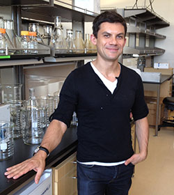 Roberto Zoncu合作论文数Biochemistry, Biophysics and Structural Biology Faculty, Department of Molecular and Cell Biology, University of California, Berkeley4
 Tarangelo Amy合作论文数4D Golovoy合作论文数3Thomas Shaler合作论文数3Siti Nur Sarah Morris合作论文数3
 Carl Ward合作论文数Univ Birmingham, Inst Canc & Genom Sci, Coll Med & Dent Sci, Birmingham, W Midlands, England3
 Thomas Maimone合作论文数Department of Chemistry, College of Chemistry, University of California, Berkeley3
 David Morgens合作论文数Dept Genet, Stanford Univ3
 Zhipeng Li合作论文数3
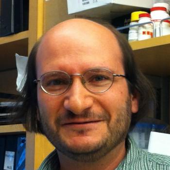 Michael Bassik合作论文数Department of Genetics, Stanford University3
 Eva Feldman合作论文数Department of Neurology, University of Michigan Medical School;Center for Cell Plasticity and Organ Design, University of Michigan;Center for Computational Medicine and Bioinformatics, University of Michigan3
 Allan Levey合作论文数Department of Neurology, School of Medicine, Emory University;Emory News Center, Emory University;Departments of Pharmacology, Emory University3Howard D. Rees合作论文数3L-S. Chin合作论文数2Emilee Easter合作论文数2Andrew He合作论文数2Dara E Leto合作论文数2Joseph M. Hendricks合作论文数2Jason Rodencal合作论文数2Ryan E. Tyler合作论文数2
 Yana Petri合作论文数Department of Chemistry, Massachusetts Institute of Technology2Margaret M. P. Pearce合作论文数2Veronica L Li合作论文数Department of Pathology, Stanford University School of Medicine, Stanford, CA 94305, USA2Sayed Hadi Hashemi合作论文数2
 Jennifer Yinuo Cao合作论文数Department of Biology, Stanford University2Jian-Ping He合作论文数2
 Uladzimir Barayeu合作论文数Univ Leipzig, Fac Chem & Mineral, Inst Bioanalyt Chem, D-04103 Leipzig, Germany2Tucker R Huffman合作论文数2Pridgeon Julia W合作论文数2Nalls Mike A合作论文数Laboratory of Neurogenetics, Intramural Research Program, National Institute on Aging2
 Zachary T. Schafer合作论文数2
 Martin Kampmann合作论文数Kampmann Lab, University of California, San Francisco2Ko Pin-Joe合作论文数2




