BACKGROUND:Osteofibrous dysplasia (OFD) is a rare non neoplastic, self-limited intracortical fibro-osseous lesion that most commonly affects the diaphysis of the tibia and fibula of children, the best treatment is still debated. Therefore we performed a retrospective study in children mostly under 10 years old with OFD aiming to determine whether early surgery is necessary and which is the best treatment.HYPOTHESIS:Symptomatic OFD lesions should be treated proactively, and appropriate treatment can achieve favorable outcome.METHODS:We retrospectively reviewed 23 patients with OFD of the tibia (n=22) and fibula. Management varied according to the severity of symptoms (deformity, pain or pathological fracture) and the extent of the lesion. Cases were divided into four groups (Gr.): Gr. 1: observation (n=4); Gr. 2: curettage and allograft (n=6); Gr. 3: curettage, allograft and elastic stable intramedullary nailing (ESIN) fixation (n=9); Gr. 4: extra-periosteal resection and bone transport (n=5). One patient received two different treatments successively (23 patients and 24 cases). All patients had regular clinical and radiographic follow-up to assess bone consolidation and complications.RESULTS:In Gr. 1, four asymptomatic patients underwent observation after open biopsy. Gr. 4 had the lowest local recurrence rate: 0 cases (0%) vs. 4 (66.7%; Gr. 2) vs. 1 (11.1%; Gr. 3) (p=0.002), incidence of deformity: 0 case (0%) vs. 0 (0%; Gr. 2) vs. 3 (50%; Gr. 3) (p=0.023), and pathological fracture: 0 case (0%) vs. 0 (0%; Gr. 2) vs. 1 (16.7%; Gr. 3) (p=0.006), although the consolidation time was longer: 6.8 vs. 2 (Gr. 2) vs. 2.7 months (Gr. 3) (p=0.017) and the rate of complication was higher than those of Gr. 2 and Gr. 3 (p<0.05). Gr. 3 had a lower local recurrence rate: 1 case (11.1%) vs. 4 cases (66.7%) (p=0.002), incidence of deformity: 0 case (0%) vs. 3 cases (50%) (p=0.023), and pathological fracture: 0 case vs. 1 case (16.7%) (p=0.006) than Gr. 2.DISCUSSION:Open biopsy is an important step in the management of patients with imaging suggestive of OFD in order to rule out Adamantinoma and other bone tumors or infection. Observation should be reserved for asymptomatic patients, while surgical treatment is indicated in patients with persistent pain, pathological fracture or significant deformity of the tibia. ESIN is a valid option to preserve the anatomical axis of the tibia in symptomatic patients with lesions of limited size; large circumferential lesions or recurrence can be managed by extra-periosteal resection and bone transport, although the complication rate can be relatively high.LEVEL OF EVIDENCE:III; case control study.
Purpose: To investigate the feasibility and short-term clinical efficacy of the arthrography-assisted joystick technique for the treatment of adolescent transitional ankle fracture. Methods: A retrospective analysis was performed in 10 adolescent transitional ankle fracture patients treated with the arthrography-assisted joystick technology. There were 7 male patients and 3 female patients with a mean age of 12.80 +/- 1.81 years (range: 10-16 years). All cases were closed fractures, and the operation was performed after the failure of manual reduction [X-ray or computed tomography (CT) showed that the fracture gap was > 2 mm]. After the operation, X-ray or CT examination was performed to assess reduction of the fracture. Ankle function and fracture healing were evaluated by assessing the imaging indexes and evaluating the American Orthopedic Foot and Ankle Society (AOFAS) ankle score system. Results: All patients were followed up for an average period of 12.00 +/- 3.40 months (range: 8-20 months). The operation time was 40.50 +/- 16.90 min (range: 25-80 min), the number of intraoperative fluoroscopy sessions was 18.70 +/- 5.91 (range: 10-30 sessions), and the intraoperative blood loss was 5.90 +/- 3.38 ml (range: 2-10 ml). X-ray examination showed that all cases achieved bone healing. The fracture healing time was 11.00 +/- 2.45 weeks (range: 8-16 weeks). Four cases showed early closure of the epiphysis. The AOFAS scoring system evaluation results of these 10 pediatric cases were excellent. Conclusion: The arthrography-assisted joystick technique for the treatment of adolescent transitional ankle fracture offers advantages, such as minimal trauma, simple operation, ideal reduction effect, and the recent curative effect is satisfactory. (c) 2022 Elsevier Ltd. All rights reserved.
背景:骨碎补的有效成分柚皮苷具有补肝肾强筋骨的传统功效,能增加骨痂厚度,提高骨折愈合质量.目的:探究载中药骨碎补有效成分柚皮苷壳聚糖/羟基磷灰石复合支架的骨传导和骨诱导性能.方法:将一定钙磷比的羟基磷灰石前体液与含柚皮苷的壳聚糖溶液在碱性条件下原位结晶、冷冻干燥,获得柚皮苷-壳聚糖/羟基磷灰石多孔支架.将15只成年SD大鼠随机分成空白组(n=5)、对照组(n=5)和实验组(n=5),建立直径5 mm颅骨骨缺损模型,空白组未填充生物材料,对照组填充壳聚糖/羟基磷灰石支架,实验组填充柚皮苷-壳聚糖/羟基磷灰石复合支架.术后4周取材,CT扫描观察颅骨修复情况,苏木精-伊红染色观察颅骨修复的形态学,骨形态发生蛋白2和血管内皮生长因子免疫组化染色后观察缺损区域局部成骨活性因子的表达.结果 与结论:①CT扫描显示,空白组大鼠颅骨未见明显骨生成,仅在缺损边缘可见少量新生骨;对照组于缺损孔隙可见新生骨形成,新生骨较少;实验组骨缺损修复良好,新生骨组织与缺损孔隙周围颅骨密度相似,大面积新生骨广泛填充了缺损孔隙.②苏木精-伊红染色显示,空白组缺损区填充以稀薄的疏松网状纤维组织,可见大量炎性反应病灶,仅在缺损边缘有少量新骨生成;对照组与实验组缺损区均可见支架材料残留,新生骨小梁及成骨细胞成簇分布于支架孔隙及缺损区边缘,周围伴有大量的毛细血管,其中以实验组表现出更强的新骨爬行替代能力.③免疫组化染色显示,实验组局部成骨活性因子骨形态发生蛋白2和血管内皮生长因子的表达高于对照组、空白组(P<0.05).④结果表明,柚皮苷-壳聚糖/羟基磷灰石复合支架可为骨缺损修复提供必要的载体,柚皮苷可创造局部成骨微环境、加速新生骨组织的生长,具有良好的骨修复性能.
目的 探讨锁定钢板内固定治疗青少年股骨干骨折内固定术后骨折不愈合的手术方法与临床疗效.方法 回顾性分析自2013-01-2019-12诊治的11例青少年股骨干骨折内固定术后骨折不愈合,9例附加锁定钢板内固定(髓内钉附加侧方钢板及双钢板技术),2例改用锁定钢板内固定.结果 11例均获得1年以上随访,随访时间平均18.5(12~23)个月.末次随访时所有患者均达到骨性愈合标准,骨折愈合时间平均7.1(3~12)个月.骨折愈合后所有患者均可完全负重并独立行走.随访期间未出现感染、螺钉松动、内固定物断裂等并发症.采用Uliana等的标准评定疗效:10例获得了良好及良好以上评分(评分为40~50分,平均48分),1例获得较好评分(20分).结论 附加或改用锁定钢板内固定治疗青少年股骨干骨折内固定术后骨折不愈合是安全有效的方法,针对骨折不愈合类型、原有内固定方式、原有内固定是否有效而个体化选择应用锁定钢板,有利于骨折断端桥接修复.
Objective:To evaluate the ultrasonography-guided percutaneous leverage reduction in the treatment of pediatric radial neck fractures.Methods:From May 2016 to May 2018, 42 patients with Judet Ⅲ or Ⅳ radial neck fracture were treated at Department of Pediatric Orthopedics, The Second Hospital of Fuzhou. They were 15 boys and 27 girls, aged from 3 to 12 years. All of them were treated by percutaneous leverage reduction and Métaizeau technique. The operative procedures were guided by ultrasonography in 20 cases and by C-arm fluoroscopy in 22 cases. Postoperative evaluation was based on Métaizeau imaging criteria, Tibone and Stoltz functional criteria; complications such as radial nerve injury were recorded.Results:There were no statistically significant differences between the ultrasonography-guided group and the fluoroscopy-guided group in preoperative general data, showing comparability ( P>0.05). There were no significant differences between the 2 groups in terms of Métaizeau imaging, Tibone or Stoltz functional scores ( P>0.05). The frequency of fluoroscopy was (2.7±0.6) times for the ultrasonography group and (14.6±3.4) times for the fluoroscopy group while the operation time averaged (25.8±5.9) min for the former and (38.1±9.2) min for the latter, showing significant differences ( P<0.05). No postoperative complications were observed in the ultrasonography group while 4 cases of radial nerve injury in the fluoroscopy group, also showing significant difference between the 2 groups ( P< 0.05). Conclusion:Ultrasonography-guided percutaneous leverage reduction is a suitable surgical treatment of radial neck fracture in children, because it is simple, effective and safe.
La dysplasie ostéofibreuse (OFD) est une lésion fibro-osseuse intracorticale bénigne qui touche les plus souvent la diaphyse du tibia et de la fibula de l’enfant, et dont le meilleur traitement n’a pas encore été déterminé. Nous avons réalisé une étude rétrospective chez les enfants de moins de 10 ans avec OFD du tibia ou de la fibula pour déterminer si la chirurgie précoce est indiquée, et quelle est la meilleure option chirurgicale. Les lésions symptomatiques doivent être traitées de façon proactive, et un traitement approprié assure des bons résultats. Nous avons analysé rétrospectivement 21 enfants avec OFD du tibia (n = 22) et de la fibula (n = 1). Le traitement a été adapté en fonction de la gravité des symptômes (déformation, douleur ou fracture pathologique) et de la taille de la lésion. Les patients ont été divisés en quatre groupes (Gr.): Gr. 1 : observation (n = 4) ; Gr. 2 : curettage et allograft (n = 6) ; Gr. 3 : curettage, allograft et enclouage centromédullaire élastique stable (ESIN) (n = 9) ; Gr. 4 : résection extrapériostée et transfert osseux (n = 5) ; un patient a été traité avec deux techniques (24 procédures au total). Tous les patients ont bénéficié d’un suivi radioclinique régulier afin d’évaluer la consolidation osseuse et le taux de complications. Les quatre patients asymptomatiques du Gr. 1, avec OFD confirmée à la biopsie, ont été observés. Le Gr. 4 avait le taux le plus bas de récidive locale : 0 cas (0 %) vs 4 (66,7 % ; Gr. 2) vs 1 (11,1 % ; Gr. 3) (p = 0,002), de déformation : 0 cas (0 %) vs 0 (0 % ; Gr. 2) vs 3 (50 % ; Gr. 3) (p = 0,023), et de fracture pathologique : 0 cas (0 %) vs 0 (0 % ; Gr. 2) vs 1 (16,7 % ; Gr. 3) (p = 0,006) ; néanmoins, le temps de consolidation : 6,8 vs 2 (Gr. 2) vs 2,7 mois (Gr. 3) (p = 0,017) et le taux de complications étaient plus élevés que dans le Gr. 2 et le Gr. 3 (p < 0,05). Par rapport au Gr. 2, le Gr. 3 avait un taux plus élevé de récidive locale : 1 cas (11,1 %) vs 4 cas (66,7 %) (p = 0,002), de déformation : 0 cas (0 %) vs 3 cas (50 %) (p = 0,023), et de fracture pathologique : 0 cas vs 1 cas (16,7 %) (p = 0,006). La biopsie osseuse est fondamentale dans la prise en charge des patients avec ODF et permet d’éliminer le diagnostic d’adamantinoma ou d’infection. L’observation est réservée aux patients asymptomatiques tandis que la chirurgie est indiquée chez les patients avec douleur, fracture pathologique ou déformation osseuse. Chez les patients symptomatiques avec une lésion de taille modérée, l’ESIN permet de préserver l’axe anatomique de l’os ; par contre, les lésions circonférentielles, ou les récidives, peuvent bénéficier d’une résection extrapériostée et d’un transfert osseux, même si le taux de complications est relativement élevé. III ; étude cas contrôle.
背景:国内外的研究表明,多种染色法联合应用有利于探究骨与软骨组织疾病的形态学表现.目前对于多种染色法探究骨骺损伤后骨桥形成过程的组织形态学表现研究较少.目的:通过苏木精-伊红染色、番红O-固绿染色、Masson染色3种染色方法观察骨骺损伤后骨桥形成过程的组织形态表现.方法:SD大鼠40只由福建中医药大学实验动物中心提供,实验方案经福建中医药大学动物实验伦理委员会批准.随机将40只SD大鼠分为2组:对照组不做处理;模型组建立胫骨近端骨骺损伤模型.分别于造模后1,7,28 d拍摄X射线片和7,28 d行MRI扫描;于造模后1,7,14,28 d麻醉大鼠后取胫骨损伤区组织,行苏木精-伊红染色、番红O-固绿染色、Masson染色,观察术后不同时间点生长板损伤区的组织形态表现.结果与结论:①X射线片显示:造模后1 d,胫骨干骺端骨骺损伤,软骨膜周围生长板间隙增宽;第7天,骨骺损伤处骨膜反应明显;第28天,骨膜反应消失,干骺端与生长板间隙关系基本恢复正常;MRI显示:造模后第28天MRI T2WI示骺板模糊,并可见一黑色线条形低信号影,提示骨桥形成;②造模后第1天,3种染色均可显示入侵的血管,Masson染色更为清晰;第7天:苏木精-伊红染色较番红固绿染色及Masson染色显示了损伤区软骨细胞的形态变化更加明显;第14天,番红O-固绿染色生长板与骨桥对比鲜明,Masson染色可观察到骨桥内尚未骨化的纤维血管组织;第28天,苏木精-伊红染色软骨细胞的周期性形态变化较明显,番红O-固绿染色骨桥和生长板形态分明,可清晰的表现损伤区生长板的压缩性改变;③结果说明,3种染色方法的联合应用能够全面、客观地探究骨骺损伤后骨桥形成过程的组织形态学表现.
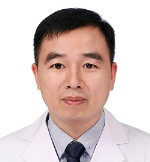 Shunyou Chen (陈顺有)合作论文数Fuzhou Second General Hospital7
 Yunan Lu合作论文数4
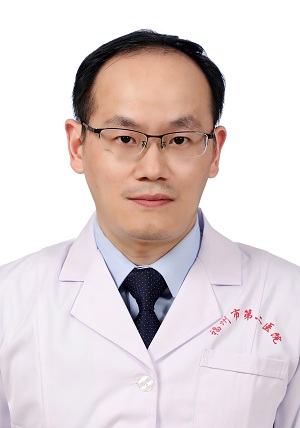 Ran Lin (林然)合作论文数Fuzhou Second General Hospital4YuanCheng Pan (潘源城)合作论文数3Federico Canavese合作论文数2Lin Binbin (林斌斌)合作论文数2
 Wentao Wang (王文涛)合作论文数School of Life Sciences, Sun Yat-sen University2Zhibin Ouyang合作论文数1yuling huang (黄玉玲)合作论文数1YueMing Guo合作论文数1Jinchen CHEN (陈晋宸)合作论文数1Gan Xu合作论文数1Song Chen合作论文数1
 Jingdi Chen (陈景帝)合作论文数Marine College, Shandong University1




