Purpose:The purpose of this study was to evaluate the accuracy of femoral tunnel location, post-operative pain management, functional rehabilitation and clinical outcomes in medial patellofemoral ligament (MPFL) reconstruction using all-arthroscopic technique. Methods:Between 2020 and 2021, 160 patients with recurrent patellar dislocation undergoing MPFL reconstruction were categorized into control (traditional surgery) and study (all-arthroscopic technique) groups. Femoral tunnel accuracy was assessed via computed tomography scans, pain management, functional rehabilitation, knee range of motion and daily activities were evaluated up to 6 months post-operatively. Knee function was assessed using Kujala and Lysholm scores at post-operative 12 months. Results:Seventy-one patients in the control group and 69 patients in the study group reached the final follow-up with no demographic differences. Follow-up duration was 12.65 ± 0.68 vs 12.77 ± 0.73 months in the control and study groups (p = 0.3145). The intra-class correlation coefficient was excellent (r = 0.97). In femoral tunnels, 93.5% in the control group and 92.4% in the study group were correctly localized. In patellar tunnels, 96.1% in the control group and 96.2% in the study group were correctly localized (p > 0.9999). Post-operative strong opioid analgesics were used 25.9 ± 31.0 versus 12.0 ± 22.2 mg/day in the control and study groups (p = 0.0016). The pain score was 3.4 ± 1.1 versus 2.7 ± 1.2 in the control and study groups (p = 0.0006) during post-operative functional rehabilitation. Time to resume daily living was 8.2 ± 0.6 versus 7.6 ± 0.6 weeks in the control and study groups (p < 0.0001). Time to resume low-intensity exercise was 12.3 ± 0.6 versus 11.7 ± 0.6 weeks in the control and study groups (p < 0.0001). In the more than 1-year follow-up, no significant difference was found in the Kujala and Lysholm scores. Conclusions:The all-arthroscopic technique for MPFL reconstruction in recurrent patellar dislocation ensures precise femoral tunnel placement. It offers advantages in early post-operative pain management and functional recovery, enabling faster rehabilitation compared to traditional non-all-arthroscopic techniques. Level of Evidence:Level III.
Abstract The effectiveness of existing tissue‐engineering cartilage (TEC) is known to be hampered by weak integration of biocompatibility, biodegradation, mechanical strength, and microenvironment supplies. The strategy of hydrogel‐based TEC holds considerable promise in circumventing these problems. Herein, a non‐toxic, biodegradable, and mechanically optimized double‐network (DN) hydrogel consisting of polyethylene glycol (PEG) and kartogenin (KGN)‐conjugated chitosan (CHI) is constructed using a simple soaking strategy. This PEG‐CHI‐KGN DN hydrogel possesses favorable architectures, suitable mechanics, remarkable cellular affinity, and sustained KGN release, which can facilitate the cartilage‐specific genes expression and extracellular matrix secretion of peripheral blood‐derived mesenchymal stem cells (PB‐MSCs). Notably, after tracing the transplanted cells by detecting the rabbit sex‐determining region Y‐linked gene sequence, the allogeneic PB‐MSCs are found to survive for even 3 months in the regenerated cartilage. Here, the long‐term release of KGN is able to efficiently and persistently activate multiple genes and signaling pathways to promote the chondrogenesis, chondrocyte differentiation, and survival of PB‐MSCs. Thus, the regenerated tissues exhibit well‐matched histomorphology and biomechanical performance such as native cartilage. Consequently, it is believed this innovative work can expand the choice for developing the next generation of orthopedic implants in the loadbearing region of a living body.
Cartilage injury of the knee joint is very common. Due to the limited self-healing ability of articular cartilage, osteoarthritis is very likely to occur if left untreated. Bone marrow mesenchymal stem cells (BMMSCs) are widely used in the study of cartilage injury due to their low immunity and good amplification ability, but they still have disadvantages, such as heterogeneous undifferentiated cells. MicroRNAs can regulate the chondrogenic differentiation ability of MSCs by inhibiting or promoting mRNA translation and degradation. In this research, we primarily investigated the effect of microRNA-210-3p (miR-210-3p) on chondrogenic and adipogenic differentiation of BMMSCs in vitro. Our results demonstrate that miR-210-3p promoted chondrogenic differentiation and inhibited adipogenic differentiation of rat BMMSCs, which was related to the HIF-3α signalling pathway. Additionally, miR-210-3p promotes mRNA and protein levels of the chondrogenic expression genes COLII and SOX9 and inhibits mRNA and protein levels of the adipogenic expression genes PPARγ and LPL. Thus, miR-210-3p combined with BMMSCs is a candidate for future clinical applications in cartilage regeneration and could represent a promising new therapeutic target for OA.
软骨损伤是一种常见的关节疾病,由于软骨缺乏血管和神经支配,其再生能力非常有限.目前,传统的临床治疗方法均不能达到满意的疗效,而组织工程技术为软骨损伤的修复提供了新的方向.水凝胶作为一种典型的生物支架材料,具有高度水化的三维交联结构、良好的生物相容性、生物降解性以及性能、结构可定制的特征,被广泛应用于软骨组织工程领域并显示出较好的软骨修复效果.本文就近些年来软骨损伤修复中常用的天然聚合物水凝胶、合成水凝胶及混合水凝胶的特点、软骨再生情况、各自优缺点以及水凝胶支架的先进制备方法、临床应用现状等进行综述,并对水凝胶在软骨再生领域的后续研究方向进行了探讨.
Purpose: To examine the indications and outcomes of medial patellofemoral ligament reconstruction (MPFLR) with or without tibial tubercle osteotomy (TTO) in treating recurrent or habitual patellar dislocation with an increased tibial tuberosity-trochlear groove (TT-TG) distance. Methods: We performed a literature search of the established medical databases Cochrane Central, PubMed-MEDLINE, EMBASE, and Web of Science. The inclusion criteria were as follows: skeletally mature patients with recurrent or habitual patellar dislocation and an increased TT-TG distance, treatment with MPFLR combined with a TTO procedure or isolated MPFLR, and reporting of clinical outcomes and complications. Each study was assessed for quality and the level of evidence. The general characteristics, indications, surgical techniques, TT-TG distance, clinical results, imaging evaluation findings, and complications of each study were recorded. Results: Nine studies consisting of 288 knees met the inclusion criteria. The average Coleman score was 71.56 (range, 5583). The threshold for an increased TT-TG distance ranged from 16 to 20 mm in the included studies. Similar good postoperative outcomes were reported in patients with an increased TT-TG distance treated with MPFLR with versus without a TTO procedure. The mean postoperative Lysholm score ranged from 75.0 to 94.7 (I2 = 87.6%) in the isolated MPFLR group and from 85.0 to 87.6 (I2 = 16.3%) in the TTO-with-MPFLR group. Similar postoperative congruence angles were reported in both groups. The postoperative redislocation rate ranged from 0% to 4.2% in the TTO-withMPFLR group, and no redislocation was found in the isolated MPFLR group. The postoperative apprehension sign was only reported in isolated MPFLR patients. Conclusions: The outcomes of MPFLR with or without TTO to treat recurrent or habitual patellar dislocation with an increased TT-TG distance appeared similar. However, this study was limited by the considerable heterogeneity, variety of techniques, variety of TT-TG distances, and variability in patella alta and trochlear dysplasia among the included studies. Level of Evidence: Level IV, systematic review of Level II to IV studies.
局灶性关节软骨缺损在运动医学领域较为常见,通常导致关节疼痛和功能障碍,由于软骨无血管、缺乏内源性修复细胞的特点,其自愈能力有限,若不及时治疗最终将发展为骨关节炎.手术一直是临床治疗关节软骨缺损的主要手段,按照外科修复技术主要分为姑息性手术、修复性手术、重建性手术和软骨替代性手术等四大类.本文主要就近年来有关以上不同外科手段治疗局灶性软骨缺损的适应证、优势、局限性及远期疗效等方面的进展情况进行综述.
Purpose: The purpose of the present study was to analyze the anatomic landmarks of Schottle's point and establish a locating method for identification. Methods: From 2013 to 2016, patients undergoing medial patellofemoral ligament (MPFL) reconstruction for patellofemoral instability were enrolled. Inclusion criteria: at least 2 episodes of patellar dislocation. Exclusion criteria: previous knee surgeries, open physes, severe trochlear dysplasia, tibial tuberosity lateralization, or patella alta. Group A: From January 2013 to December 2013, preoperative 3-dimensional computed tomography (3D-CT) images were obtained. Anatomic features of Schottle's point were measured on the 3D-CT images. A Schottle's point locating method with 2 distinct landmarks was established. Group B: From January 2014 to January 2016, consecutive MPFL reconstructions were performed. The placement of Schottle's point was following the established method without fluoroscopy. The accuracy of femoral tunnel positions was assessed on the 3D-CT images postoperatively. Results: CT images of 53 knees were obtained in group A. Forty-seven MPFL reconstructions were performed in group B. No significant difference was found between the 2 groups regarding to demographic characteristics. The intraclass correlation coefficients were excellent for all measures (r = 0.97). In group A, Schottle's point was 8.1 +/- 0.2 mm (95% confidence interval [CI], 7.7-8.5) distal to the apex of the adductor tubercle and 8.0 +/- 0.3 mm (95% CI, 7.4-8.6) anterior to the posterior edge. Apex of the adductor tubercle was defined as the most convex point, and posterior edge was defined as the edge of the posteromedial cortex in the transition area between the medial condyle and femoral shaft. In group B, 44 of 47 femoral tunnels (93.6%) were considered localized in the proper zone. Conclusions: Schottle's point was approximately 8 mm distal to the apex of the adductor tubercle and 8 mm from the posterior edge. Schottle's point locating method without fluoroscopy had high accuracy.
 Jiakuo Yu (余家阔)合作论文数Beijing Tsinghua Changgung Hospital;Peking University Third Hospital7
 You-Rong Chen合作论文数5Yi-Fan Song合作论文数5Fu-Zhen Yuan合作论文数4Bao-Shi Fan合作论文数3Jing Ye合作论文数3
 Haijun Wang (王海军)合作论文数Department of Sports Medicine, Peking University Third Hospital3Xinjie Wang合作论文数2Bingbing Xu合作论文数2
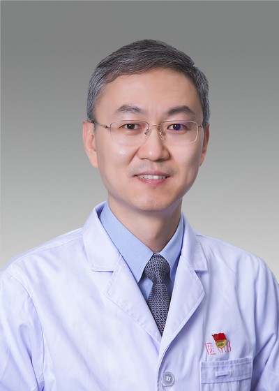 Fei Wang (王飞)合作论文数Heibei Medical University Third Hospital2Jiying Zhang (张吉英)合作论文数2Zi-Mu Mao合作论文数1Ze-yi CHEN (陈则亦)合作论文数1WANG Xing (王星)合作论文数1Yang Liu合作论文数1YANG Meng (杨梦)合作论文数1Shi-Tang Song合作论文数1Meng Yang合作论文数1Songbo Han合作论文数1Ming-Ze Du合作论文数1Jian Guan合作论文数1Zijie Xu (许梓杰)合作论文数1YongJian Wang (王永健)合作论文数1
 Decheng Wu (吴德成)合作论文数Department of Biomedical Engineering, Southern University of Science and Technology1
 Lin Lin (林霖)合作论文数Department of Sports Medicine, Peking University Third Hospital1
 Xing Wang (王兴)合作论文数College of Life Science and Technology, Beijing University of Chemical Technology1
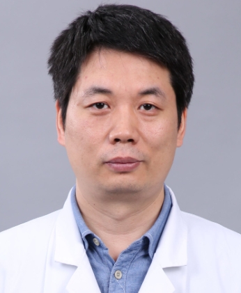 Jian Wang (王健)合作论文数Institute of Sports Medicine, Peking University Third Hospital1




