目的:以病理组织学检查为金标准,探讨超声声衰减成像(attenuation imaging,ATI)技术无创评估肝脂肪变性严重程度的诊断效能.方法:收集2020年9月—2022年6月于复旦大学附属中山医院就诊并进行肝脏活组织病理学检查或者肝脏良性肿瘤手术的肝脂肪变性患者97例,其中男性56例,女性41例.分析脂肪变性不同病理学等级患者ATI测值的稳定性及差异.采用多元线性回归分析ATI测值的影响因素.采用受试者工作特征(receiver operating characteristic,ROC)曲线评估ATI对肝脂肪变性的诊断效能.结果:分析发现脂肪变性不同病理学等级患者ATI测值组内相关系数(intra-class correlation coefficient,ICC)均大于0.90.不同组间ATI测值差异有统计学意义(P<0.05),但S2组与S3组ATI测值差异无统计学意义(P>0.05).Pearson相关分析显示ATI测值和病理肝脂肪变性程度呈显著正相关(r=0.75,P<0.01),多元回归分析发现肝脂肪变性病理学程度是ATI测值的独立影响因素(b=0.13,t=9.12,P<0.001).以病理组织学检查结果为金标准,ATI测值诊断肝脂肪变性程度≥S1、≥S2和≥S3的曲线下面积(area under curve,AUC)分别为0.966(95%CI 0.895~0.987)、0.931(95%CI 0.876~0.978)和0.708(95%CI 0.655~0.818).结论:肝脂肪变性程度是ATI测值的独立影响因素.ATI技术在不同程度的肝脂肪变性的评估中均表现出良好的诊断效能,特别是对较低水平的肝脂肪变性.
目的:探讨基于超声剪切波弹性成像(shear wave elastography,SWE)及剪切波频散成像(shear wave dispersion,SWD)技术检测健康成人肝脏黏弹性的最优测量方法及医学参考值范围.方法:选择2020年5月至10月复旦大学附属中山医院的健康体检者50例,行肝脏SWE及SWD检测,分析不同肝脏检测部位、不同取样面积及年龄、性别对测值的影响,进一步确定健康人群的肝脏黏弹性的参考值范围.结果:肝右叶S5/S6段切面检测的成功率高于肝左叶最大切面(100%v s 76%),结合肝右叶测值内部一致性和离散程度均优于肝左叶测值,肝右前叶为测量的最佳位置;不同取样面积(直径10 m m及20 mm)的肝脏SWE及SWD测值差异无统计学意义;不同年龄组及不同性别组间肝脏SWE及SWD测值差异无统计学意义.结论:应用剪切波黏弹性技术检测肝脏黏弹性成功率高,稳定性好,肝右前叶为最佳测量部位,不同取样面积、性别及年龄对测值无明显影响.推算肝脏SWE参考值范围为3.93~8.75 kPa;肝脏SWD参考值范围为8.80~14.99 m/(s·M Hz).
1 资 料 患者,男性,23岁.因"体检发现左肾占位3个月"入院治疗.实验室检查各项指标及各血清学肿瘤标志物水平均在正常范围.超声检查显示左肾中上部有1个3.1 cm×2.8 cm的稍高回声团块,向外突出,内见多枚强回声团块,边界欠清,形态欠规则,彩色多普勒血流成像(color Doppler flow imaging,CDFI)示周边点状彩色血流,脉冲多普勒(pulsed Doppler,PD)测及动静脉频谱,阻力指数(resistance index,RI)为0.56(图1).
Objective:To explore the diagnostic performance of ultrasound attenuation imaging (ATI) in grading the degree of hepatic steatosis in metabolic dysfunction-associated fatty liver disease (MAFLD).Methods:The liver gray-scale ultrasound and ATI examinations were performed on 212 subjects who were treated in Zhongshan Hospital Affiliated to Fudan University from August 2020 to March 2021. The attenuation coefficient(AC) values among different degrees of hepatic steatosis were analyzed and the diagnostic performance of ATI was evaluated. Relationships between AC values and clinical characteristics were assessed by Pearson′s correlation analysis.Results:The AC values for normal liver, mild, moderate and severe fatty liver were (0.56±0.05)dB·cm -1·MHz -1, (0.68±0.09)dB·cm -1·MHz -1, (0.82±0.09)dB·cm -1·MHz -1, (0.94±0.09)dB·cm -1·MHz -1, respectively. There were significant differences in AC values among different hepatic steatosis divisions( P<0.008). There was highly significant correlation between AC values and the degree of hepatic steatosis( r=0.860, P<0.01), moderate correlation between AC values and BMI( r=0.425, P<0.01), weak correlation between AC values and HDL-C( r=-0.237, P=0.029), no correlations between AC values and age, TC, TG, LDL-C ( r=0.083, 0.055, 0.133, -0.039, all P>0.05) .The areas under the receiver operating characteristics curve of ATI for mild fatty liver and above, moderate fatty liver and above, severe fatty liver and above were 0.958, 0.962, 0.918; the sensitivity were 90.1%, 95.8%, 94.9%, the specificity were 96.1%, 87.1%, 73.9%, and the cut-off values were 0.666 dB·cm -1·MHz -1, 0.719 dB·cm -1·MHz -1, 0.803 dB·cm -1·MHz -1, respectively. Conclusions:ATI is a reliable and convenient method for evaluating the degree of hepatic steatosis in MAFLD.
Objective To evaluate the value of contrast-enhanced ultrasound (CEUS) in preoperative classification for hilar cholangiocarcinoma. Methods Forty-six patients with 46 hilar cholangiocarcinoma were diagnosed by surgical pathology in Zhongshan Hospital of Fudan University from January 2007 to April 2013. The echogenicity difference on conventinal ultrasound and CEUS were compared with chi-square test. The accuracy of conventinal ultrasound and CEUS for evaluating invaded bile duct, detective rates for portal vein invasion and displaying rate of metastatic hilar lymph nodes were compared with chi-square test or Fisher’s Exact test according to the golden standard of operative exploration. Results On CEUS, 82.6%(38/46) and 91.3%(42/46) hilar cholangiocarcinoma were hypoechoic in portal vein phase and delayed phase respectively, while 63.0%(29/46) hilar cholangiocarcinoma were isoechoic on conventinal ultrasound with vague margin. The clearly displaying rates were 37.0%(17/46), 84.8%(39/46) and 91.3%(42/46) in conventinal ultrasound, portal vein and delayed phase of CEUS and the echogenicity was signiifcantly different. The evaluation accuracy of hilar cholangiocarcinoma invading bile duct was improved from 80.4%(37/46, conventinal ultrasound) to 100%(46/46, CEUS) significantly (χ2=7.882,P=0.005). Portal vein invasion were found in 9 cases during operative exploration and the detective rates on conventinal ultrasound and CEUS were 78%(7/9) and 89%(8/9) without signiifcant difference (P=1.000). Metastatic hilar lymph nodes were found in 8 cases and the displaying rates on conventinal ultrasound and CEUS were the same (75%, 6/8) without signiifcant difference (P=1.000). Conclusions CEUS could signiifcantly improve the clearly displaying rate of hilar cholangiocarcinoma and improve the evaluation accuracy for invaded bile duct comparing with conventinal ultrasound.
Objective:To explore the correlation of intima-media thickness(IMT) of carotid artery(CCA) and the enhanced intensity(EI) of carotid plaque contrast-enhanced ultrasound(CEUS) in patient with carotid atherosclerosis.Methods:CEUS were performed on 135 patients from 2009 to 2011.Bilateral atherosclerotic lesions were detected in 32 patients.Therefore,a total of 167 carotid arteries with plaques were documented and analyzed.Sonographic examinations were performed with ultrasound instruments equipped with low mechanical index contrast imaging software.IMT of CCA was measured with QLAB software.Contrast-enhancement within the plaque was categorized as grade 0 to grade 4 by visual interpretation and evaluated by quantitative analysis.EI of carotid plaque and the ratio of EI within the plaque to that in the lumen of the carotid artery were calculated.The correlation of EI and ratio with IMT of CCA was analyzed.Results:IMT was not measured on 6 carotid arteries.In 161carotid arteries,there was no significant difference in IMT among patients with different grades of intraplaque enhancement on CEUS(P = 0.994).IMT was not associated with EI(P = 0.687) and ratio(P = 0.852).Conclusions:EI of carotid plaque is not associated with the IMT of CCA.Appropriate screening and follow-up means should be selected according to the specific circumstances in clinic.
OBJECTIVE:To observe the inhibition of neovascularisation in the oxygen-induced retinopathy (OIR) mice by a stromal cell-derived factor 1 (SDF-1) antagonist.METHODS:Experimental study. Fifty-eight 7-day-old C57BL/6 mice were divided into 3 groups randomly, the control group (n = 17), the test group (n = 17) and the medication group (n = 24). According to the dosage of AMD3100, the medication group (n = 24) were divided into low dose group, high dose group, low dose control group, and high dose control group (each group n = 6). Each group (19-day-old) was sacrificed to perform ADPase staining, paraffin sections and immunohistochemical staining (anti-VEGF and anti-SDF-1). The average positive staining area percentage (APSAP) was measured as the outcomes and processed with the Students' t-test.RESULTS:Real-time PCR showed expression of both VEGF mRNA (0.080 ± 0.022 vs. 0.123 ± 0.032) and SDF-1 mRNA (0.731 ± 0.099 vs.0.544 ± 0.108) in retinas from the control group and test group, respectively. The expression of these factors in the test group was significantly higher (t = 2.488, P = 0.038;t = 2.864, P = 0.021). The number of neovascular endothelial nuclear that broke through the retinal internal limiting membrane in the paraffin section in the high dose group and the low dose group was significantly less than that in the self-control group (t = -9.507, P = 0.000; t = -10.761, P = 0.000). The appearance of ADPase staining sections in the medication group was more similar to the simple control group than that of the test group. Immunohistochemical staining sections showed that VEGF and SDF-1 expressed in neuroepithelial cells in each group. APSAP in the high dose group and the low dose group was significantly lower than that in the self-control group (VEGF: t = -7.249, P = 0.000; t = -9.02, P = 0.000; SDF-1: t = -5.246, P = 0.000; t = -5.216, P = 0.000).CONCLUSION:These results indicate that AMD3100 block the SDF-1 receptor to reduce the effect of SDF-1, decrease the production of VEGF protein and inhibite neovascularization.
Objective: To establish a retinal vein occlusion (RVO) animal model and observe the therapeutic effect of a Chinese herbal composition (Fufang Xueshuantong Capsule, FXC) in ischemic retinal disease. Methods: Fifteen adult male Sprague-Dawley rats underwent laser photothrombosis to induce RVO on their right eyes and were subsequently randomized to receive FXC (the intervention group, n=7) or placebo treatment (the control group, n=8). Fundus fluorescein angiography was performed after 2, 4 and 8 weeks of treatment. Real-time reverse transcription-PCR was used to quantify the mRNA expression of vascular endothelial growth factor (VEGF) and stromal cell-derived factor-1 (SDF-1). The main outcomes were the mRNA copies of VEGF and SDF-1 and the counts of RVO signs. Results: Laser photothrombosis procedure induced typical lesions of RVO, including hemorrhage, leakage, retinal detachment, capillary non-perfusion, filling defect of retinal vessels, and lateral circulation/dilation of small vessels. The retinal lesions were associated with an increased expression of VEGF (P<0.05). No significant change of SDF-1 expression was noticed. Compared with the control group, the intervention group had numerically fewer RVO lesions at week 2 (1.71 +/- 0.76 vs. 3.50 +/- 1.51, t=-2.82, P<0.05). The benefit of intervention remained at weeks 4 and 8. Conclusions: A rat model of laser photothrombosis-induced RVO was established and an increase in the VEGF expression was observed in the retinal lesion. The FXC had therapeutic benefit in improving retinal lesions in the rat model of RVO.
目的 分析超声造影显示颈动脉粥样硬化斑块增强强度与患者临床症状之间的关系.方法 颈动脉粥样硬化患者58例,分为有临床症状及无症状2组.常规超声评价颈动脉斑块回声类型,超声造影观察斑块内增强强度并进行分级.分析斑块回声、斑块内增强情况与患者临床症状的关系.结果 有症状组69.4%的斑块超声造影出现增强且2级以上增强者达63.3%,而在无症状组58.3%的斑块超声造影表现为不增强,2级以上增强者仅为29.2%,2组间有显著性差异.结论 实时超声造影可敏感显示斑块内的动态增强过程,反应斑块内的新生血管形成,与患者的临床症状具有相关性.
Objective To evaluate contrast-enhanced ultrasound imaging of carotid atherosclerosis with different stenostic degree as a clinical tool to study intraplaque neovascularization.Methods Fifty-eight patients suspected of carotid stenosis with 73 plaques were studied by standard and contrast-enhanced ultrasound.Plaque echogenicity at standard ultrasound were evaluated.Contrast enhancement within the plaques was categorized as 0 - 4 degree and compared to the stenosis degree and the plaque echogenicity.The degree of stenosis was determined by intravenous digital subtraction angiography.Results In the group with stenosis less than 90% ,stenosis degree was not associated with the grade of contrast enhancement (P =0.358),while the grade of enhancement was significantly higher in the group with sever stenosis(>90%).The grades of enhancement were significantly different between plaques with different echogenicity (P =0.000).Conclusions Contrast-enhanced real-time ultrasound imaging can demonstrate the enhancement of carotid plaques non-invasively,which is helpful for assessing intraplaque neovascularization and provide valuable information for plaque risk stratification
视网膜新生血管形成是眼部多种疾病共有的病理特点,其引发的疾病已成为目前致盲的重要原因.VEGF家族在新生血管形成中扮演着重要的角色,其通过影响细胞增生、细胞迁移、诱导毛细血管腔形成,从而促进新生血管的生成和生长.本文以早产儿视网膜病变为例,介绍VEGF家族及其受体,在早产儿视网膜病变的病理过程中的作用和针对其作用机制可采取的相应防治方法.
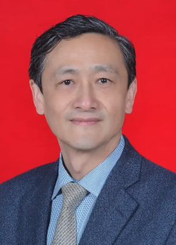 Wenping Wang (王文平)合作论文数Zhongshan Hospital, Fudan University8
 Yu-Li Zhu合作论文数3BAO Jing-wen (包静文)合作论文数3Beijian Huang (黄备建)合作论文数复旦大学3Wanyuan He合作论文数3Kun Wang (王坤)合作论文数3Chaolun Li (李超伦)合作论文数3Pei-Li Fan合作论文数3Zhiyuan Yuan (原致远)合作论文数2JiaYing Cao (曹佳颖)合作论文数2Qing Yu (俞清)合作论文数2Yi Dong (董怡)合作论文数Huashan Hospital, Fudan University2Qiannan Zhao (赵倩楠)合作论文数1Hong Han合作论文数1Ju-li ZHANG (张菊莉)合作论文数1Jia Yu合作论文数1HanTao Wang (汪瀚韬)合作论文数1Qin Si (司芩)合作论文数1LiYang Wang (王历阳)合作论文数1
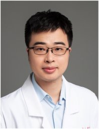 Mingfeng Xia合作论文数Fudan University, Fudan University1Lei Li合作论文数1Yuan Haixia合作论文数1
 Fei Yuan合作论文数Department of Electrical and Computer Engineering, Ryerson University1Feng Mao (毛枫)合作论文数1




